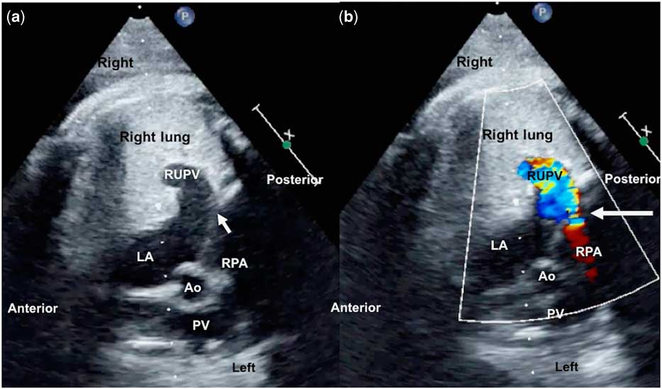
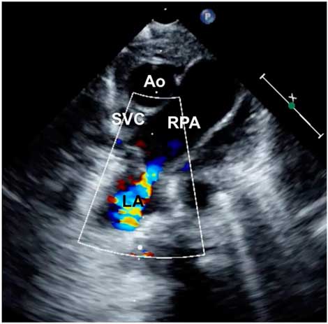
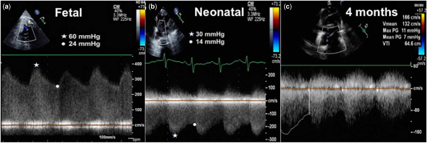
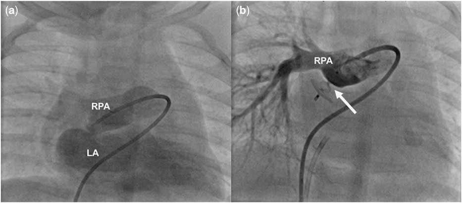
A second-trimester fetal echocardiogram showed an abnormal fistulous communication from the right pulmonary artery to the left atrium and dilated right pulmonary veins owing to a high-velocity jet through the fistula caused by high fetal pulmonary artery pressures and fetal pulmonary resistance (Fig 1, Supplementary movie 1). Echocardiogram after birth delineated the track from the undersurface of the right pulmonary artery to the left atrium with lower gradients owing to postnatal fall in pulmonary artery pressures (Fig 2, Supplementary movie 2). The pressure gradient further reduced at 4 months, but cyanosis persistedReference Chowdhury, Kothari, Airan, Subramaniam and Venugopal 1 (Fig 3). This fistula was closed at 4 months with a 6-mm Amplatzer Duct Occluder II device (StJude Medical, Plymouth, Minnesota, United States of America) (Fig 4, Supplementary movies 3 and 4). Follow-up echocardiogram and CT after 3 months confirmed occlusion of the fistula and remodelling of the dilated right pulmonary veins (Supplementary file).
Fetal echocardiography. B mode ( a ) and color Doppler imaging ( b ) of the fistula from the right pulmonary artery (RPA) to the left atrium (LA) with dilated right pulmonary veins (RUPV) shown on fetal echocardiography done at 28 weeks’ gestation. Arrow points to the communication between RPA to LA. Ao=aortic root; PV=pulmonary valve.

Postnatal imaging. Imaging from the suprasternal window demonstrates the fistulous communication between the undersurface of the right pulmonary artery (RPA) and the left atrium (LA). Ao=aortic root; SVC=superior vena cava.

Changing gradients. Serial Doppler tracings from fetal ( a ), neonatal ( b ), and at 4 months of age ( c ) showed reducing gradients owing to progressive fall in pulmonary vascular resistance.

Catheter intervention. After right pulmonary artery (RPA) angiogram ( a ) of the fistula, the communication was closed ( b ) with an occlude. LA=left atrium.

The embryonic dorso-ventral looping of the heart tube brings the right pulmonary artery derived from distal right sixth arch in proximity to the posterior wall of the primitive atria, in which the common pulmonary vein gets incorporated. Unroofing in this region of proximity explains this fistula. These fistulae cause cyanosis and are often identified on agitated saline contrast echocardiography. The pressure gradient across the fistula progressively falls as the fetal pulmonary vascular resistance falls postnatally. In fetal life with high pulmonary vascular resistance, the pressure difference between the right pulmonary artery and the left atrium leads to a high-velocity jet directed often towards the right pulmonary veins. This leads to dilated pulmonary veins, which may become aneurysmal in some instances. This is the first report of a fistula tracked from fetus to postnatal life with serial documentation of haemodynamic changes until its final interventional closure.Reference Yang, Zheng and Zhou 2
Acknowledgements
None.
Financial Support
This research received no specific grant from any funding agency, commercial or not-for-profit sectors.
Conflicts of Interest
None.
Ethical Standards
All procedures performed in studies involving human participants were in accordance with the ethical standards of the institutional and/or national research committee and with the 1964 Helsinki declaration and its later amendments or comparable ethical standards.
Supplementary material
To view supplementary material for this article, please visit https://doi.org/10.1017/S1047951118000616