Case presentation
A 2-month-old male infant weighing 3.5 kg was referred to our hospital for a cardiac evaluation and surgery. He was prenatally diagnosed with asplenia, single right ventricle, and pulmonary stenosis, and the diagnosis was confirmed after birth with echocardiography. Because of the severe atrioventricular valve regurgitation, an early Glenn procedure was desirable, and cardiac catheterisation was performed to evaluate the indications. Transthoracic echocardiography revealed right-sided abdominal inferior vena cava turning to the left just below the atrial connection.
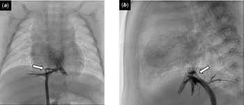
Cardiac catheterisation was performed for a pre-operative evaluation. Because it was difficult to advance the 5-Fr wedge catheter via the inferior vena cava into the right atrium, we used a Radifocus 0.021 guidewire (Terumo, Tokyo, Japan) to insert the catheter into the atrium without any unusual resistance. We assumed that this difficulty was because the angle of the curved inferior vena cava was steeper than previously expected. The catheterisation was continued, and the catheter was inserted into the atrium multiple times in the same manner. Inferior vena cava angiography at the end of the examination revealed local stenosis, flexion, and connection to the right hepatic vein (Fig 1). No extravasation was observed at the time of angiography, and the patient went back to the paediatric ward.
Anterior ( a ) and lateral ( b ) views of inferior vena cava angiography. The inferior vena cava was locally stenosed and flexed and was connected to the right hepatic vein (open arrow).

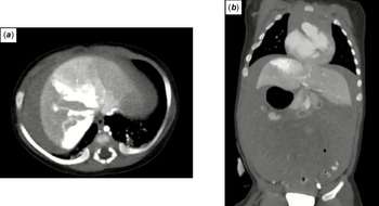
Six hours later, he showed abdominal distention, hypotension, and impaired consciousness. He was admitted to the ICU. Contrast-enhanced CT revealed hepatic injury with intrahepatic extravasation and massive ascites (Fig 2). The diagnosis was revised to polysplenia instead of asplenia based on the tracheal morphology and presence of spleens.
Axial ( a ) and coronal ( b ) views of contrast-enhanced CT. Hepatic injury with intrahepatic extravasation and massive ascites were observed.

Vascular injury due to catheter or guidewire manipulation in the stenosed and flexed part of the inferior vena cava was suspected as the cause of the bleeding complication. His condition was stabilised with transfusion, and we treated him conservatively. Contrast-enhanced CT 7 days later showed no intrahepatic extravasation, and the ascites had disappeared. He required 8 days of mechanical ventilation and 13 days in the ICU but was discharged home without any sequelae after 36 days.
Discussion
A wide variety of inferior vena cava anomalies have been reported. Reference Petik1 Patients with heterotaxy syndrome, especially polysplenia, are known to have a high percentage of inferior vena cava anomalies. Reference Uemura, Ho, Devine and Anderson2 Bleeding complications associated with cardiac catheterizations can be life-threatening. The following three examinations are essential in order to avoid the complications observed in the present case: echocardiography with precise knowledge of the vascular anomaly, contrast-enhanced CT or cardiac MRI before cardiac catheterisation for patients with indications, and inferior vena cava angiography when there is unusual resistance with catheter manipulation.
The inferior vena cava embryologically develops through anastomosis and regression of veins. Abnormalities at each stage of complex development result in inferior vena cava anomalies, so there are numerous anatomical variations. Reference Petik1 “Crossing IVC,” in which the inferior vena cava ascends across the vertebral body before the atrial inflow, is a typical anomaly Reference Freedom, Mawson, Yoo and Benson3 and was suspected based on echocardiography findings before cardiac catheterisation in the present case. Cases of atrial inflow with narrowing of the inferior vena cava, similar to the present case, have also been reported. Reference Cullu, Yeniçeri, Deveer and Tetiker4,Reference Agrawal, Das and Gupta5 Being aware of this type of inferior vena cava anomaly is important in preliminary echocardiography to avoid complications with cardiac catheterisation.
Contrast-enhanced CT or cardiac MRI is useful for diagnosing inferior vena cava anomalies and should be considered prior to cardiac catheterisation. CT requires radiation exposure and the use of contrast agents, while MRI often requires sedation, especially in children. Therefore, it is not practical to perform either test on all patients. In heterotaxy syndrome, there is a high incidence of abnormal systemic venous connections, Reference Petik1 and it is important to diagnose the relationship of the atria, ventricles, and vessels, and the position and morphology of the bronchi, lungs, and abdominal organs. Furthermore, if the diagnosis of Asplenia is made based on the morphology of the spleen, appropriate infection prophylaxis measures can be taken. For this reason, it is reasonable to routinely perform contrast-enhanced CT or cardiac MRI in patients with heterotaxy syndrome, especially polysplenia. In the present case, contrast-enhanced CT after the occurrence of haemorrhagic shock provided substantial valuable information, including information on the venous system, tracheal morphology, and the presence of a spleen, although it required radiation exposure (17.601 mSV). The procedure might have been different if we could have obtained this information before performing cardiac catheterisation.
During cardiac catheterisation, inferior vena cava angiography should be performed without hesitation to confirm the morphology when there is unexpected resistance with catheter or guidewire manipulation. In the present case, it was unusual that we could not advance the catheter to the atrium without the help of a guidewire. However, since we were not aware of the stenosis and abnormal connection of the inferior vena cava, we continued the examination. We recognised the inferior vena cava anomaly after the angiography at the end of the examination. If we had performed angiography on encountering unusual difficulty advancing the catheter into the atrium, we might have avoided the complication.
In conclusion, we report an infant with a single ventricle who had intra-abdominal haemorrhaging 6 hours after cardiac catheterisation. A precise diagnosis of the vascular anomaly before the cardiac catheterisation is crucial, especially in patients with heterotaxy syndrome. Contrast-enhanced CT or MRI should be performed before cardiac catheterisation for patients with heterotaxy syndrome, especially those with polysplenia.
Acknowledgements
None.
Financial support
This research received no specific grant from any funding agency, commercial, or not-for-profit sectors.
Conflicts of interest
None.