Introduction
Carcinoma oesophagus is the seventh most common cancer worldwide with more than 50,000 new cases in 2018. Reference Bray, Ferlay, Soerjomataram, Siegel, Torre and Jemal1 Also, it is the sixth most common cause of cancer-related deaths. Two major histological subtypes are oesophageal adenocarcinoma and oesophageal squamous cell carcinoma. Environmental and genetic factors play a major role in the causation of oesophageal cancer. Squamous cell histology is the predominant subtype in China, while adenocarcinoma is more common in North America and Europe. Reference Lepage, Rachet, Jooste, Faivre and Coleman2
Treatment of carcinoma oesophagus requires a multimodality approach which includes surgery, radiotherapy (RT) and chemotherapy. RT is an important treatment modality in carcinoma oesophagus in various settings. It is used as external beam radiotherapy (EBRT) or brachytherapy or both. Despite the inherent radio sensitivity of oesophageal tumours, curative doses are difficult to achieve because of close proximity of the target to vital organs.
By exploiting the inverse-square law resulting in rapid dose fall-off, brachytherapy becomes an effective means of delivering high dose to tumour while delivering much lower doses to surrounding organs at risk (OARs) such as the lung, heart and spinal cord. High-dose rate (HDR) technique allows treatment to be delivered in 5 to 10 minutes by an afterloading catheter introduced through the nose into the primary tumour site under fluoroscopic guidance.
It requires expertise to perform oesophageal brachytherapy. Fear of perforation of the oesophageal wall during dilation and the requirement of special infrastructure make it a less used modality for treatment in oesophageal cancers. Considering the good results obtained by brachytherapy in sites such as cervix, breast, rectum, head and neck, it has been tried in oesophageal carcinoma in different settings, such as curative, palliative and adjuvant. More sophisticated technology has allowed us to tailor the treatment as per requirement.
Different modes of brachytherapy used in carcinoma oesophagus are intraluminal, seed brachytherapy and intraoperative. Both seed and intraoperative brachytherapy are lesser used modes of brachytherapy and has not been reviewed. There has been recent advancement in applicators which addresses the shortcoming of previous applicators. This paper comprehensively reviews three modes of brachytherapy and recent developments of applicators used for carcinoma oesophagus over the past three decades.
Search strategy
We performed a comprehensive search in PubMed, Embase, Web of Science and Scopus database for English language sources from 1990 onwards, using the keyword as oesophageal cancer brachytherapy. There were 504 results in PubMed, 622 in Embase, 385 in Web of Science and 557 in Scopus. All the articles were imported in Endnote X9 reference manager, and the articles were selected for this review based on their content and impact on treatment strategy. The limitation of the study is most of the studies selected are older. There are limited studies in present era due to availability of advanced conformal planning techniques which has resulted in lesser use of brachytherapy for carcinoma oesophagus.
Intraluminal radiation therapy (ILRT)
When one or several radioisotope sources contained in appropriate applicator devices is/are placed in geometrical manner in lumen of hollow organs in and around cancerous growth, it is called intraluminal radiation therapy (ILRT). It is most routinely used form of brachytherapy for carcinoma oesophagus. It can be used in definitive, adjuvant or palliative settings. Definitive indications of ILRT are superficial tumour (T1) with curative intent where it can be used as the sole treatment modality. In adjuvant settings, ILRT can be used in combination with EBRT to boost the primary disease. If the tumour remission achieved by EBRT has not been sufficient or in patients with the local recurrence, ILRT can be given with the hope to increase the local effect. One of the recent indications is patients with interstitial lung disease, who are unable to receive a full dose of EBRT where the deficit of EBRT dose is compensated by brachytherapy. Good candidates for ILRT in adjuvant settings as per American Brachytherapy Society (ABS) guidelines (1997) Reference Gaspar, Nag, Herskovic, Mantravadi and Speiser3 include unifocal primary tumour ≤ 10 cm in length, tumour confined to oesophageal wall, mid-thoracic location and no evidence of intra-abdominal or metastatic disease. Poor candidates are extra oesophageal extension, tumour >10 cm in length, regional nodal involvement or tumour involving gastro-oesophageal junction or cardia. Relative contraindications are tracheal/bronchial involvement, cervical oesophagus growth or non-negotiable stenosis. ILRT is useful with a palliative intent in patients with advanced oesophageal carcinoma or recurrence after definitive RT with following indications: to improve dysphagia in cases of tumour recurrence after definitive EBRT, to prevent rapid tumour regrowth after clearing the stenosis keeping in mind the risk of perforation. In advanced stenosis, self-expandable metallic stent (SEMS) is used for providing quick relief and considered good alternative to ILRT. However, in comparison with SEMS, prolonged duration of relief of dysphagia is observed with ILRT, coupled with lesser complications and better quality of life. Reference Homs, Steyerberg and Eijkenboom4
Since the introduction of high linear activity sources such as iridium-192 (192Ir) in afterloading brachytherapy, source placement has become possible using small diameter applicators in advanced obstructive tumours. Using large diameter applicator results in lower contact doses to mucosa and a less steep dose gradient over oesophageal wall. These applicators are single catheters, with diameters that vary between 1·7 and 20 mm, allowing the treatment of cigar-shaped planning target volume (PTV).
The larger applicators are used in a wide oesophageal lumen, since it is difficult to dilate the lumen mechanically in large obstructive tumours; hence, small diameter bougie applicators are designed to mechanically dilate the oesophageal lumen. Mechanical dilation must be carried out by trained physician to minimise the chances of perforation and bleeding.
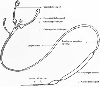
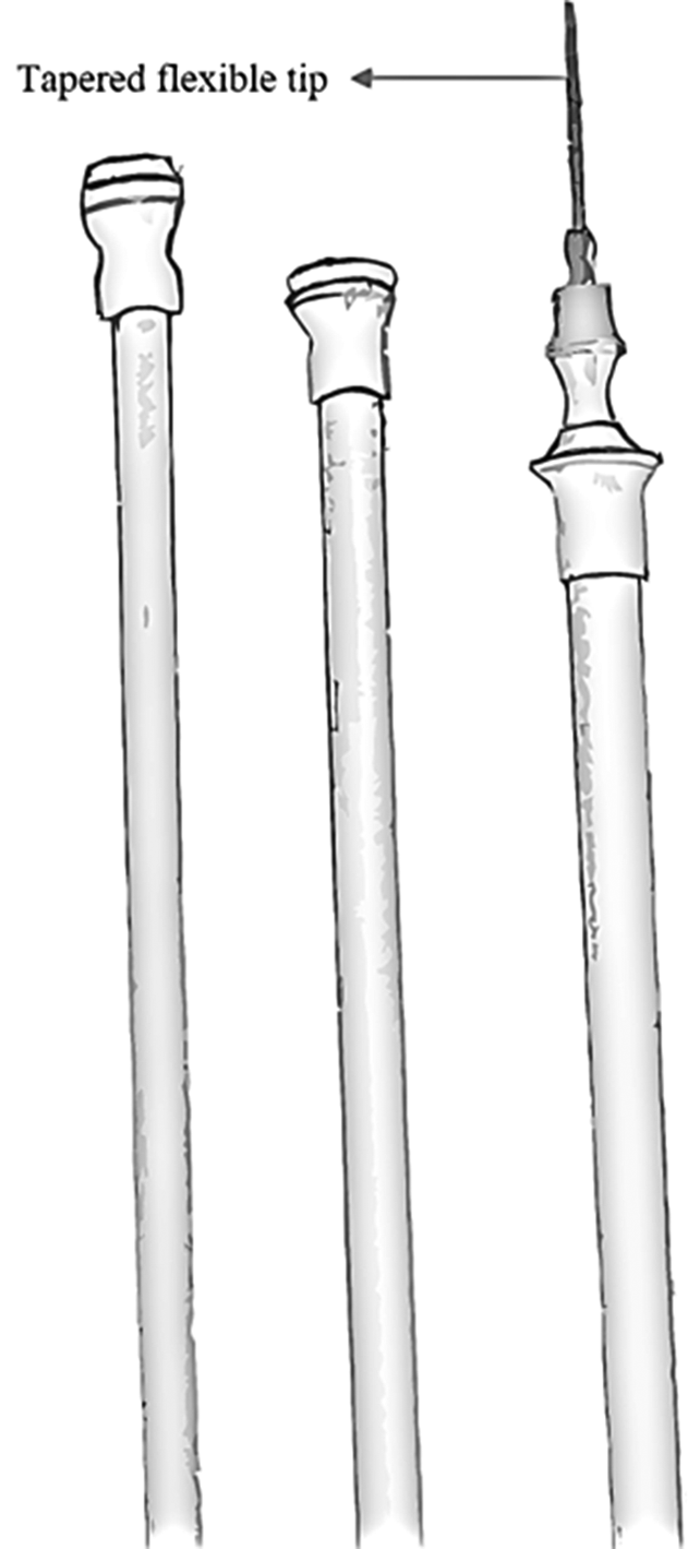
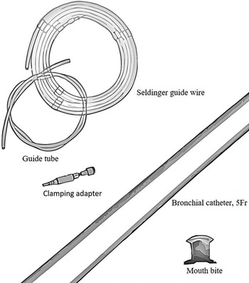
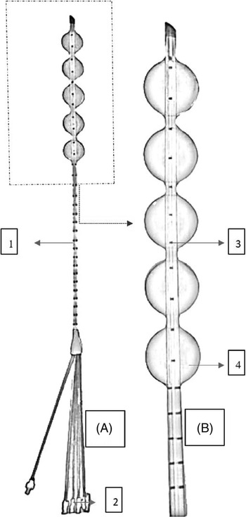
Some centres may not have commercially available applicators, hence in such scenario, applicators such as Sengstaken–Blakemore tube (Figure 1) may be modified to allow insertion of radiopaque catheters. Bonvoisin-Gérard esophageal applicator set (Figure 2) is a special dilation applicator designed to assist in the treatment of an obstructive oesophageal growth. It has a specially designed tapered flexible tip, which allows access of bouginage to treatment region. The bougie tube also assists in centring the catheter which ensures reduced dose to mucosal layer and simultaneously allowing optimal depth dose penetration of the oesophageal wall. Radiopaque markers on the bougie tube aids in fluoroscopic placement and localisation of the applicator. Applicator set connects directly to the after-loader, and hence no transfer tube is required additionally. Preliminary clinical data suggest decreased local side effects. Reference Raval and Zouain5 Oesophageal single-channel applicator set (Figure 3) is designed for the direct insertion into oesophagus. It is the most widely used applicator and can be placed under endoscopic or fluoroscopic guidance. An additional layer is provided to support applicator integrity and reduce dose to mucosal layer. Varian Medical Systems provides plastic-tipped applicator which are compatible with 1·5 and 3 Tesla Magnetic Resonance Imaging (MRI). Transoral balloon centering esophageal applicator (Figure 4) is designed to address mainly three limitations of conventional oesophageal applicators, which are non-centred catheter, displacement of catheter and mucosal injury of nasopharynx due to applicator insertion. The applicator is equipped with five independently inflatable balloons distally. These balloons filled with radiocontrast material allows the catheter to place centrally and maintain distance from healthy tissue. Inflatable balloon makes it a self-anchoring applicator, and the position of catheter can be confirmed with a plain radiograph and necessary adjustments can be made by deflating/re-inflating balloons until a satisfactory position is achieved. This accurate and central position of the distal part of applicator due to the inflatable balloons allows increase in therapeutic ratio and dose optimisation. The applicator is placed via transoral route, preventing mucosal injury to nose and nasopharynx. Reference Buzurovic, Hansen and Bhagwat6 A preliminary study of 33 patients found complete response in 58·6% of patients, and Grade II and III oesophageal toxicities 38 and 0·03%, respectively. Reference Taggar, Pitter and Cohen7 LumenCare® Azure multichannel applicator set consists of 10 5 Fr plastic tubes which are equidistantly mounted around an 8-mm plastic catheter. The inner bore diameter is 6·5 mm, through which a paediatric endoscope can be inserted to guide the optimal placement of the applicator. Multi-catheters allows more freedom of dose optimisation and dose escalation. Reference Berger, Schmid and Wisgrill8 A trial (NCT03335813) using six-channel balloon repositioning applicator is ongoing.
Diagrammatic depiction of Sengstaken–Blakemore tube.

Distal end of Bonvoisin-Gérard oesophageal applicator.

Diagrammatic representation of intraluminal applicator set.

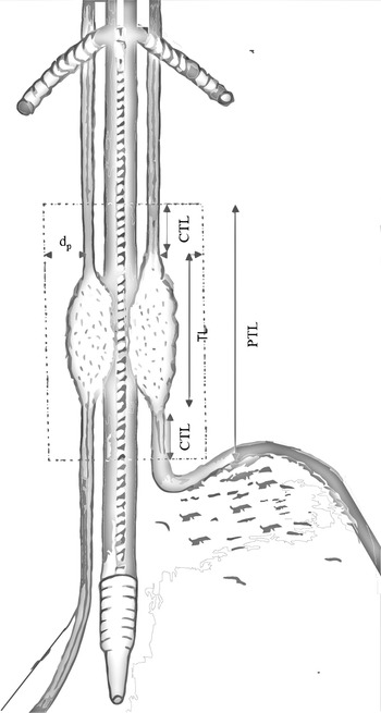
Diagrammatic representation of oesophageal growth with intraluminal applicator depicting various target volumes – target length (TL), clinical target length (CTL), planning target volume (PTV), prescribed depth from applicator surface (dp), reference volume length (RVL) – in red dashed lines.

ILRT – treatment planning
The application is usually performed on an outpatient basis, except in cases of low-dose rate (LDR) treatment lasting longer than 6 h. It can be done under fluoroscopic guidance alone or by an endoscope. The patient is asked to fast overnight and intravenous access is secured. Lignocaine is sprayed over oral cavity, oropharynx and hypopharynx to facilitate the entry of applicator and endoscope. Sedatives and vagolytic agents can be used in overanxious patients. For fluoroscopy-assisted applications, fresh barium swallow images should be available. The target volume is defined according to diagnostic findings from oesophagoscopy, endoscopic ultrasound (EUS), barium swallow, radiological imaging and findings from the procedure. Macroscopic tumour extent in longitudinal direction is incorporated by adding 2–3 cm margin at the proximal and distal end of the tumour to cover submucosal microscopic spread and applicator movement uncertainties. Laterally, it includes the oesophageal wall, and the thickness is dependent on tumour extension and the filling condition. The placement of applicator in target region should be eccentric, and diameter of applicator should be maximum to avail good dosimetry and lesser toxicities. The application should preferably be conducted in a dedicated endoscopy room. The patient lies on the left side as usual for oesophageal endoscopy. The surface of endoscope is covered with local anaesthesia gel and then introduced between two fingers placed into oropharynx to lead the tip of endoscope towards the proximal aperture of the oesophagus, and the endoscope is gently introduced into the oesophagus with the patient swallowing. The upper end of growth is identified and scope is gently negotiated further to identify lower end. The distance in centimetre from the teeth is noted and correlated with pre-therapeutic findings. A flexible guide wire, usually twice the length of the oesophagoscope, is advanced through the biopsy channel and placed beyond the distal tumour margin. The oesophagoscope is then withdrawn over the guide wire which remains in place, and the applicator is then introduced via guide wire. In fluoroscopy-assisted application, the applicator is placed between two fingers and gently placed in the oropharynx to guide the tip of the applicator towards hypopharynx. With the patient swallowing, the applicator is gently advanced avoiding any significant pressure against resistance. After the applicator has been placed correctly, the length of the applicator from incisors is noted and marked. If not able to negotiate the obstruction, endoscope-assisted application must be attempted rather than taking more attempts (may result in life-threatening perforations). A fluoroscopic simulation image is taken after the placement of applicator to verify the quality of placement of applicator. If satisfied, target volumes are identified taking into account the pre-therapeutic examinations. The length of PTV (PTL) is ascertained by taking into consideration the tumour length, margins for subclinical extension, clinical target length and positional uncertainties like displacement of catheter. The treated length includes PTL at the depth of prescription point (dp). The reference volume length includes PTL at dP (intersection with the 90% isodose) (Figure 5). The dp is usually taken at an arbitrary distance related to the applicator surface (e.g., 5–8 mm) in any applicator or the source axis (at 10 mm) in case of small diameter applicators.
Transoral balloon centring esophageal applicator (BCEA). (A) Full view of applicator and (B) magnified view of distal end of applicator, (1) catheter with markings, (2) inflatable port, (3) markings and (4) inflatable balloons.

There is no consensus as to how the dose should be recorded and reported. According to ICRU 58 recommendations, reference depth for reporting is specified in the central plane at 10 mm from the source axis for small applicators. However, this recommendation is related to the source and not to the individual application and the anatomy in patient. The ABS guidelines states that the dose should be always prescribed at 10 mm from mid-source position, while the Japanese recommends at 5 mm from the applicator surface. There is a need of standardised procedure for recording and reporting the dose of intraluminal brachytherapy independent of the prescription strategy chosen.
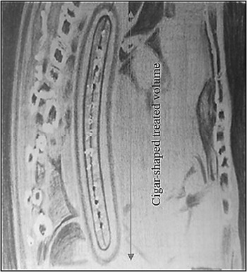
Fluoroscopic planning (2D planning) is the most widely used method, but the dP is taken by approximation and there is no dose–volume histogram (DVH) available to optimise doses to target volume and OARs. The dP can be delineated precisely using CT or MRI. With this conformal 3D planning, we have convenience of DVH to optimise dose. MRI has better visualisation of the tumour as compared to CT, but there is more possibility of artefacts in imaging due to diaphragmatic motion, which can be minimised by T2-weighted fast-recovery fast spin-echo sequences. Reference Reci, Lundgren, Förnvik and Engelholm9 Usually, the dose distribution around a linear source with constant linear activity results in cigar-shaped treated volume (Figure 6), narrowing at the end of the PTL. Total dose, dose rate, fractionation and treatment time of brachytherapy depend on the intent of treatment and institutional practice. If used with EBRT, the recommended doses as per ABS guidelines Reference Gaspar, Nag, Herskovic, Mantravadi and Speiser3 are HDR 10 Gy in two fractions of 5 Gy per week or LDR 20 Gy in a single course at 0·4–1 Gy/hour. Brachytherapy should follow EBRT and should not be given concurrently with chemotherapy. Japanese studies recommend biological dose equivalent to 3 Gy (BED3) of 119·3 or 123·3 Gy to mucosal surface after 50 Gy in 25 fractions of EBRT to minimise mucosal toxicity and maximum local control. Reference Yorozu and Dokiya10 Brachytherapy dose comes out to be 12 Gy in two fractions or 15 Gy in three fractions specified at 10 mm from the mid-axis. A palliative approach consists of 35–45 Gy (conventional fractionation) EBRT followed by two brachytherapy applications of 5 to 6 Gy in each fraction. The total brachytherapy dose is 15–25 Gy, given in three to four brachytherapy applications. There should be a gap of at least 1–2 weeks between EBRT and ILRT to help in recovery from acute reactions of EBRT. The key step during oesophageal brachytherapy is correct positioning of the applicator. Sedatives and vagolytics may be helpful in allaying discomfort. Adequate fixation of catheter by micropore must be done. Mouth piece in case of LDR applicators is absolutely necessary to prevent the displacement of catheter. External markers help in checking the tube position before brachytherapy and matching with post-placement images of catheter.
Diagrammatic representation of intraluminal applicator showing isodose curves with cigar-shaped treated volume.

ILRT – results
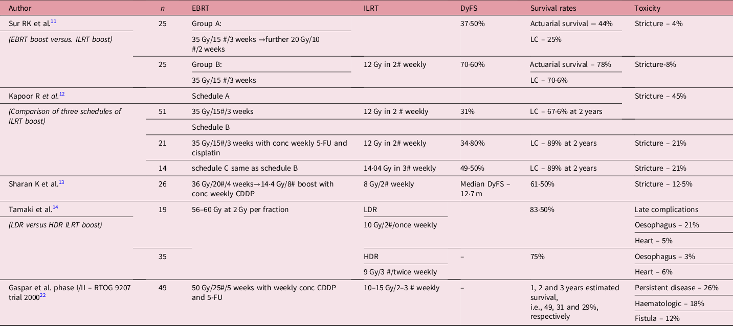
Various studies have been conducted over last three decades to establish the role of brachytherapy in oesophageal carcinoma (Table 1). A study done in 1992 compared HDR brachytherapy with EBRT boost. There was a marked improvement in dysphagia and local control at the end of 1 year, though not statistically significant. Reference Sur11 In the same institute, a retrospective study compared different radiation schedules with HDR brachytherapy and showed that concurrent chemoradiation followed by HDR brachytherapy is associated with the better 2-year local control and dysphagia-free survival (DyFS). Reference Kapoor, Bansal, Kocchar, Kumar and Sharma12 Another study also concluded that the ILRT boost after the completion of concurrent chemoradiotherapy is effectively tolerated and potentially improves outcome in terms of DyFS and disease-free survival. Reference Sharan, Fernandes, Prakash Saxena, Banerjee and Sathian13
Selected clinical series employing intraluminal radiotherapy in radical settings

EBRT, external beam radiotherapy; ILRT, intraluminal radiotherapy; DyFS, dysphagia-free survival; LC, local control; HDR, high-dose rate; conc, concurrent: 5-FY, 5-Flouro uracil; CDDP, Cis-DiammineDichloroPlatinum(II).
HDR versus LDR boost was studied by Tamaki et al. Reference Tamaki, Ishikawa and Takahashi14 They found no statistical difference in survival rates, however reactions such as oesophageal ulcers were found to be more common in LDR arm (21 versus 3%, p = 0·03). According to them, HDR ILRT is preferred over LDR due to shorter treatment time and better compliance.
The best timing for ILRT boost is yet to be determined. Most commonly, boost is given after EBRT to potentiate the dose to tumour bed. However, it becomes difficult to identify tumour bed post-EBRT. Also, the insertion of applicator in the irradiated region increases the risk of perforation. Vuong et al. Reference Vuong, Szego and David15 staged HDR boost at the start of the treatment with the hypothesis that inserting the tube and dilating are relatively easy in normal tissue and allow easier identification of the tumour bed. It also allows rapid relief in dysphagia due to high dose to the tumour bed. Brunner et al. Reference Brunner, Rupp, Melzner, Grabenbauer and Sauer16 delivered HDR brachytherapy sandwiched with EBRT with the hypothesis that a better dose coverage is expected due to decreased tumour thickness.
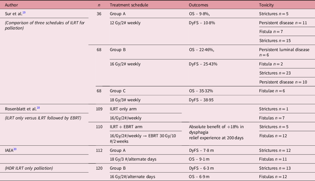
Few studies optimised the dose of HDR brachytherapy in advanced oesophageal carcinoma for palliative settings (Table 2). International Atomic Energy Agency (IAEA) conducted a multicentre randomised phase III trial evaluating whether the addition of EBRT improves dysphagia and overall survival. Preliminary results did not find any improvement in DyFS and overall survival (OS). Reference Sur, Donde and Conrad17 In continuation of this trial, another study was conducted with sufficient power to show that DyFS significantly improved with combination therapy, for an absolute benefit of 18% after median follow-up of 197 days. Reference Rosenblatt, Jones and Sur18 A dose optimisation study suggested that 16 Gy in two fractions or 18 Gy in three fractions are optimal doses for good palliation. Reference Sur, Donde, Levin and Mannell19 A follow-up multicentric, prospective randomised study conducted by International Atomic Energy Agency (IAEA) randomised patients to either receive 18 Gy in three fractions on alternate days (6 Gy per fraction, Group A) or 16 Gy in two fractions on alternate days (8 Gy per fraction, Group B). The DyFS and the OS were significantly more in Group A. The incidence of strictures and complications like fistulas were similar in both groups. Reference Sur, Levin, Donde, Sharma, Miszczyk and Nag20 A systemic review and meta-analysis reinforced that brachytherapy is highly effective and safer treatment alternative than metal stent placement. Reference Fuccio, Mandolesi and Farioli21
Selected clinical series employing ILRT (intraluminal radiotherapy) in palliative settings

EBRT, external beam radiotherapy; ILRT, intraluminal radiotherapy; HDR, high-dose rate; DyFS, dysphagia-free survival; OS, overall survival.
Majority of the patients suffer from acute oesophagitis, confirmed by oesophagoscopy; however, perforation and bleeding are rare. Long-term side effects such as chronic oesophagitis, ulceration (up to 30%), stenosis/stricture (5–30%) and fistula (5–10%) occur after radical combination treatment. RTOG 9207 reported life-threatening toxicity or treatment-related deaths in 24 and 10% cases, respectively. Treatment-related oesophageal fistulas occurred in 12% of patients when brachytherapy was used as a boost. Reference Gaspar, Winter, Kocha, Coia, Herskovic and Graham22
Seed brachytherapy
Loco-regional recurrence is the most common cause of death in oesophageal carcinoma. Reference Isono, Onoda, Ishikawa, Sato and Nakayama23 Among loco-regional recurrences, regional lymph nodes are one of the major sites. Reference Shimada, Kitabayashi and Nabeya24 Antecedent use of surgery, chemotherapy and RT limits the treatment options for nodal recurrences. Seed brachytherapy is the use of radioisotope, permanently implanted into a malignant node. 125I is one of the radioisotope of iodine which sends out soft gamma radiation and X-rays with a maximum energy of about 35 KeV along with conversion, and auger electrons are also emitted. The killing radius of 125I seed is 1·7 cm, ensuring no to minimal dose to normal tissues, since the intensity is inversely correlated with the square of the radius. The 125I radiation can efficiently inhibit the proliferation and repair of tumour cells, while the surrounding normal tissue receives less than 25% of the dose. The slow emission of radiation from the 125I seed also allows the surrounding normal tissue receiving sublethal or potentially lethal dose of radiation to have sufficient time to repair and recover. It is used in special cases where after curative treatment there is a nodal recurrence and other modalities of treatment are already exhausted. The 125I seed is covered with a cylindrical titanium body with length of 4·5 mm inside a silver column. Its radioactivity is 0·8 mCi with the average energy of 27–35 KeV and half-life of 59·6 days. Percutaneous insertion of brachytherapy needles under CT fluoroscopy guidance is used to place 125I seeds. Another approach is via EUS, where it is inserted into oesophageal lumen, and metastatic nodes are identified. EUS is considered more accurate than CT in the nodal staging of oesophageal cancer. Reference Ziegler, Sanft, Friedrich, Stein, Haring and Riecken25
Seed brachytherapy – treatment planning
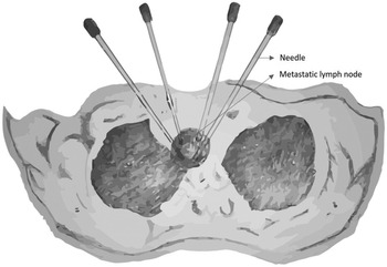
Patient preparation is done in similar fashion as described for ILRT. PTV is defined as gross tumour volume with 1·5 cm uniform expansion. PTV edge should be covered by 80–90% of isodose curves. CT images with 3 mm of section thickness are obtained and target volume is carefully delineated in every CT slice. Matched peripheral dose (MPD) is calculated and 125I seed is chosen based on its activity. With CT fluoroscopic guidance, four to six catheters of 18 G are inserted into a lymph node and positioned symmetrically after local anesthesia (Figure 7). A clip implant is then attached to the applicator for implantation. Every 125I seed should be placed at a distance of 0·5–1·0 cm from each other. In order to achieve a prescribed MPD of 100–140 Gy based on three perpendicular diameters within the target volume, treatment planning system generates a DVH and isodose curves of different percentages. The coordinates of brachytherapy applicator are calculated along with dose and number of implanted seeds. A Computed Tomography (CT) scan is usually performed within a month to verify the intensity and position of 125I seeds. In tumours showing insufficient radioactivity, more 125I seeds can be implanted.
Seed brachytherapy – percutaneous insertion of seeds.

Percutaneous insertion of catheter can result in pneumothorax or local haematoma. Seed migration is also a possibility which can cause radiation pneumonitis. In a study by Fei et al. Reference Gao, Li, Gu, Huang and Wu26 study, out of 16 patients, 2 had local hematoma, 2 had pneumothorax, 1 had minor displacement after a follow-up of 15 months and none had radiation pneumonitis. Lah et al. Reference Lah, Kuo, Chang and Nguyen27 reported no immediate or long-term complications.
Seed brachytherapy – results
There is limited data on seed brachytherapy in carcinoma oesophagus. In a retrospective study by Fei et al. Reference Gao, Li, Gu, Huang and Wu26 , effectiveness of CT-guided 125I brachytherapy was evaluated in 16 patients with mediastinal metastatic lymph nodal recurrence. The LC were 75, 50, 42·9 and 33·3% after 3, 6, 10 and 15 months, respectively. Lah et al. Reference Lah, Kuo, Chang and Nguyen27 discussed a case where CT was unable to detect nodal recurrence near crux of diaphragm, but EUS-guided needle aspiration confirmed the recurrence. Under EUS guidance, 125I seeds were inserted for a lifetime dose of 110 Gy. At 8 months, EUS evaluation showed complete resolution of treated nodes. Novel concept by Guo et al. Reference Guo, Teng and Zhu28 attempted SEMS loaded with 125I seeds and compared with SEMS. The 2-month DyFS and survival rates were significantly better in the irradiated stent group.
Intraoperative radiotherapy (IORT)
Intraoperative radiotherapy (IORT) is the delivery of high-dose single-fraction RT to tumour bed perioperatively. Surgical dissection of lymphatics in the upper left mediastinum is technically difficult with considerable morbidities. IORT can provide effective tumouricidal dose to such sites of relapse. Tissue within surgical territories possess rich vascularisation, resulting in radiosensitivity owing to oxygen effect. IORT eliminates the risk of geographical miss of tumour bed by delivering brachytherapy perioperatively. The dose to OARs can be minimised by displacing the adjacent structures or placing lead shield. Local dose escalation can be easily achieved and organ function can be preserved. IORT is a well-established option in breast and pancreatic cancer. However, there is a paucity of literature on the utility of IORT for oesophageal carcinoma.
Presently, it is used to boost upper abdominal lymph node region. Mobile linear accelerators delivering megavoltage electron such as MOBETRON® or NOVAC7® or kilovoltage photon-generating linear accelerator such as INTRABEAM® can be used to deliver IORT.
IORT – treatment planning
Standard surgical preparation is required. IORT is performed immediately after oesophageal resection in the perioperative period. Since it is directly delivering the dose to tumour bed with no chances of geographical miss, no margins are required. Earlier, perioperatively, metallic surgical clip used to be placed at the medial portion of the subclavian artery and the right main bronchus as a landmark. The patient is then carried to the radiation therapy room, and the electron beam applicator is inserted onto the marked area and the dose delivered. But after the advent of mobile linear accelerators, IORT can be delivered on the surgical table without transferring the patient to RT facility. In order to cover the tumour bed adequately, the energy of therapeutic electrons (9–12 MeV) is determined, depending on the depth. A single fraction dose of 20–30 Gy high-energy electrons or kilovoltage photons are used. Routine post-operative care is required. Usual complications of IORT include wound infection, pneumonia, anastomotic leaks or stenosis. Incidence rates of these complications were not significantly different between two groups in Tamaki et al. Reference Tamaki, Sasaki and Ejima29 study. Most prominent late complication seen in Arimoto et al study Reference Arimoto, Takamura, Tomita, Suzuki, Hosokawa and Kaneko30 was tracheal ulceration. No complications related to IORT were noted in Kuroda et al study. Reference Murakami, Kuroda and Nakajima31
IORT – results
A report by Arimoto et al. Reference Arimoto, Takamura, Tomita, Suzuki, Hosokawa and Kaneko30 in 1993 showed the prevention of mediastinal lymph node metastasis by IORT targeted at the upper mediastinum successfully. In 1999, Murakami et al. Reference Murakami, Kuroda and Nakajima31 showed the reduction in rate of abdominal lymphatic recurrences with IORT without survival benefit. In a study by Tamaki et al., Reference Tamaki, Sasaki and Ejima29 117 patients of thoracoabdominal oesophageal carcinoma were randomised to IORT versus non-IORT group. Perioperatively, a single-fraction dose of 20–30 Gy, encompassing the upper abdominal lymph node area, was delivered with high-energy electrons to IORT group. The 5-year local control rate in abdomen was significantly higher in the IORT group (89·2 versus. 72·9%; p = 0·022) with no significant improvement in OS. No obvious perioperative complication was attributed to IORT. So, to conclude, IORT is effective in securing good local control targeted to the upper abdominal lymph node region with some limitations.
Conclusion
Oesophageal brachytherapy is less commonly used due to requirement of high expertise, lack of established evidence, risk of life-threatening complications and lack of interest in brachytherapy per se. But still it remains an attractive option due to superior dosimetry which may result in high therapeutic ratio. It offers quick and useful palliation for a prolonged period, along with the good quality of life. In the past three decades, most studies have also shown its utility in adjunct settings, though life-threatening toxicity remains an issue. Recent development of novel applicators may allow dose escalation and lower toxicity. Seed brachytherapy is also emerging as a promising option in nodal recurrences. As per present scenario, oesophageal brachytherapy will remain a tangible part of treatment of oesophageal cancer.
Acknowledgements
None.
Declaration of Conflicting Interests
The authors declared no potential conflicts of interest with respect to the research, authorship and/or publication of this article.
Funding
The authors received no financial support for the research, authorship and/or publication of this article.