





Introduction
Intracytoplasmic sperm injection (ICSI) is commonly used in assisted reproductive technology and involves the microinjection of a single sperm cell directly into a mature oocyte; it is primarily used to address severe cases of male factor infertility. This technique is also useful for achieving fertilization when previous in vitro fertilization (IVF) attempts have been unsuccessful (Palermo et al., Reference Palermo, Joris, Devroey and Van Steirteghem1992; Van Steirteghem et al., Reference Van Steirteghem, Nagy, Joris, Liu, Staessen, Smitz, Wisanto and Devroey1993; Lv et al., Reference Lv, Zhang, He, Chen, Li, Ding and Zhang2020). Although ICSI results in fertilization rates of 70–80%, it does sometimes fail (Montag et al., Reference Montag, Köster, van der Ven, Bohlen and van der Ven2012; Lv et al., Reference Lv, Zhang, He, Chen, Li, Ding and Zhang2020), due to either oocyte abnormalities or, most commonly, activation deficiency (Dozortsev et al., Reference Dozortsev, De Sutter and Dhont1994; Tesarik et al., Reference Tesarik, Sousa and Testart1994; Lu et al., Reference Lu, Chen, Gao, Ma, Li, Hu and Li2006; Nasr-Esfahani et al., Reference Nasr-Esfahani, Razavi, Mardani, Shirazi and Javanmardi2007).
Oocyte activation is an intricately regulated process that is triggered by intracellular calcium (Ca2+) oscillations from endoplasmic reticulum stores after the sperm enters the ooplasm (Tesarik et al., Reference Tesarik, Sousa and Testart1994). Sperm-specific phospholipase C-zeta (PLCz), a sperm factor located in the peri-nuclear theca of spermatozoa, is the primary factor responsible for inducing intracellular Ca oscillations via an inositol-1,4,5-triphosphate (IP3)-mediated pathway (Saunders et al., Reference Saunders, Larman, Parrington, Cox, Royse, Blayney and Lai2002; Kashir et al., Reference Kashir, Heindryckx, Jones, De Sutter, Parrington and Coward2010; Ramadan et al., Reference Ramadan, Kashir, Jones and Coward2012). When IP3 binds to receptors on the surface of the endoplasmic reticulum, it triggers the release of Ca2+ from the internal stores of the cell (Berridge Reference Berridge2009). Consequently, the primary reason for unsuccessful fertilization and the failure of oocyte activation is often considered a deficiency in the PLCz cascade related to sperm (Yoon et al., Reference Yoon, Eum, Lee, Lee, Kim, Han, Won, Park, Shim, Lee, Fissore, Lee and Yoon2012; Nomikos et al., Reference Nomikos, Kashir, Swann and Lai2013).
Ca2+ activation is important for fertilization, as it triggers downstream processes including the activation of CAMKII and inactivation of MPF, leading to meiotic resumption and pronucleus formation. Subsequent Ca2+ oscillations are also important for the first mitotic division and later embryologic development processes (Neri et al., Reference Neri, Lee, Rosenwaks, Machaca and Palermo2014). Therefore, these oscillations, which are regulated by changes in the intracellular Ca2+ concentration in the oocyte, are responsible for controlling the processes that enable a smooth transition from oocyte to embryo development (Putney Reference Putney1986; Malcuit et al., Reference Malcuit, Kurokawa and Fissore2006; Saunders et al., Reference Saunders, Swann and Lai2007; Martinez et al., Reference Martinez, Durban, Santaló, Rodríguez and Vassena2021).
Researchers have attempted to improve clinical outcomes in patients with fertilization failure or low fertilization rates using artificial oocyte activation (AOA), including Ca2+ ionophores (e.g. ionomycin or calcimycin [A23187; CultActive]), as well as strontium chloride, electrical and mechanical activation methods (Zhang et al., Reference Zhang, Yao, Zhang, Hu, Yang, He, Qina, Fan, Bai and Sun2023). According to studies and meta-analyses, Ca ionophore (Ca-I) application significantly improves the overall fertilization rates in patients who experience fertilization failure (Murugesu et al., Reference Murugesu, Saso, Jones, Bracewell-Milnes, Athanasiou, Mania, Serhal and Ben-Nagi2017; Shebl et al., Reference Shebl, Trautner, Enengl, Reiter, Allerstorfer, Rechberger, Oppelt and Ebner2021; Chen et al., Reference Chen, Zhao, Lv, Dong, Zhao, Sui, Cui, Liu and Wu2022). Positive effects of Ca-I application have been detected at various embryonic development stages (Zhang et al., Reference Zhang, Yao, Zhang, Hu, Yang, He, Qina, Fan, Bai and Sun2023). Although male factor infertility and fertilization failure history are the main indications for Ca-I, its use also improves fertilization success and embryo development in patients with infertility for various other reasons, including cases of persistent developmental embryo arrest (Ebner et al., Reference Ebner, Oppelt, Wöber, Staples, Mayer, Sonnleitner, Bulfon-Vogl, Gruber, Haid and Shebl2015; Lv et al., Reference Lv, Zhang, He, Chen, Li, Ding and Zhang2020; Xu et al., Reference Xu, Yao, Niu, Fan, Ma, Shi, Jin, Song and Sun2021; Tsai et al., Reference Tsai, Lin, Lian, Li, Vitale, Mikuš, Su, Tsai, Tsui and Lin2022). In a recent study, an AOA group had better embryo quality than a ICSI group in women with advanced maternal age and diminished ovarian reserve, with higher rates of cell cleavage and top-quality day 3 embryos (Tsai et al., Reference Tsai, Lin, Lian, Li, Vitale, Mikuš, Su, Tsai, Tsui and Lin2022).
Time-lapse imaging (TLI) have been developed to objectively observe the progression of embryo development, enabling embryologists to continuously observe embryos while analysing their dynamic and morphokinetic events. This allows for more comprehensive analyses of embryo development (Sciorio Reference Sciorio2021). With conventional incubators, many morphological changes are frequently overlooked, including irregular cell division, blastocyst collapse and re-expansion, and the formation and absorption of fragments. However, TLI can detect and analyse these changes (Zaninovic et al., Reference Zaninovic, Irani and Meseguer2017). In addition, time lapse parameters have been studied for their potential to assess the risk of aneuploidy based on embryo grading (Basile et al., Reference Basile, del Carmen Nogales, Bronet, Florensa, Riqueiros, Rodrigo, García-Velasco and Meseguer2014; Patel et al., Reference Patel, Shah, Kotdawala, Herrero, Rubio and Banker2016). By combining TLI with PGT-A, the selection of euploid embryos with the greatest potential for implantation may be increased (Zaninovic et al., Reference Zaninovic, Irani and Meseguer2017). TLI also enhances the criteria for selecting blastocysts and can accurately predict blastocyst formation (Motato et al., Reference Motato, de los Santos, Escriba, Ruiz, Remohí and Meseguer2016).
Although many studies have investigated the effect of Ca-I after ICSI on fertilization, few have investigated the impact of Ca-I on embryomorphokinetic parameters and clinical pregnancy outcomes (Martinez et al., Reference Martinez, Durban, Santaló, Rodríguez and Vassena2021; Shebl et al., Reference Shebl, Trautner, Enengl, Reiter, Allerstorfer, Rechberger, Oppelt and Ebner2021). There is also limited information on the use of Ca-I in various patient groups. Therefore, we investigated the effects of oocyte activation with Ca-I on embryomorphokinetic parameters and clinical pregnancy outcomes in a large cohort including patients with various causes of infertility.
Materials and methods
Patients
A total of 412 patients underwent ICSI/ICSI + AOA at the Izmir University of Economics IVF Unit, Medical Park Hospital between 2018-2020. ICSI + AOA was used in 225 patients while ICSI only was used in 187. The cycle results were compared retrospectively.
The main criteria for Ca-I use were male factor infertility, previous fertilization failure, low fertilization rates (<30%) and poor embryological development pattern in previous cycles. PGT-A cases and azoospermia patients were not included in the analysis.
Oocyte stimulation
Gonadotropins (follitropin alfa or menotropin 150–450 IU) were administered starting on day 3 of the menstrual cycle, with 0.25 mg GnRH antagonist cetrorelix acetate (Cetrotide, Merck, Germany) on cycle day 7. Human chorionic gonadotropin (hCG) was used to trigger ovulation once the dominant follicle reached 18 mm and oocyte pick-up was performed 35 h after triggering.
Oocyte collection, fertilization, and time lapse embryo culture
Oocyte–cumulus complexes were selected in Quinn’s Advantage™ Medium with HEPES plus HSA (Sage, USA) and surplus cumulus was dissected from around the oocytes using a stereomicroscope in a class II hood with a heated stage. This preliminary mechanical reduction was performed to ensure a more controlled and efficient enzymatic denudation process and minimize oocyte exposure to enzymatic agents, thereby preserving oocyte integrity. The oocytes were washed and cultured in SAGE 1 Step™, with HSA (Origio, Denmark) covered with liquid paraffin oil (Origio, Denmark) for between 2 and 4 h in an atmosphere of 5.5% CO2 and 5.0% O2 in a Sanyo MCO-19 M (Panasonic) incubator. After incubation, the cumulus cells were denudated with 15–20 IU/mL cumulase (Origio, Denmark) and the corona radiata was completely removed. Oocytes with an obvious first polar body (1st Pb) were identified as mature (MII), and used for ICSI. To prepare Ca-I (GM508 CultActive), the ionophore medium was first incubated in a standard incubator at 37°C with 5.5% CO2 for 4 h before the microenjeciton procedure. Then, the microinjected oocytes were immediately placed into the pre-equilibrated calcimycin solution (GM508 CultActive) for 15 min for Ca2+ activation. Following this 15-minute activation period, the oocytes were washed several times in fresh culture media to ensure the complete removal of the ionophore. Subsequently, they were placed individually on a pre-equilibrated Embryoslide (Vitrolife, Denmark) containing 12 wells holding 1.2 mL liquid paraffin oil (Origio, Malov, Denmark). The oocytes were incubated in the time lapse monitoring system (EmbryoScope; Unisense FertiliTech, Denmark) in an atmosphere of 5% O2, 5.5% CO2, at 37°C until embryo transfer.
Time-lapse monitoring embryo culture system
EmbryoViewer software (Unisense Fertilitech Aarhus) was used to inspect the embryo images to elucidate the embryo growth after microinjection over time (hours). After microinjection, oocyte development was evaluated in the time-lapse system, including pronuclear fading (Pnf), the moment when both pronuclei disappear, first cleavage, and the times at which the zygote divides into 2–8 cells (t2 to t8, respectively). The second embryonic cell cycle length (EMCC2 = t3–t2) is the time of embryo cleavage from two blastomeres into three blastomeres. The time from this cleavage to a four-blastomere embryo is the second synchrony (s2 = t4–t3). The time for the three-blastomere embryo to split into a five-blastomere embryo is the prolongation of the third cell cycle (cc3 = t5–t3). A previous study determined the optimal morphological values for the best implantation rate for t5 (t5 = 48.8–56.6 h), s2 (s2 < 0.76 h), and cc2 (cc2 < 12 h, 20.5<t5–t2 < 29, and 11<cc3[t5–t3]<18) (Basile et al., Reference Basile, del Carmen Nogales, Bronet, Florensa, Riqueiros, Rodrigo, García-Velasco and Meseguer2014). We also considered these optimal values.
Embryo grading
Embryo quality was graded A, B, C, or D based on morphokinetic parameters as follows: embryos with a duration of t5–t2 > 20.5 h were defined as A or B, while those outside this optimal range were defined as C or D. Embryos with a duration of cc3 between 11 and 18 h were classified as A or C, while those beyond the optimal range were classified as B or D, depending on t5–t2 (Basile et al., Reference Basile, del Carmen Nogales, Bronet, Florensa, Riqueiros, Rodrigo, García-Velasco and Meseguer2014).
Embryo transfer
Embryos were transferred between the second and fifth days of development with a Sure-Pro Ultra Embryo Replacement Catheter with an Obturator (Cooper Surgical Company, The Netherlands) and a medium Sage One Step.
All patients received luteal support with the daily use of a 400 mg vaginal progesterone tablet (Progestan vaginal capsule 400 mg) and 30 mg oral dydrogesterone (Duphaston tablet 10 mg; Abbott Laboratories, Eczacibasi, Turkey).
A biochemical pregnancy is characterized by a serum level of β-hCG>20 IU/I on day 12 following embryo transfer. Clinical pregnancy is established when the activity of the heart of the fetus is detected through ultrasonography 8 weeks post-embryo transfer. Any pregnancy loss that occurs after clinical pregnancy confirmation is classified as a miscarriage.
Statistical analysis
All data statistical analysis were performed using Statistical Package for Social Sciences 26.0 studio programme and p < 0.05 was considered statistically significant. Continuous data were shown as the mean and standard deviation. Independent sample T-test was used for the analysis of the baseline parameters and the embryomorphokinetic parameters. Categorical variables (fertilization, embryo grade A, clinical pregnancy rates) were presented as percentages and were evaluated using the Pearson’s chi-square test. Embryomorphokinetic parameters were additionally analysed using a Linear Mixed Model.
Power analysis
The G*Power 3.1.9 package programme was employed for the power analysis of the study. Following the collection of data, a post-hoc power analysis based on pregnancy outcomes revealed that the study had a power of 0.89 for 412 patients.
Results
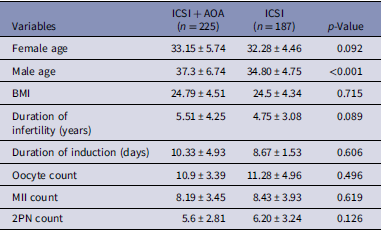
In this retrospective cohort study, a total of 412 patients were included. ICSI + AOA group consisted of 225 patients and ICSI group included 187 patients. When female age, male age, BMI, duration of infertility, duration of induction(days), oocyte count, MII count and 2PN count were examined, there were no significant differences between groups, except for male age (Table 1).
Clinical parameters

Table 1. Long description
Baseline clinical characteristics of 225 patients in the ICSI+AOA group and 187 patients in the ICSI group. Variables include female age, male age, body mass index, duration of infertility, duration of ovarian stimulation, oocyte count, MII oocyte count, and 2PN count. Male age was significantly higher in the ICSI+AOA group (p < 0.001),while there were no significant differences between the groups for the remaining variables (p > 0.05).
Note: Independent sample T-test. Data were presented as mean ± standard deviation.
AOA = Artificial Oocyte Activation, BMI = Body Mass Index, n = number of patients included in each group.
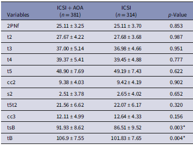
When the durations from 2PN to the blastulation time (tB) were examined, no significant differences were found between the two groups in terms of t2, t3, t4, or t5. When the optimal time lapse parameters (t5, cc2, S2, cc3, and t5-t2) were evaluated no significant relation was found between the probability of optimal time lapse parameters and the use of Ca-I (Table 2). The use of Ca-I was found to extend (p < 0.05) time to start of blastulation (Figure 1) and blastulation times (tB) (p < 0.05) (Figure 2). The embryo morphokinetic data were analysed at the embryo level. Since more than one embryo could originate from the same patient and, in some cases, double embryo transfer was performed, observations were not fully independent. To account for intra-patient clustering, we performed an additional analysis using a Linear Mixed Model, with patient identity included as a random effect.
Time to start of blastulation in the ICSI + AOA and ICSI groups.

Blastulation times in the ICSI + AOA and ICSI groups.

Embryomorphokinetic parameters

Table 2. Long description
Embryomorphokinetic parameters were compared between 381 embryos in the ICSI+AOA group and 314 embryos in the ICSI group. Early developmental parameters, including 2PNf, t2, t3, t4, t5, cc2, s2, t5–t2 and cc3, were similar between groups. Time to start of blastulation (tsB) and blastulation time (tB) were significantly longer in the ICSI+AOA group (p<0.05).
* p < .05.
Note: Independent sample T-test. Data were presented as mean ± standard deviation.
t2: time to the appearance of the 2-cell stage, t3: time to the appearance of the 3-cell stage t4: time to the appearance of the 4-cell stage, t5: time to the appearance of the 5-cell stage, cc2: second cell cycle duration, s2: Synchronization time of the second cell cycle, t5t2: Time interval between the 5-cell stage and the 2-cell stage, cc3: third cell cycle duration, tsB: time to start of blastulation tB: Blastulation time, n = number of embryos analysed in each group.
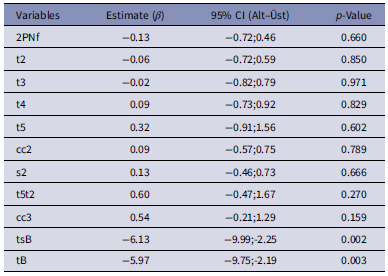
According to this analysis, no statistically significant differences were observed between the ICSI + AOA and only ICSI groups in early morphokinetic parameters, including t1–t5, cc2, s2, t5t2, and cc3 (all p > 0.05). In contrast, significant delays were detected in later developmental parameters, particularly time to start of blastulation (tsB) (p = 0.002) and time to blastocyst formation (tB) (p = 0.003) (Table 3).
Mixed model results for morphokinetic parameters (ICSI + AOA vs. ICSI)

Table 3. Long description
To account for intra-patient clustering, embryomorphokinetic parameters were additionally analyzed using a linear mixed model with patient identity included as a random effect. No significant differences were observed in early morphokinetic parameters between the ICSI+AOA and ICSI groups, whereas significant delays were detected in time to start of blastulation (tsB; p = 0.002) and blastulation time (tB; p = 0.003) in the ICSI+AOA group.
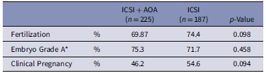
According to the fertilization results, there were no significant differences between the groups that received and that did not receive AOA treatment (69.87% vs. 74.4%, respectively). When it comes to embryo grades, the grade A embryo rates were not significantly different between ICSI + AOA and ICSI groups (75.3% vs. 71.7%, respectively). There were also no significant differences in the clinical pregnancy outcomes (46.2% vs. 54.6%, respectively) (Table 4).
Pregnancy outcomes

Table 4. Long description
Fertilization rate, Grade A embryo rate, and clinical pregnancy rate were compared between the ICSI+AOA and ICSI groups. No statistically significant differences were observed for fertilization (69.9% vs 74.4%), Grade A embryos (75.3% vs 71.7%), or clinical pregnancy (46.2% vs 54.6%) (all p>0.05).
Note: Pearson’s chi-square test. Data were presented as mean ± standard deviation. Embryo grades were determined using the morphokinetic parameters described by Basile et al.*.
Discussion
We investigated the effects of AOA application with Ca-I on embryomorphokinetic parameters, fertilization, and clinical pregnancy outcomes. To the best of our knowledge, our sample population is the largest of any study conducted on this subject to date. Some clinical studies and meta-analyses have reported that AOA may help improve fertilization, embryonic development, and clinical pregnancy outcomes in many clinical conditions (Murugesu et al., Reference Murugesu, Saso, Jones, Bracewell-Milnes, Athanasiou, Mania, Serhal and Ben-Nagi2017; Chen et al., Reference Chen, Zhao, Lv, Dong, Zhao, Sui, Cui, Liu and Wu2022). In line with our findings, the Ca ionophore group appeared to exhibit a tendency toward a poorer baseline prognosis, although most differences did not reach statistical significance. For example, male age was significantly higher in the Ca ionophore group, while female age and duration of infertility were also numerically higher, albeit without statistical significance. These baseline differences may have contributed, at least in part, to the observed outcomes. Also, because changes in intracellular Ca2+ levels influence the entire development process from oocyte to embryo transition, we examined the correlations between intracellular Ca2+ (in the form of Ca-I) and morphokinetic parameters.
The use of AOA is controversial, as synthetic activating agents do not fully replicate the natural Ca2+ signalling and physiological mechanisms observed in mammalian zygotes (Vanden Meerschaut et al., Reference Vanden Meerschaut, D’Haeseleer, Gysels, Thienpont, Dewitte, Heindryckx, Oostra, Roeyers, Van Lierde and De Sutter2014; Martinez et al., Reference Martinez, Durban, Santaló, Rodríguez and Vassena2021). Activation of the embryo by Ca-I may lead to variations in cellular kinetic patterns and division patterns that differ from normal processes. Therefore, this technique is still experimental, and gathering data from a larger number of patient embryos could yield valuable insights.
A study has evaluated the relationship between AOA and the morphokinetic behaviour of the embryo. Martinez et al. reported that AOA had no effect, with the exception of second polar body extrusion (tPB2) and time to second cell division (t3); however, they concluded that these changes did not seem to affect the morphokinetic pattern of the preimplantation embryo and ascribed the findings to a rapid transient nonphysiological increase in free intracytoplasmic Ca2+ (Martinez et al., Reference Martinez, Durban, Santaló, Rodríguez and Vassena2021). Similarly, Shebl et al. performed a sibling oocyte study in patients with previous fertilization problems. Half of the oocytes were treated with calcimycin. They found that ionophore-treated oocytes underwent significantly earlier pronuclei transformation and had a better synchronized third cell cycle (Shebl et al., Reference Shebl, Trautner, Enengl, Reiter, Allerstorfer, Rechberger, Oppelt and Ebner2021). Ca-I treatment significantly improved the overall rates of fertilization and blastocyst formation. In contrast to these findings, in our study pronuclei transformation or third cell cycle synchronization were not different and we found a prolonged tB in AOA cycles. The patient populations evaluated in the aforementioned studies primarily consisted of couples with fertilization failure and severe male factor infertility. However, our cohort was more heterogeneous, including not only cases with male-factor fertilization failure but also patients with low fertilization rates (< 30%) and poor embryological developmental patterns in previous cycles. Therefore, the differences between our findings and previous reports may reflect differences in patient populations. A possible explanation for these discrepant findings is that calcium ionophores may affect early and late developmental events differently. Although previous studies have reported accelerated or improved early cleavage events in certain patient populations, altered calcium signalling dynamics may have different effects on later developmental processes, such as blastulation.
Research on humans has highlighted the potential of tB as a determining factor for selecting embryos, surpassing other components of embryo morphological grading (Ho et al., Reference Ho, Arrach, Rhodes-Long, Salem, McGinnis, Chung, Bendikson, Paulson and Ahmady2018; Moustafa et al., Reference Moustafa, Rosen, Boylan and Mersereau2020; Huayhua et al., Reference Huayhua, Rodríguez, Vega, Briones, Rodriguez-Alvarez and Mellisho2023). Lee et al. and Moustafa et al. noted delayed tB in embryos with chromosome abnormalities (Lee et al., Reference Lee, Chen, Huang, Cheng, Chen, Ho and Lee2019; Moustafa et al., Reference Moustafa, Rosen, Boylan and Mersereau2020). The clinical importance of these changes in embryo morphokinetic parameters is not clear. The ICSI + AOA process results in non-physiological Ca2+ signalling patterns within the oocyte. In normal physiological processes, oocyte activation is characterized by recurrent Ca2+ oscillations from internal stores, orchestrated by sperm-borne PLCzeta over several hours. However, in AOA via Ca-I induces a single, transient, high-amplitude Ca2+ peak. This situation may be related to the faster expulsion of the second polar body in the initial fertilization and faster cleavage stages (t3) mentioned in previous reports, while the lack of long-lasting oscillations may be associated with the prolongation of the tsB and tB periods (Putney Reference Putney1986; Yoon et al., Reference Yoon, Eum, Lee, Lee, Kim, Han, Won, Park, Shim, Lee, Fissore, Lee and Yoon2012). Previous studies and our study results may suggest the importance of taking caution when applying Ca-I and the prolonged tsB and tB durations may serve as a warning for future clinical practice. It would be more appropriate to consider its utilization in cases where there is a clear indication rather than opting for elective use.
Morphokinetic behaviour has also been used to predict euploidy rates in different studies (Basile et al., Reference Basile, del Carmen Nogales, Bronet, Florensa, Riqueiros, Rodrigo, García-Velasco and Meseguer2014; Patel et al., Reference Patel, Shah, Kotdawala, Herrero, Rubio and Banker2016) because aneuploidy can potentially occur at any stage from the disjunction of chromatids to the mitotic divisions within the embryo (Hassold and Hunt Reference Hassold and Hunt2001; Wang and Sun Reference Wang and Sun2006; Chavez et al., Reference Chavez, Loewke, Han, Moussavi, Colls, Munne and Reijo Pera2012; Campbell et al., Reference Campbell, Fishel, Bowman, Duffy, Sedler and Hickman2013). We used the morphokinetic embryo grading system that predicts aneuploidy devised by Basile et al., and did not find any differences between the two groups. The literature contains little information on embryo aneuploidy and Ca-I activation. However, our results are in line with a recent study that found that Ca-I application did not increase the risk for aneuploidy and there were no developmental differences in live births as a result of transfers made with Ca-I administration (Zhang et al., Reference Zhang, Yao, Zhang, Hu, Yang, He, Qina, Fan, Bai and Sun2023). Thus Ca2+ ionophores have the capability to bind Ca2+ cations, and due to their hydrophobic properties, they form a complex at the lipid bilayer of the membrane. This structure enables ionophores to transfer Ca2+ across the membrane and release them into the cytosol (Brasseur et al., Reference Brasseur, Notredame and Ruysschaert1983). Consequently, ionophores themselves may not necessarily enter the oocyte, potentially explaining the absence of a detectable effect of ionophores on chromosomal segregation (Capalbo et al., Reference Capalbo, Ottolini, Griffin, Ubaldi, Handyside and Rienzi2016). Reports have indicated that implanted embryos after IVF, using Ca-I as an artificial oocyte activator, do not exhibit any chromosome abnormalities (Nakagawa et al., Reference Nakagawa, Yamano, Moride, Yamashita, Yoshizawa and Aono2001; Lu et al., Reference Lu, Chen, Gao, Ma, Li, Hu and Li2006). Moreover, no perinatal morbidity or mortality was reported among the patients involved in these studies. Comparable outcomes have also been observed in children aged 3–10 years, who were born following ICSI cycles using Ca-I. No differences have been noted compared to their peers (Vanden Meerschaut et al., Reference Vanden Meerschaut, D’Haeseleer, Gysels, Thienpont, Dewitte, Heindryckx, Oostra, Roeyers, Van Lierde and De Sutter2014; Deemeh et al., Reference Deemeh, Tavalaee and Nasr-Esfahani2015).
Furthermore, ionomycin and calcimycin may induce different intracellular Ca2+ signalling dynamics and embryological responses (Quintana-Vehí et al., Reference Quintana-Vehí, Martínez, Zamora, Rodríguez, Vassena, Miguel-Escalada and Popovic2023). Since calcium oscillation patterns regulate key events in embryo development (Malcuit et al., Reference Malcuit, Kurokawa and Fissore2006; Neri et al., Reference Neri, Lee, Rosenwaks, Machaca and Palermo2014), these differences may influence embryo morphokinetics and developmental competence. In the present study, calcimycin (CultActive) was used for oocyte activation. Previous studies using calcimycin (A23187)-based protocols, including CultActive, have reported effects on fertilization, embryo developmental outcomes, and embryo morphokinetics, as well as no increased risk of aneuploidy or adverse developmental outcomes (Capalbo et al., Reference Capalbo, Ottolini, Griffin, Ubaldi, Handyside and Rienzi2016; Murugesu et al., Reference Murugesu, Saso, Jones, Bracewell-Milnes, Athanasiou, Mania, Serhal and Ben-Nagi2017; Shebl et al., Reference Shebl, Trautner, Enengl, Reiter, Allerstorfer, Rechberger, Oppelt and Ebner2021; Tsai et al., Reference Tsai, Lin, Lian, Li, Vitale, Mikuš, Su, Tsai, Tsui and Lin2022; Zhang et al., Reference Zhang, Yao, Zhang, Hu, Yang, He, Qina, Fan, Bai and Sun2023). Although our study did not demonstrate differences in early cleavage parameters, the prolonged tsB and tB observed in the AOA group may further support the potential influence of calcimycin on embryo developmental kinetics. Likewise, ionomycin-based studies have also demonstrated alterations in embryo morphokinetics without adverse long-term developmental outcomes (Vanden Meerschaut et al., Reference Vanden Meerschaut, D’Haeseleer, Gysels, Thienpont, Dewitte, Heindryckx, Oostra, Roeyers, Van Lierde and De Sutter2014; Deemeh et al., Reference Deemeh, Tavalaee and Nasr-Esfahani2015; Martinez et al., Reference Martinez, Durban, Santaló, Rodríguez and Vassena2021).
The principal limitation of our study lies in its retrospective design. Consequently, definitive conclusions are challenging to establish. However, it is important to acknowledge that this limitation is mitigated by our substantial sample size. In addition, due to the retrospective design and incomplete follow-up, live birth outcomes were not available; thus, findings should be interpreted within the scope of embryomorphokinetic outcomes. Our findings may help understanding the embryomorphokinetic behaviour and embryo selection in ICSI + AOA cycles in future applications.
Conclusion
In our study, we did not find significant differences in most of the embryomorphokinetic parameters except prolongation of tB and time to start of blastulation. However, the clinical significance of these changes is controversial because of the lack of correlations between these parameters and clinical outcomes. More studies with larger populations are needed on this subject.
Data availability statement
The data that support the findings of this study are available from the corresponding author(F.G) upon reasonable request.
Acknowledgements
We would like to thank Selin Madran (Nursing) and Buket Ekmekçi (Biostatistics) for their contributions. The grammar check was carried out using TextCheck (Scientific and Technical Editing Service).
Author contributions
Funda Gode: Data curation – Lead, Formal analysis – Lead, Investigation – Lead, Project administration – Lead, Software – Lead, Supervision – Lead, Writing – original draft-Lead, Writing – review & editing – Lead; İbrahim Pala: Data curation – Equal, Methodology – Equal; Burcu Tamer: Data curation – Equal; Ahmet Zeki Işık: Supervision – Equal.
Competing interests
The authors have no relevant financial or non-financial interests to this close
Funding information
None.
Ethical standards
Institutional Review Board approval was obtained from Bahçeşehir University Faculty of Medicine before the beginning of the study.



 Open access

