Introduction
In the muscle post mortem, some proteins are oxidized and this process has an impact on meat tenderization (Maltin et al., Reference Maltin, Balcerzak, Tilley and Delday2003). Increased proteases oxidation reduces the functional properties of these proteases and decreases their efficiency in the meat protein maturation process (Rowe et al., Reference Rowe, Maddocks, Lonergan and Huff-Lonergan2004a and Reference Rowe, Maddocks, Lonergan and Huff-Lonergan2004b). Consequently, it can be hypothesized that some in vivo oxidation processes in the muscles may have an impact on the enzymes activities and protein structure both in vivo and post mortem (Stadtman and Levine, Reference Stadtman and Levine2000). To counteract these oxidation processes, many reactive oxygen species (ROS) scavengers exist within the cells (Fang et al., Reference Fang, Yang and Wu2002). Among these ROS, the water-soluble antioxidants (some antioxidant enzymes, peptides or vitamins soluble in water) are of major significance to counteract protein oxidation and carbonyl formation (Hipkiss et al., Reference Hipkiss, Preston, Himsworth, Worthington, Keown, Michaelis, Lawrence, Mateen, Allende, Eagles and Abbott1998 and Reference Hipkiss, Brownson and Carrier2001).
Among the various stress affecting ruminants, the seasonal variations in forage availability during extensive rearing affect protein metabolism with as a result, for example, compensatory growth (Hoch et al., Reference Hoch, Begon, Cassar-Malek, Picard and Savary-Auzeloux2003). The alteration of the growth pattern during compensatory growth is due to significant modifications in both protein synthesis and protein breakdown (Bruce et al., Reference Bruce, Ball and Mowat1991; Van Eenaeme et al., Reference Van Eenaeme, Evrard, Hornick, Baldwin, Diez and Istasse1998; Hornick et al., Reference Hornick, Van Eenaeme, Gérard, Dufrasne and Istasse2000). The increase in protein anabolism efficiency during refeeding after food restriction, a highly beneficial adaptation of animals to undernutrition, requires a large amount of energy (Lobley, Reference Lobley2003) and stimulates the mitochondrial redox chain, which is one of the sources of ROS (Aurousseau, Reference Aurousseau2002; Dröge, Reference Dröge2002; Griffiths, Reference Griffiths2002). Such oxidation processes occurring during these adaptation periods, which happen simultaneously with alterations in the food supply, have not been studied in farm animals.
The present study aimed to assess, in growing lambs, the effects of compensatory growth on protein oxidation and on some water-soluble antioxidants in muscles since they are partly responsible for an alteration of meat tenderness. The rapid increase in body weight observed during the compensatory growth process can be explained by two related mechanisms which are an increased capacity for dietary intake (‘nutritional effect’) associated with an increased efficiency for nutrients utilization for muscle protein anabolism (‘metabolic effect’) (Hoch et al., Reference Hoch, Begon, Cassar-Malek, Picard and Savary-Auzeloux2003). The separation, within the compensatory growth process, of the ‘metabolic’ and ‘nutritional’ effects by use of pair-feed animals allowed the assement of the mechanism(s) responsible for the alteration of the equilibrium existing between the oxidation processes and the antioxidant status.
Material and methods
Animals and diets
Sixteen male lambs (breed: Ile de France × INRA 401) of 3 months of age weighing 26.3 ± 0.2 kg were fed ad libitum with a 10% hay and 90% commercial concentrate diet (wheat: 12%, barley: 22%, soybean meal: 9%, wheat feed: 8%, wheat bran: 15%, sugar beet pulp: 15%; and vitamins and minerals (vitamin A: 6000 IU/kg, vitamin E: 20 IU/kg) from THIVAT, St Germain des Salles, France). Animals were housed in individual stalls with free access to drinking water and salt licks and under continuous lighting throughout the entire experiment. After 3 weeks of adaptation to ad libitum feeding, animals were randomly assigned to four treatments (n = 4 animals per group). One treatment group was fed ad libitum for 21 days (Control group, C) and slaughtered. One treatment group was restricted to 60% of the intake of the C group for 21 days (Restricted group, R) and slaughtered. Although this level of food restriction was a drastic decrease in intake in order to induce large effects on protein oxidation, it still allowed weight gain. In order to clearly identify which effect on oxidation/antioxidants can be attributed to the increased intake and/or to the adaptation of the animal metabolism to previous undernutrition, the last two treatment groups of lambs were either restricted similarly to the R group during the first 21 days, refed ad libitum for 12 days and slaughtered (Restricted-refed Ad Libitum group, RAL), or restricted similarly to the R treatment group for 21 days and refed with the same amount of food as the C group during the following 12 days and slaughtered (Restricted-refed Pair Fed to C group, RPF). To conduct the pair-feeding and avoid any Period effect, the animals were randomly assigned to one of four blocks with one animal per treatment group within each block.
Sample analysis
At the end of the experimental periods, animals of the C, R, RAL and RPF groups were slaughtered in the experimental slaughterhouse of the Research Institute. The animals had access to water and food until their slaughter. Just before they were slaughtered, the animals were transported to the slaughterhouse (3 min drive between the sheepfold and the slaughterhouse). The animals were then immediately stunned prior to bleeding. The supraspinatus (SP), longissimus dorsi (LD) and semitendinosus (ST) muscles were removed as quickly as possible (less than 10 min after slaughter for all muscles), minced and stored immediately at −80°C before analysis.
Carbonyl content
Protein oxidation level in tissues was measured by an estimation of the carbonyl groups (Levine et al., Reference Levine, Williams, Stadtman and Shacter1994). Briefly, 1 g of tissue sample was homogenized in 10 ml of KCl solution (0.1 M, pH 7.4) using a homogenizer. A solution of butylated hydroxytoluene 2% w/v was added to the sample to avoid oxidation during the sampling process. The proteins from two aliquots of the homogenate were precipitated with trichloroacetic acid (TCA) 10% (w/v, final concentration) after centrifugation at 5000 r.p.m. for 5 min. One pellet was resuspended in a 2 M HCl solution and the other pellet in a solution of 2 M HCl with 2,4-dinitrophenylhydrazine (DNPH) (0.2%, w/v), resulting in the formation of a stable 2,4-dinitrophenyl hydrazone product. Both samples were incubated for 1 h at room temperature and stirred every 10 min. The proteins in both samples were reprecipitated with 10% TCA (w/v, final concentration) and the pellets were washed carefully four times with ethanol : ethyl acetate (1 : 1). After the last wash, the pellets were dried under nitrogen flow and resuspended in a 6 M guanidine with 20 mM potassium phosphate buffer (pH, 2.3) and heated in a boiling bath for 5 min. The insoluble fragments present in the solutions were removed by centrifugation (10 000 r.p.m. for 5 min). Sample absorbance (blank and DNPH-treated aliquots) was measured at 370 nm and the blank value was subtracted from the DNPH treated value. The carbonyl content was expressed as nmol of DNPH fixed/mg proteins using an absorption coefficient of 22 mM−1 cm−1.
Glutathione concentration
To measure total glutathione concentration, frozen powdered tissues (obtained after milling pieces of frozen muscle in a mill (IKA, Fisher Bioblock, Illkirch, France)) were homogenized in a solution containing 1% picric acid. The homogenate was then centrifuged for 15 min at 3000 × g. The supernatant was filtered through a gauze and assayed for GSH + GSSG content using a standard enzymatic recycling procedure as described by Malmezat et al. (Reference Malmezat, Breuillé, Pouyet, Patureau Mirand and Obled1998) according to the method developed by Tietze (Reference Tietze1969).
Carnosine and anserine concentrations
Tissues (1 g) were homogenized in a 3% perchloric acid w/v solution containing homocarnosine (Sigma, France) (1 mmol/l final concentration) as an internal standard. The sample was centrifuged at 10 000 r.p.m. for 15 min and the supernatant was stored at −80°C for further analysis. Carnosine and anserine concentrations in the tissues were measured using a high-performance liquid chromatography (HPLC) method according to Van Eijk et al. (Reference Van Eijk, Rooyakkers and Deutz1993) and Maynard et al. (Reference Maynard, Boissonneault, Chow and Bruckner2001). The HPLC system used was a Series 200 (Perkin Elmer, Courtaboeuf, France) coupled with a fluorescence detector. The thawed supernatant was filtered using a 0.45 μm filter and 90 μl of the filtered sample was derivatized with 90 μl of an ortho-phtalaldehyde (OPA) reagent (Sigma, France). After 5 min of reaction time, the derivatized sample was injected onto the column (C18 HDO column (250 mm × 46 mm), Interchim, Montlucon, Cedex, France) balanced with 100% solvent A at a flow rate of 0.9 ml/min. During the 60 min run, the gradient varied according to the following pattern: 70% A and 30% B for 30 min, 60% A and 40% B for 15 min and 100% B for 5 min and 100% A for 10 min. Solvent A was made of 92.5% 50 mM sodium acetate buffer, pH 5.7 (pH adjusted with glacial acetic acid), 5% methanol and 2.5% tetrahydrofuran (THF) and solvent B was made of 97.5% methanol and 2.5% THF. The temperature of the column was maintained at 40°C. The excitation wavelength used was 335 nm and the emission wavelength was 440 nm. Carnosine and anserine retention times and quantification were determined using pure carnosine and anserine (Sigma, France) standards. Data were expressed as μmol of carnosine and anserine/g tissue.
Total antioxidant status
As stated by Girard et al. (Reference Girard, Madani, El Boustani, Belleville and Prost2005), the total antioxidant status (TAS) measurement takes into account the possible synergic effects of antioxidants against oxidant attacks and thus is highly complementary to the measurement of the specific antioxidant activity.
Tissue samples were homogenized in a phosphate buffer (10 mM, pH 7.4) using a homogenizer and centrifuged at 1200 × g for 10 min. The supernatant was filtered through a gauze and stored at −80°C for further analysis. TAS was determined by the method based on the absorbance of the ABTS+ cation (2,2′-azinobis-(3-ethylbenzothiazoline-6-sulfonic acid)) described by Miller et al. (Reference Miller, Rice-Evans, Davies, Gopinathan and Milner1993) and adapted to ruminant samples in the laboratory. Briefly, the production of ABTS+ was initiated by adding 450 μM hydrogen peroxide to 1 ml quartz cuvette containing 12 μl tissue extract, 500 μM ABTS and 48.8 μM metmyoglobin diluted in degassed phosphate buffered saline (PBS). Absorbance at 732 nm was measured immediately (A t0) and 3 min after addition of hydrogen peroxide (A t3) at 37°C using a double-beam spectrophotometer Uvikon 923 (Kontron Analysis Division, Zurich, Switzerland). Absorbance of a blank solution (i.e. containing degassed PBS instead of plasma) and of a homogenate was also determined to calculate the inhibition percentage of the reaction as follows:

Inhibition percentages were converted into Trolox Equivalent Antioxidant Capacity (TEAC) using a trolox standard curve ranging from 0 to 2.5 mM.
Antioxidant enzyme activity
Frozen tissue samples (250 mg) were homogenized in 3 ml of a phosphate buffer (50 mM, pH 7), centrifuged at 1000 × g for 15 min at 4°C and filtered through a gauze (Mercier et al., Reference Mercier, Gatellier and Renerre2004).
Total superoxide dismutase (SOD) activity was assayed according to Marklund and Marklund (Reference Marklund and Marklund1974). The capacity of SOD to inhibit pyrogallol autoxidation was assayed in a basic medium (Tris 50 mM, cacodylic acid 50 mM, diethylenetriaminepentaacetic acid 1 mM, pH 8.2). The activity was expressed as the inhibition percentage of pyrogallol autoxidation with one SOD unit corresponding to 50% inhibition.
Glutathione peroxidase (GPx) activity was measured by reducing the GSH regenerated by NADPH oxidation by glutathione reductase according to Agergaard and Thode Jensen (Reference Agergaard and Thode Jensen1982). NADPH disappearance at 366 nm was measured and GPx activity was expressed as μmol of oxidized NADPH min−1.
Statistical analysis
The results were analyzed by ANOVA using treatments (T) and blocks as main factors. The interaction between blocks and treatments was never significant and was withdrawn from the model. The analysis was carried out using the general linear model procedure of Statistica version 5.5 (1984–2000; Statsoft, Tulsa, OK, USA). Means were compared by contrasts (contrast 1: C v. R, contrast 2: RAL v. RPF and contrast 3: R v. RAL). Differences were considered as significant when P < 0.05 and as a tendency for 0.05 < P < 0.1.
Results and discussion
The growth rates as well as the dry matter (DM) and nitrogen (N) intakes of the four groups of animals are detailed in Table 1. As expected, the growth rate of the animals was reduced after the food restriction and re-increased during refeeding. As also expected, the feed intake (DM and N intake) is significantly inferior in the RPF v. RAL animals and it is similar between RPF and C animals.
Live weight at slaughter, growth rate and intakes in four groups of four growing lambs offered different intake levelsFootnote aFootnote b

a C = Control group (ad libitum); R = Restricted group; RAL = Restricted-refed Ad Libitum group; RPF = Restricted-refed Pair Fed to C group.
b Growth rate, dry matter intake and nitrogen intake for RAL and RPF groups correspond to the 12 days of refeeding (ad libitum or pair fed) and do not take into account of the restricted feeding period.
Protein oxidation
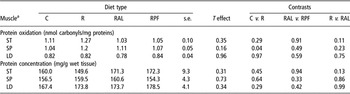
Muscle protein oxidation levels in the C animals (Table 2), as indicated by the carbonyl content in muscle proteins, were consistent with the results previously found in ruminant muscles (Martinaud et al., Reference Martinaud, Mercier, Marinova, Tassy, Gatellier and Renerre1997).
Mean protein carbonyl content (index of protein oxidation) and protein concentration in three muscles in growing lambs offered different intake levelsFootnote b

a ST = semitendinosus; SP = supraspinatus; LD = longissimus dorsi.
b C = Control group (ad libitum); R = Restricted group; RAL = Restricted-refed Ad Libitum group; RPF = Restricted-refed Pair Fed to C group.
No significant effect of food intake on protein carbonyl contents in lamb muscles was detected (T effect = 0.35, 0.16 and 0.96 for ST, SP and LD muscles, respectively; Table 2). It is now well known that the level of carbonyl groups in proteins is increased by many metabolic alterations such as aging in rats (Reznick and Packer, Reference Reznick and Packer1994) or humans (Fano et al., Reference Fano, Mecocci, Vecchiet, Belia, Fulle, Polidori, Felzani, Senin, Vecchiet and Beal2001) exercise (Leeuwenburgh and Heinecke, Reference Leeuwenburgh and Heinecke2001), and various diseases (Himmelfarb et al., Reference Himmelfarb, McMonagle and McMenamin2000; Pennathur et al., Reference Pennathur, Ido, Heller, Byun, Danda, Pergola, Williamson and Heinecke2005). However, the effects of the diet on protein oxidation and carbonyl contents in the literature show variable results. Indeed, it has now clearly been shown that a drastic (a reduction to 60% of the ad libitum food intake level) or a long-lasting (several months) food restriction decreased the carbonyl content in human plasma (Dandona et al., Reference Dandona, Mohanty, Ghanim, Aljada, Browne, Hamouda, Prabhala, Afzal and Garg2001) and in the muscles or in the liver of rodents (Nagai et al., Reference Nagai, Takahashi and Goto2000; Pamplona et al., Reference Pamplona, Portero-Otin, Requena, Gredilla and Barja2002). By contrast, other more specific alterations of the diet composition such as high protein content in the diet of rats (Petzke et al., Reference Petzke, Elsner, Proll, Thielecke and Metges2000) or supplementation with eicopentaenoic acid and decosahexaenoic acid (Wander and Du, Reference Wander and Du2000) in humans had no effect on plasma protein carbonyls. In cattle finished on pasture (rich in polyunsaturated fatty acids) and compared with animals fed high concentrate diets, there was a similar (Mercier et al., Reference Mercier, Gatellier and Renerre2004) or an increased (Savary-Auzeloux et al., 2001) content in protein carbonyls in some muscles. Although the level of restriction of the present animals was similar to that previously tested in the literature (60% of the ad libitum level), food restriction lasted only for 3 weeks. Moreover, the present lambs were polygastric animals whereas the rodents or humans used in the literature were monogastrics, adults or old, so that antioxidants status and metabolisms differed in both cases. Lastly, most studies measured carbonyl contents at the muscle level in mitochondrial proteins, which, due to their proximity to significant synthesis processes of free radical and oxygen species, were more susceptible to oxidation.
Consequently, these results showed that a decreased protein synthesis after food restriction followed by an increased protein synthesis during refeeding (Hoch et al., Reference Hoch, Begon, Cassar-Malek, Picard and Savary-Auzeloux2003) did not significantly alter protein oxidation in muscles. This means that the metabolic stress created by food restriction followed by refeeding was not sufficient to modify the oxidation level of proteins in animals at slaughter. Consequently, in our study, an alteration of protein oxidation during the compensatory growth process in vivo cannot be responsible for an altered protease activity during meat aging in animals undergoing compensatory growth (Maltin et al., Reference Maltin, Balcerzak, Tilley and Delday2003; Rowe et al., Reference Rowe, Maddocks, Lonergan and Huff-Lonergan2004a and Reference Rowe, Maddocks, Lonergan and Huff-Lonergan2004b). However, proteins (and hence the proteases) still undergo oxidation during maturation, its intensity being highly dependent on the antioxidants present in the muscles and meat (Martinaud et al., Reference Martinaud, Mercier, Marinova, Tassy, Gatellier and Renerre1997; Mercier et al., Reference Mercier, Gatellier, Viau, Remignon and Renerre1998; Rowe et al., Reference Rowe, Maddocks, Lonergan and Huff-Lonergan2004a).
Antioxidant status
Global antioxidant defense
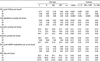
As stated by Warner et al. (Reference Warner, Dunshea, Ponnampalam and Cottrell2005) in live animals, the antioxidant capacity level and its interaction with oxidizable moieties were of great significance at animal slaughter. In the present study, even if the restricted feeding had no effect on muscle protein oxidation, it induced a strong decrease in total antioxidant capacity in muscles (−51%, −43%, P < 0.01 C v. R, for ST and LD muscles, respectively) (Table 3). Total antioxidant capacity in the most oxidative muscles such as SP was less sensitive to restricted feeding as the decrease observed was not significant (P = 0.15). More interestingly, total antioxidant capacity in muscles was recovered after 12 days of ad libitum refeeding (R and RAL groups significantly different for ST and LD muscles (P < 0.05) and tend to be different for SP muscle (P = 0.08) for R v. RAL) but not with the RPF animals in the same muscles (RAL and RPF groups significantly different for ST, LD and SP muscles P < 0.05). Consequently, the intake level during recovery had to be considered to be of major significance after restricted feeding since, in this case, a 12% decrease between RPF and RAL in N intake during recovery (Table 1) induced a lack of restoration of the antioxidant capacity in muscles. This lack of restoration of the antioxidant capacity level in RPF animals could be detrimental to animal health if another oxidative stress occurred following this type of restriction/refeeding pattern.
Mean total antioxidant status (TAS), total glutathione, carnosine and anserine concentrations, glutathione peroxidase (GPx) and superoxide dismutase (SOD) activities in three muscles of growing lambs offered different intake levelsFootnote b

a ST = semitendinosus; SP = supraspinatus; LD = longissimus dorsi; TEAC = Trolox Equivalent Antioxidant Capacity.
b C = Control group (ad libitum); R = Restricted group; RAL = Restricted-refed Ad Libitum group; RPF = Restricted-refed Pair Fed to C group.
This finding has not been previously reported in the literature in which effects of diseases or of specific antioxidants such as vitamin E, vitamin C, plant extracts tested on humans and/or animals were related (Liu et al., Reference Liu, Lanari and Schaefer1995; Fano et al., Reference Fano, Mecocci, Vecchiet, Belia, Fulle, Polidori, Felzani, Senin, Vecchiet and Beal2001; Alia et al., Reference Alia, Horcajo, Bravo and Goya2003, etc.). In ruminants, the effects of various antioxidants and especially vitamin E, a liposoluble antioxidant, have been tested in animals fed diets enriched in unsaturated lipids (for review, see Liu et al., Reference Liu, Lanari and Schaefer1995; Wood and Enser, Reference Wood and Enser1997; Wood et al., Reference Wood, Richardson, Nute, Fisher, Campo, Kasapidou, Sheard and Enser2003; Durand et al., Reference Durand, Scislowski, Gruffat, Chilliard and Bauchart2005). However, no effect of the alteration of the diet associated with a production system had been investigated so far.
As an effect of the restriction/refeeding pattern on total antioxidant capacity has been demonstrated, the next step consisted in determining which specific antioxidant was altered. For this purpose, some specific water-soluble antioxidants, which mostly could interact with proteins, were studied. So, the activities of some antioxidant enzymes and the concentrations of antioxidant peptides especially found in muscles such as glutathione, carnosine and anserine were measured. Glutathione is one of the most powerful antioxidants found in animal tissues (Liu and Eady, Reference Liu and Eady2005). Carnosine and anserine, two intracellular antioxidants involved in 50% of the cell buffering capacity, could influence two factors involved in meat maturation: intracellular pH and protease activity (Maltin et al., Reference Maltin, Balcerzak, Tilley and Delday2003). The antioxidant effect of carnosine in muscles and meat (Mei et al., Reference Mei, Cromwell, Crum and Decker1998) as well as the interaction of carnosine with carbonyl groups is well documented (Hipkiss et al., Reference Hipkiss, Preston, Himsworth, Worthington, Keown, Michaelis, Lawrence, Mateen, Allende, Eagles and Abbott1998 and Reference Hipkiss, Brownson and Carrier2001). Because of their antioxidant action, these peptides have been suggested to be natural enhancers of antioxidant defenses in meat consumers (Liu and Eady, Reference Liu and Eady2005; Park et al., Reference Park, Volpe and Decker2005). Other components of the water-soluble antioxidant equipment in animal tissues are the antioxidant enzymes (Chan and Decker, Reference Chan and Decker1994), which directly interact with the ROS such as SOD (dismutation of the superoxide anion radical) or catalase (destruction of H2O2) or which interfere with the glutathione redox system such as the glutathione peroxidase (GPx).
Glutathione
The glutathione concentrations reported in the literature are in accordance with the values observed in the present study (0.6 μmol/g in ST muscle of C animals) (Malmezat et al, Reference Malmezat, Breuillé, Pouyet, Patureau Mirand and Obled1998). Glutathione, which is a tripeptide (glutamic acid, cysteine and glycine), is a substrate for specific enzymes such as GPx or is direct scavenger for free radicals (Masella et al., Reference Masella, Di Benedetto, Vari, Filesi and Giovannini2005). It is present at a concentration of 0.7 μmol/g in rat muscles (Potter and Tran, Reference Potter and Tran1993). Interestingly, glutathione concentration in the ST muscles exhibited the same pattern of change as that of the TAS with a decreased concentration of glutathione in ST muscles after food restriction, compared with the C group (P < 0.05), and a recovery of the glutathione concentration only in the RAL group (RAL and RPF significantly different, P < 0.05). Consequently, the reduced global antioxidant decrease and the recovery previously observed could at least partially be explained by the glutathione status. A similar but not significant pattern of changes was observed in the other muscles. A decrease in glutathione concentration after food restriction has already been reported in rats (Cho et al., Reference Cho, Sahyoun and Stegink1981) and was explained by a decreased availability of the substrates such as cysteine needed for glutathione synthesis (Malmezat et al., Reference Malmezat, Breuillé, Capitan, Patureau Mirand and Obled2000). Methionine and its transmethylation and transsulfuration product, cysteine, are one of the most limiting amino acids in ruminant diets (Rulquin et al., Reference Rulquin, Vérité, Guinard-Flament and Pisulewski2001). Food restriction in our animals, and thus the reduction in methionine and cysteine supply, was long enough and of sufficient severity to induce a decrease, which was significant in the ST muscles in glutathione concentrations.
Carnosine and anserine
The carnosine and anserine concentrations in the muscles of the C group (Table 3) were similar to those described in the literature for similar animals (Chan and Decker, Reference Chan and Decker1994), resulting in a carnosine/anserine ratio close to 1 irrespective of the considered muscle (Chan and Decker, Reference Chan and Decker1994). Our results clearly showed that carnosine and anserine concentrations differed with the metabolic characteristics of the muscle. The carnosine concentrations observed in the SP muscles of the C animals, for instance, were smaller (11.4 v. 35.6 or 44.7 μmol/g wet tissues, P < 0.05, Student’s t test) than the values obtained in ST and LD muscles, respectively; a similar pattern was observed for anserine. Similar observation can be made between the SP and the other muscles in the R, RPF and RAL animals. In lambs, the SP muscle is considered as a slow red muscle (high oxidative capacity), whereas LD and ST muscles are fast red and fast white muscles (high glycolytic capacity), respectively (Briand et al., Reference Briand, Talmant, Briand, Monin and Durand1981a and Reference Briand, Talmant, Birnad, Monin and Durand1981b). This was in agreement with the reported findings in the literature where carnosine and anserine concentrations have been demonstrated to increase in muscles with intense glycolytic activity in many species (Sewell et al., Reference Sewell, Harris, Marlin and Dunnett1992; Aristoy and Toldra, Reference Aristoy and Toldra1998; Cornet and Bousset, Reference Cornet and Bousset1999). Moreover, their buffering role (Maltin et al., Reference Maltin, Balcerzak, Tilley and Delday2003) and its impact on pH stabilization during anaerobic contraction in glycolytic muscles may explain their higher concentration in muscles with high glycolytic capacities.
No significant effect of the dietary management could be demonstrated on carnosine or anserine concentrations in muscles (Table 3). This result confirmed the previous studies in which muscle carnosine and anserine concentrations seemed quite stable throughout the time, even during meat maturation where significant protein oxidation along with peptide proteolysis and degradation occur (Savary-Auzeloux et al., unpublished results; Moya et al., Reference Moya, Flores, Aristoy and Toldra2001). The only studies showing some alterations of the carnosine and anserine concentrations in muscles were undertaken in very drastic conditions such as a histidine-free diet, which induced a drop in carnosine and anserine concentrations in rat muscles (Tamaki et al., Reference Tamaki, Tsunemori, Wakabayashi and Hama1977). Similarly, high levels of carnosine in the diet (1.8%) were shown to increase the carnosine concentration in the soleus muscle of rats (Maynard et al., Reference Maynard, Boissonneault, Chow and Bruckner2001). However, even in that case, only the 1.8% enriched diet and not the 0.1% diet induced an effect on carnosine and anserine concentrations in the soleus muscle but not on the gastrocnemius, white and red vastus lateralis muscles. Thus, a non-physiological dietary enrichment/depletion in carnosine or anserine precursors is needed to result in some modifications of the carnosine and anserine concentrations in muscles. Furthermore, as stated by Mei et al. (Reference Mei, Cromwell, Crum and Decker1998) and Maynard et al. (Reference Maynard, Boissonneault, Chow and Bruckner2001), muscles rich in slow oxidative fibers seem to be more responsive to alterations in dietary histidine or carnosine concentrations although the mechanisms are still unknown. So, with the animals on a 60% ad libitum food restriction with a 50 g/day weight gain, it appeared that the histidine depletion (which occurred simultaneously with the other amino acid depletion) was not strong enough to induce a significant decrease in the carnosine and anserine concentrations in muscles.
Antioxidant enzymes
No effect of restriction or refeeding was observed on the two antioxidant enzymes studied (SOD, GPx) in muscles. Consequently, and unlike glutathione, an alteration of the activities of these enzymes cannot account for the alteration of the total antioxidant capacity demonstrated above. GPx and SOD activities were measured in various species (rats, chicks, pigs, humans, etc.), tissues and organs (erythrocytes, muscles, kidney, liver, etc.), situations of oxidative stress (diabetes, ethanol, saturated/unsaturated fatty acids feeding, etc.), and following different antioxidant treatments (vitamin E, C, polyphenols, diverse plant extracts, etc.) (Gladine et al., Reference Gladine, Morand, Rock, Gruffat, Bauchart and Durand2007) and the activities of those enzymes are consistent with the literature (Gladine et al., Reference Gladine, Morand, Rock, Gruffat, Bauchart and Durand2007). The experiments led to conflicting results. In apparently comparable situations, Alia et al. (Reference Alia, Horcajo, Bravo and Goya2003) demonstrated that the activities of the antioxidant enzymes were unchanged, increased or decreased depending on the studies. Consequently, the lack of effect of the present dietary management on SOD and GPx activities is difficult to compare with any other study in the literature. The fragmented knowledge concerning the exact role and mechanisms of action of these enzymes and their high connection with the other numerous antioxidant systems (endogenous and/or exogenous) may explain the apparent lack of coherence in the interpretation of the data concerning these enzymes (Masella et al., Reference Masella, Di Benedetto, Vari, Filesi and Giovannini2005).
Conclusion
The present study clearly showed that a restriction/refeeding pattern had no effect on protein oxidation in muscles. It showed that the overall protein oxidation status remained quite stable throughout time. However, such observations did not apply for the total antioxidant capacity, which drastically decreased after food restriction and re-increased only after ad libitum refeeding. Thus, an inappropriate feeding level after a nutritional stress such as restricted feeding may have consequences on the animal’s capacity to cope with future oxidative processes as well as on the oxidation level of the meat components during the maturation process. The proteins and other substrates susceptible to oxidation, such as lipids, could be more sensitive to free radical attacks occurring post mortem.
Among the specific antioxidants involved in the alteration of the total antioxidant capacity observed in our animals, glutathione is a potent candidate. However, it is probable that other components such as other exogenous liposoluble or water-soluble antioxidants (vitamin E, vitamin C, polyphenols, etc.) could explain the variations in total antioxidant capacity. An extensive study of the alteration of the main liposoluble antioxidants associated with a measurement of lipid concentration and oxidation in the muscles of animals in similar conditions would be of interest.
Acknowledgments
The authors are grateful to the staff of the animal experimental unit for their excellent maintenance and care of the animals and to C. Cossoul and A. Thomas for their skilled technical assistance.