Introduction
Buffaloes are important livestock resources that provide milk, meat and draft power in many ecologically disadvantaged agricultural systems due to their high performance in these areas. Oocytes are unique cells that alone can give rise to whole embryos. Oocyte-specific genes play important roles in regulating ovarian development and the timely progression of oogenesis and folliculogenesis. Cumulus cells play an important role in cell signalling, leading to maturation and fertilization of oocytes.
Parthenogenetic activation is a form of asexual reproduction that involves the activation of the oocyte without sperm to produce a nonviable blastocyst. Parthenogenetic embryos carry only maternal chromosomes and can be generated by inducing oocytes to resume meiosis without fertilization. Methods such as electrical stimulus, use of chemical agents such as calcium ionophores, ethanol, strontium chloride, phorbol ester, thimerosal and phospholipase zeta have been used successfully to activate bovine oocytes (Ross et al., Reference Ross, Beyhan, Lager, Yoo, Malcuit, Schellander, Fissore and Cibelli2008). The parthenogenetic activation of buffalo oocytes with zona or without zona by ethanol or calcium ionomycin followed by 6-DMAP treatment has been reported previously (Gasparrini et al., Reference Gasparrini, Boccia, Rosa, Palo, Campanile and Zicarelli2004; Shah et al., Reference Shah, George, Singh, Kumar, Chauhan, Manik, Palta and Singla2008; Singh et al., Reference Singh, Kaushik, Garg, Sharma, George, Singh, Manik, Palta, Singla and Chauhan2012).
Mammalian oocytes are arrested at metaphase II after ovulation or in vitro maturation due to high levels of maturation promoting factor (MPF). Oocytes need to resume meiosis for further development. Final maturation occurs following fertilization, due to a rise in intracellular Ca2+ in the form of a series of oscillations that lasts many hours, followed by inactivation of MPF (Wu et al., Reference Wu, Ignotz, Currie and Yang1997). Although sperm provide the natural stimulus for oocyte activation, oocytes may also be activated parthenogenetically by a variety of physical and chemical stimuli. Several chemicals have been used to induce Ca2+ oscillations and increase in Ca2+ concentration by parthenogenetic activation of oocytes with or without treatment with 6-dimethylaminopurine (6-DMAP). These chemicals activate oocytes by a mechanism similar to that used by sperm for inducing higher intracellular Ca2+ levels. Ionomycin and 6-DMAP treatment has been reported for activation of zona-intact somatic cell nuclear transfer buffalo embryos (Shi et al., Reference Shi, Lu, Wei, Cui, Yang, Wei and Liu2007) and zona-free hand-made cloned embryos (George et al., Reference George, Shah, Sharma, Palta, Singla, Manik and Chauhan2011).
Major events in mammalian preimplantation embryonic development are as follows: degradation of oocyte transcripts, transcriptional activation of the zygotic genome during cleavage, compaction, and establishment of the inner cell mass and trophectoderm lineages. The initiation of mRNA synthesis in the embryo, which is called embryonic genome activation (EGA) or maternal to embryonic transition (MET), is perhaps the most important barrier that has to be crossed for successful early embryonic development. Failure to cross this barrier, which occurs at the 8- to 16-cell stage in cattle and 2-cell stage in mice (Telford et al., Reference Telford, Watson and Schultz1990), results in a ‘developmental block’. Understanding the mechanisms and factors associated with MET are critical for improving methods and conditions for in vitro embryo production.
There have been very few comparative studies on the efficacy of different methods for activating buffalo A + B and C + D quality oocytes (Gasparrini et al., Reference Gasparrini, Boccia, Rosa, Palo, Campanile and Zicarelli2004; Mishra et al., Reference Mishra, Misra and Sharma2008; Shah et al., Reference Shah, George, Singh, Kumar, Chauhan, Manik, Palta and Singla2008; George et al., Reference George, Shah, Sharma, Palta, Singla, Manik and Chauhan2011). This study was carried out to compare the efficacy of different methods to activate buffalo A + B and C + D quality oocytes parthenogenetically and to study the in vitro developmental competence of oocytes and expression of some important genes in the different developmental stages of parthenotes.
Materials and methods
All chemicals and media were purchased from Sigma Chemical Co. (St. Louis, MO, USA) unless otherwise indicated. Disposable cell culture grade plasticware was purchased from Becton Dickinson and Co. (Lincoln Park, NJ, USA) or Nunc (Roskilde, Denmark). Syringes were purchased from Henke Saas Wolf GmbH (Tuttingen, Germany), whereas the 0.22-µm filters were from Millipore Corp. (Bedford, MA, USA). Fetal bovine serum (FBS) and research vitro cleave (RVCL) medium were purchased from Hyclone (Logan, Utah, USA) and Cook (Australia) respectively.
In vitro maturation, parthenogenetic activation and culture of oocytes
In vitro maturation of buffalo oocytes was carried out for parthenogenetic activation. Briefly, buffalo ovaries were transported to the laboratory within 6 h of slaughter of animals from a nearby slaughter house. Buffalo ovaries were washed 3–5 times with warm isotonic saline (32–37°C) containing 400 IU ml–1 penicillin and 500 µg ml–1 streptomycin. Follicles of 2–8 mm in diameter of oocytes were aspirated with a 18-gauge needle attached to a 10-ml syringe containing aspiration medium [TCM-199 + 0.3% bovine serum albumin (BSA) + 50 µg ml–1 gentamicin sulfate] and searched under a zoom stereomicroscope. After searching, the oocytes were then washed 4–6 times with washing medium (TCM-199 + 10% FBS + 0.81 mM sodium pyruvate + 50 µg ml–1 gentamicin sulfate) and divided in two categories based on their cumulus cell layer. These were: (i) A + B, which had an unexpanded cumulus mass of ≥2 layers of cumulus cells and an homogenous evenly granular ooplasm; and (ii) C + D, oocytes partially or wholly denuded or with expanded or scattered cumulus cells or with an irregular ooplasm. Oocytes of both qualities were processed separately for in vitro maturation (IVM). After washing three times with IVM medium (TCM-199 + 10% FBS + 5 μg ml–1 pFSH + 1 μg ml–1 estradiol-17β + 0.81 mM sodium pyruvate + 50 μg ml–1 gentamicin sulfate), groups of 15–20 cumulus–oocyte complexes (COCs) were cultured in 100-μl droplets of IVM medium, overlaid with sterile mineral oil in 35-mm Petri dishes, and cultured for 24 h in a humidified CO2 incubator (5% CO2 in air with 90–95% relative humidity) at 38.5°C.
For production of embryos, both (A + B) and (C + D) grade oocytes were activated with three artificial activation reagents: (i) 7% ethanol; (ii) CaI; or (iii) electrical pulse. These activated oocytes were cultured in three types of medium: (i) modified Charles–Rosenkrans + amino acids-2 (mCR2); (ii) modified synthetic oviductal fluid (mSOF); and (iii) RVCL medium (RVCL®, Cook, Australia) and were compared in this study. Briefly, oocytes were denuded of cumulus cells by incubation in 0.2% hyaluronidase in Dulbecco’s phosphate-buffered saline (PBS) for 2 min after 24 h IVM. The denuded oocytes with a prominent polar body were activated by subjecting them to one of the three following treatments: (i) 7% ethanol for 7 min; (ii) 5 µM CaI A23187 for 5 min; or (iii) electrical pulse (double DC pulse of 1.2 kV cm–1 or 2.1 kV/cm or 3.3 kV cm–1 for 20 µs each) using a BTX Electrocell Manipulator 200 (BTX, San Diego, CA, USA), followed by incubation with 2 mM 6-dimethyl aminopurine in respective culture media for 4 h in a CO2 incubator (5% CO2 in air, 90–95% relative humidity) at 38.5°C.
At the end of oocyte incubation with DMAP, the activated oocytes were washed separately several times with mCR2, mSOF or RVCL medium, respectively. The presumptive parthenotes were shifted to in vitro culture (IVC) medium [mCR2aa + 0.6% BSA + 10% FBS, mSOF with 1% fatty acid-free (FAF) BSA and RVCL supplemented with 1% FAF-BSA] and cultured in 100-µl droplets of these media on original beds of granulosa cells for up to 8 days post activation in a humidified CO2 incubator (5% CO2 in air) at 38.5°C. The medium was replaced with 50% of fresh IVC medium every 48 h.
Assessment of blastocyst quality
Cleavage and blastocyst rate were recorded on day 8 of in vitro culture and were taken as a measure of embryo development. To examine the health of the embryos produced from both grade oocytes, total cell numbers of trophectoderm (TE) and inner cell mass (ICM) of day 7 blastocysts were determined by differential staining, as described by Thouas et al. (Reference Thouas, Korfiatis, French, Jones and Trounson2001), with few modifications. Briefly the blastocysts were washed with DPBS for 10 to 15 s and transferred to 500 µl of solution I (5 µg/ml Hoechst 33342) and incubated for 40 min at 37°C. Then blastocysts were washed with DPBS and immediately transferred to 500 µl of solution II (0.04% Triton X-100) for 1 min, after that blastocysts were washed again with DPBS, followed by incubation in 25 µg/ml propidium iodide for 40 s. The number of nuclei was counted using an inverted microscope (Nikon Diaphot) fitted with a UV lamp and excitation filters (excitation wavelength: 330–380 nm; barrier filter: 420 nm). ICM cells were stained blue while troph ectodermal cells stained a red colour.
Terminal deoxynucleotidyl transferase dUTP nick end labelling (TUNEL) assay
To examine the quality of the embryos, the total cell number and the number of apoptotic nuclei in the blastocyst produced from A + B and C + D grade oocytes were determined using TUNEL staining. TUNEL was carried out using an in situ cell death detection kit (cat. no. 11 684 795 910; Roche, Mannheim, Germany) as described previously by Mohapatra et al. (Reference Mohapatra, Sandhu, Singh, Singla, Chauhan, Manik and Palta2015a). Briefly, day 8 blastocysts were collected, washed three times with PBS + 0.3% polyvinyl alcohol (PVA) and fixed in 4% paraformaldehyde for 1 h at room temperature. Fixed blastocysts were permeabilized by incubation with 0.5% Triton X-100 for 1 h. The blastocysts were then incubated with FITC-conjugated dUTP and terminal deoxynucleotidyl transferase (TdT) enzyme for 1 h at 37°C in the dark. The treated blastocysts were added to RNase (50 μg/ml) and were stained with Hochst 33342 (5 μg/ml) for 5 min at 37°C in the dark. For the positive control, blastocysts were treated with DNase solution (100 U/ml) for 20 min at 37°C prior to incubation with FITC-conjugated dUTP and TdT. The stained blastocysts were washed with DPBS (Ca2+- and Mg2+-free) and were mounted on glass slides in 3 μl droplets of antifade solution and were flattened with a cover slip. Cell counting was performed from the digital images obtained on an inverted Nikon fluorescence microscope. Apoptotic index = (number of TUNEL-positive nuclei/total number of nuclei in blastocyst) × 100.
Quantitative real-time polymerase chain reaction (PCR)
Relative mRNA abundance of developmental genes was determined by quantitative real-time PCR (RT-qPCR) as described previously (Singh et al., Reference Singh, Kaushik, Mohapatra, Garg, Ameshbabu, Singh, Palta, Manik, Singla and Chauhan2014; Mohapatra et al., Reference Mohapatra, Sandhu, Neerukattu, Singh, Selokar, Singla, Chauhan, Manik and Palta2015b). Briefly, total RNA was isolated from immature or in vitro matured oocytes, embryos at 2-, 4- and 8- to 16-cell, morula and blastocyst stages (n = 10 each), using the RNAqueous Micro Kit (Ambion Inc., The RNA Company, Austin, TX, USA) in accordance with the manufacturer’s instructions, but with some modifications. Briefly, the oocytes/embryos were lysed with lysis buffer, after which RNA was eluted from the column after several washes with the wash solution 1 and wash solution-2/3. Genomic DNA contamination was removed by DNase treatment at 37°C for 20 min. Before cDNA synthesis, RNA concentration was measured and set at 20 ng/µl. cDNA was synthesized using first-strand cDNAs Superscript III, cDNA synthesis kit (Invitrogen) in accordance with the manufacturer’s instructions. For cDNA synthesis, 2 µl total RNA (40 ng), 1 µl dNTP mix (10 mM) and 1 µl oligodT (50 µM) were heated at 65°C for 5 min, after which 2 µl RT-buffer (10×), 4 µl MgCl2 (25 mM), 2 µl DTT (0.1 M), 1 µl RNase Out (40 U/µl) and 1 µl RT enzyme (200 U/µl) were added and the mixture was incubated at 50°C for 50 min and then at 85°C for 5 min. After that, 1 µl RNase H (2 u/µl) was added and the mixture incubated at 37°C for 20 min. Nuclease-free water was then added to make the volume up to 20 µl in a 200 µl tube. The synthesized cDNA was stored at −80°C until use for RT-qPCR.
Quantification of mRNA was carried out using the CFX96 real-time system (Bio-Rad, Hercules, CA, USA). For RT-qPCR, a relative quantification method was used. For this, cDNA from all samples were diluted at a 1:3 ratio. Genes were quantified using a CFX 96 thermocycler (Bio-Rad) and detected using the Maxima_SYBR Green/ROX qPCR Master Mix (2×) (Fermentas). All reactions were run in triplicate, and three biological replicates were carried out. Relative levels of expression were determined using the 2–ΔΔCt method, where ΔCt = Ct (target gene) − Ct (internal reference), and ΔΔCt = ΔCt sample − ΔCt calibrator. Glyceraldehyde 3-phosphate dehydrogenase (GAPDH) served as the internal reference gene. Initially the expression study was performed with housekeeping genes such as beta-actin, H2A, 18S rRNA and GAPDH in buffalo oocytes/embryos. Some reference genes were not stable in different stages but GAPDH was found to be stable in all embryo stages. The 2-cell stage for each gene served as the calibrator in developmental stages. RT-qPCR reactions were performed using the PCR conditions used for all genes, and were as follows: initial denaturation at 95°C for 3 min, 40 cycles (denaturation at 95°C for 10 s, annealing at 60°C for 10 s, and extension at 72°C for 10 s), and melting cycle starting from 65°C up to 95°C with a 0.5°C s–1 transition rate. The annealing temperatures for all genes are listed in Table S1. Relative mRNA expression was expressed as n-fold mRNA expression relative to the calibrator. The specificity and integrity of PCR products was ensured through melt curve analysis. No PCR products were obtained when reverse transcriptase was omitted from cDNA synthesis or when DNA templates were omitted from the PCR reaction.
Statistical analysis
Statistical analysis was carried out using Sigma Stat version 3.1 software (Aspire Software International, VA, USA). Datasets were analyzed using Student’s t-test or one-way analysis of variance (ANOVA) followed by Holm–Sidak test. Differences were considered to be statistically significant at P < 0.05. Data were presented as the mean ± standard error of the mean.
Results
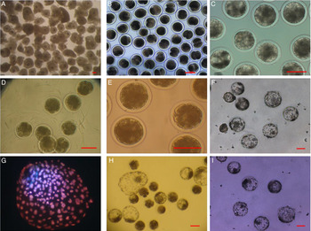
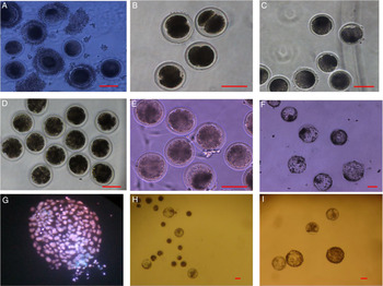
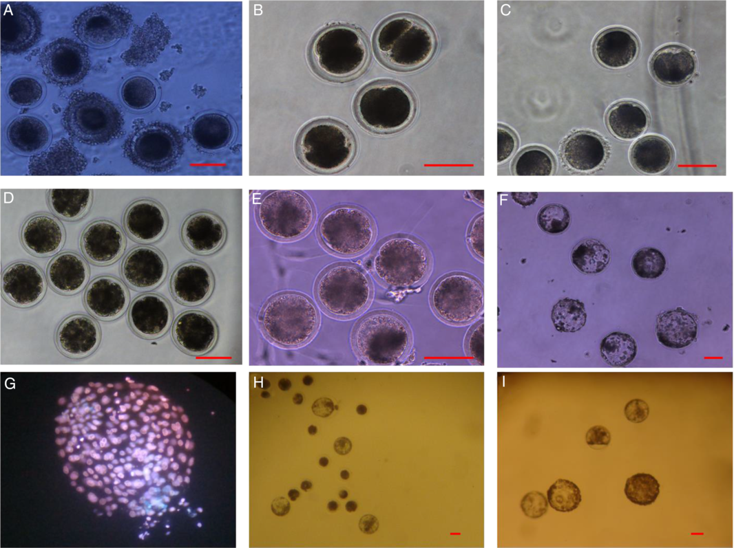
The morphology of A + B grade oocytes and different stages of parthenotes generated by ethanol activation and cultured on RVCL medium are shown in Fig. 1. Blastocysts generated by CaI and electric pulse and cultured on mCR2 and mSOF media are also shown in Fig. 1(H, I). The morphology of C + D grade oocytes and different stages of parthenotes generated by ethanol activation and cultured on RVCL medium are shown in Fig. 2(H, I). Blastocysts generated by CaI and electric pulse and cultured on mCR2 and mSOF media are also shown in Fig. 2. In the present study, the percentage of A + B oocytes (62.16 ± 5.06%, range 53.8–71.3%) was significantly higher (P < 0.001) compared with that of C + D oocytes (37.8 ± 5.00%, range 28.6–46.1%).
Morphology of buffalo A + B grade oocytes and embryos. (A) A + B grade oocytes, (B) 2-cell stage, (C) 4-cell stage, (D) 8–16-cell stage, (E) morula, (F) blastocysts, (G) differential staining of blastocyst generated by ethanol activation and cultured on RVCL medium, (H) blastocysts generated from CaI and cultured on RVCL medium, (I) blastocysts generated from electric pulse and cultured on RVCL medium. Scale bars, 100 µm.

Morphology of buffalo C + D grade oocytes and embryos. (A) C + D grade oocytes, (B) 2-cell stage, (C) 4-cell stage, (D) 8–16-cell stage, (E) morula, (F) blastocysts. (G) Differential staining of blastocyst generated by ethanol activation and cultured on RVCL medium. (H) Blastocysts generated from CaI and cultured on RVCL medium. (I) Blastocysts generated from electric pulse and cultured on RVCL medium. Scale bars, 100 µm.
Parthenogenetic activation of buffalo oocytes
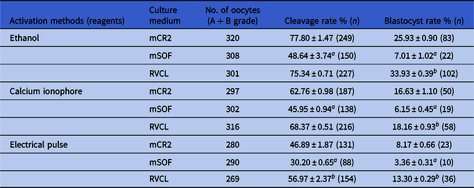
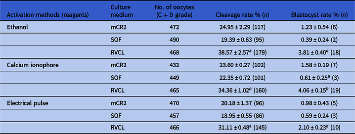
The developmental competence of A + B and C + D grade oocytes after parthenogenetic activation with three chemicals and subsequent culture in three media was examined in the present work. Table 1 summarizes the developmental competence of A + B grade oocytes. After ethanol or CaI or electrical pulse activation, a significant increase (P < 0.05) in cleavage rate and blastocyst rate was found in all three treatment groups when activated oocytes were cultured in mCR2 and RVCL media compared with that of culture in mSOF medium. Similar results were found for C + D grade oocytes after activation with three chemicals and subsequent culture in three media (Table 2). Among all combinations, ethanol activation followed by RVCL culture gave the highest cleavage and blastocyst yield for both A + B and C + D grade oocytes.
Effect of culture medium and activation methods on cleavage rate and blastocysts rate of parthenogenetically activated embryos derived from (A + B) quality oocytes*

*Data from five trials.
a,b Values having different superscripts along columns, differ significantly (P < 0.05).
Effect of culture medium and activation methods on cleavage rate and blastocysts rate of parthenogenetically activated embryos derived from (C + D) quality oocytes*
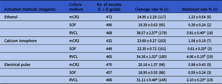
*Data from five trials.
a,b Values having different superscripts along columns, differ significantly (P < 0.05).
In the present study, the developmental competence of blastocysts derived from A + B grade oocytes activated with ethanol and cultured in RVCL were compared with blastocysts derived from C + D grade oocytes activated with ethanol and cultured in RVCL. The quality assessment of blastocyst produced through parthenogenetic activation of both grade oocytes was carried out on the basis of total cell number (TCN) and ICM/TE ratio using differential staining and apoptotic index using TUNEL assay. TCN of the blastocysts produced from A + B grade oocytes was observed to be 171.32 ± 2.64, while ICM to trophectoderm ratio was found to be 0.16 ± 0.006 (Fig. 1 G) and the TCN of the blastocysts produced through C + D grade oocytes was observed to be 163.04 ± 3.83, while ICM to trophectoderm ratio was found to be 0.10 ± 0.086 (Fig. 2 G). TCN, ICM/TE and apoptotic index of A + B group blastocysts were significantly different (P < 0.05) from that of their C + D counterpart (Table 3).
Comparison of TCN, ICM/TE ratio and apoptotic index between blastocysts derived from A + B and C + D grade oocytes

a,b Values having different superscripts along columns, differ significantly (P < 0.05).
Expression level of the cumulus–oocyte complex, fertilization, developmental competence and apoptosis-related genes in parthenogenetically produce embryos
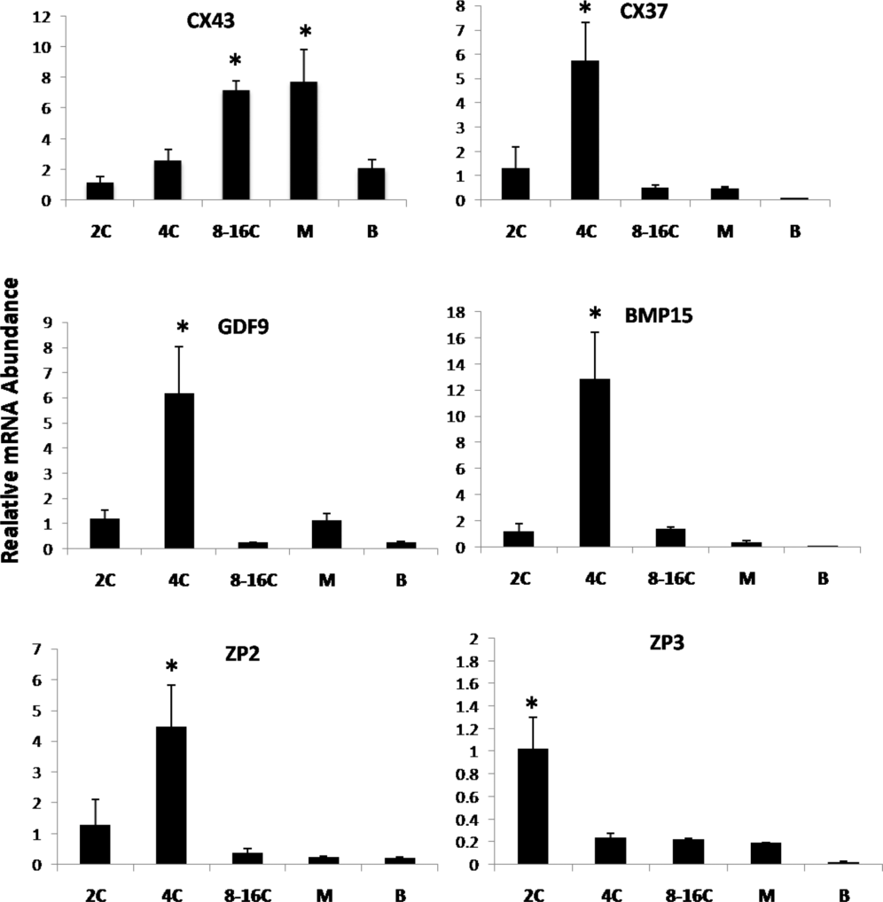
The expression of developmentally regulated genes was studied in the parthenogenetically produced buffalo embryo at different stages. The relative mRNA abundances of CX37, GDF9, and BMP15 genes were significantly (P < 0.05) higher in 4-cell stage embryos and relatively lower in the other stages such as 2-cell, 8–16-cell, morula and blastocysts (Fig. 3). CX43 expression was significantly (P < 0.05) higher in the morula and 8–16-cell stage embryos compared with that in 4-cell stage and with embryos at all the stages examined (Fig. 3).
Relative mRNA abundance of some cumulus–oocyte complex and fertilization related important genes in different developmental stages (2C, 2-cell stage; 4C, 4-cell stage; 8–16C, 8–16 cell stage; M, morula stage; B, blastocyst stage) of parthenogenetically activated buffalo embryos. Bars with different superscripts differ significantly (P < 0.05). Ten embryos at each stage were used and the experiment was repeated three times with each run in triplicates.
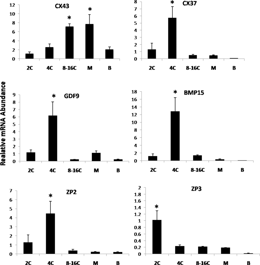
Among the genes that played a significant role in fertilization, expression of zona pellucida (ZP) ZP2 and ZP3 genes was analyzed. Relative mRNA abundance of ZP2 was significantly (P < 0.05) higher in 4-cell stage embryos compared with the 2-cell stage; expression was found to be lower in 8–16-cell stage embryos, morula and blastocysts (Fig. 3). ZP3 was found to be significantly (P < 0.05) higher in 2-cell stage embryos and its expression was found to be the same in 4-cell and 8–16-cell stages, and morula and blastocysts (Fig. 2).
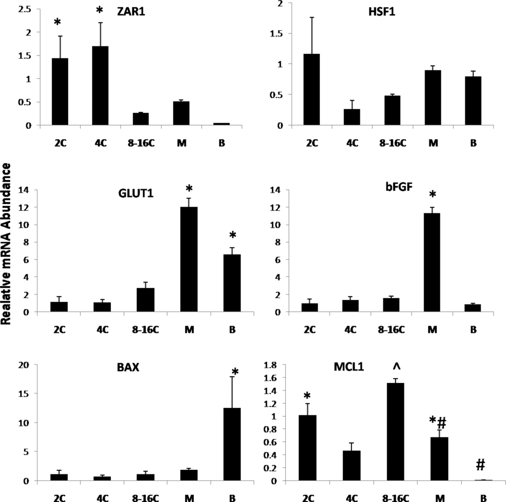
The mRNA abundance of the ZAR1 gene was found to be significantly (P < 0.05) higher in 4-cell and 2-cell stage embryos and lower in 8–16-cell stage, morula and blastocysts (Fig. 4). The expression of the HSF1 gene was the same in all stages; therefore there was no significant difference (P < 0.05) among any developmental stage (Fig. 4). GLUT1 expression was found to be significantly (P < 0.05) higher in morula and blastocysts and lower in abundance in 2-cell, 4-cell and 8–16-cell stage embryos (Fig. 4). bFGF was expressed more in the morula and its expression was the same in other examined stages (Fig. 4).
Relative mRNA abundance of some apoptosis and developmental competence related important genes in different developmental stages (2C, 2-cell stage; 4C, 4-cell stage; 8–16C, 8–16 cell stage; M, morula stage; B, blastocyst stage) of parthenogenetically activated embryos. Bars with different superscripts differ significantly (P < 0.05). Ten embryos at each stage were used and the experiment was repeated three times with each run in triplicates.

The pro-apoptotic gene BAX showed relatively higher expression in blastocysts and its expression was the same in all examined stages (Fig. 4). Expression of the anti-apoptotic gene MCL1 was higher in the 8–16-cell stage compared with that 2-cell and 4-cell stages and morula. Very low expression was observed in blastocysts (Fig. 4).
Discussion
Parthenogenetic activation has been used as a tool to study mechanisms involved in the initiation of embryonic development. To our knowledge, this is the first report on parthenogenetic activation of buffalo A + B and C + D grade oocytes. In the present study, buffalo oocytes of both grades could be activated successfully and developed parthenogenetically up to the blastocyst stage by treatment with CaI, ethanol or electrical activation followed by 6-DMAP treatment and cultured in three different media. Some studies on parthenogenetic activation of buffalo oocytes by ethanol or ionomycin followed by 6-DMAP treatment have been reported previously (Gasparrini et al., Reference Gasparrini, Boccia, Rosa, Palo, Campanile and Zicarelli2004; George et al., Reference George, Shah, Sharma, Palta, Singla, Manik and Chauhan2011; Singh et al., Reference Singh, Kaushik, Garg, Sharma, George, Singh, Manik, Palta, Singla and Chauhan2012, Reference Singh, Kaushik, Mohapatra, Garg, Ameshbabu, Singh, Palta, Manik, Singla and Chauhan2014) but no report was available to compare different culture media in two different groups of oocytes.
The use of chemical agents and electrical stimuli led to an increase in cytoplasmic Ca2+ levels, but most of these failed to generate Ca2+ oscillations, which are the hallmark of sperm-mediated oocyte activation. Instead, they caused just a single, long rise in Ca2+ (Swann and Ozil, Reference Swann and Ozil1994), which was not sufficient to completely downregulate MPF activity. Thimerosal and strontium ions are the only known chemical agents capable of causing sustained Ca2+ oscillations (Swann, Reference Swann1991; Kline and Kline, Reference Kline and Kline1992). However, thimerosal caused the destruction of meiotic spindle in oocytes (Cheek et al., Reference Cheek, McGuinness, Vincent, Moreton, Berridge and Johnson1993). In the view of their inability to mimic Ca2+ oscillations, chemical agents are used in combination with a protein synthesis inhibitor such as cycloheximide or a protein kinase inhibitor such as 6-DMAP that prevent MPF re-accumulation (Alberio et al., Reference Alberio, Zakhartchenko, Motlik and Wolf2001), yet these agents also contribute to reduction in the developmental competence of parthenogenetically activated oocytes. Whereas exposure to 6-DMAP caused a high frequency of chromosomal abnormalities in parthenogenetic cattle embryos (De La Fuente and King, Reference De La Fuente and King1998), cycloheximide seemed to prolong the completion of the first cell cycle, resulting in lower cleavage rates (Holm et al., Reference Holm, Booth and Callesen2003).
The present study has clearly shown that high efficiency of oocyte activation in the buffalo can be achieved by sequential treatment with either CaI or ethanol followed by incubation in 6-DMAP. Our results are in agreement with previous findings on a combination treatment of chemical agents (Gasparrini et al., Reference Gasparrini, Boccia, Rosa, Palo, Campanile and Zicarelli2004). We compared two groups of oocytes (A + B and C + D grade) activated with three different reagents and cultured in three different media. The A + B grade oocytes activated with ethanol and CaI had significantly (P > 0.05) higher cleavage rates and blastocyst production when these activated oocytes were cultured with RVCL and mCR2 media respectively. Oocytes activated with electric pulse also had higher cleavage and blastocyst development rates in RVCL and mCR2 media, respectively, which were lower than that of ethanol and CaI. The oocytes activated with ethanol, CaI and electric pulse were comparatively lower in terms of cleavage and blastocyst production rate when cultured in mSOF medium. Previous studies have also reported that the parthenogenetic activation of buffalo oocytes (with and without zona) with ethanol and CaI produced a better cleavage and blastocyst development rate when cultured in RVCL medium (George et al., Reference George, Shah, Sharma, Palta, Singla, Manik and Chauhan2011). Shah et al. (Reference Shah, George, Singh, Kumar, Chauhan, Manik, Palta and Singla2008) found the compaction of buffalo zona-free embryos and their subsequent development to the blastocyst stage in RVCL and mSOF media but not in mCR2 media. Mishra et al. (Reference Mishra, Misra and Sharma2008) reported that the combination of 7% ethanol + DMAP gave better cleavage and blastocyst production rates in buffalo. Saikhun et al. (Reference Saikhun, Kitiyanant, Songtaveesin, Pavasuthipaisit and Kitiyanant2004), found that ethanol and CaI with DMAP had significantly (P < 0.05) higher cleavage and blastocysts rates compared with those activated by electric pulse + DMAP. The parthenogenetic activation of sheep oocytes also had superior cleavage and blastocyst development rates in CaI and ethanol (Loi et al., Reference Loi, Ledda, Fulka, Cappai and Moor1998; Nandedkar et al., Reference Nandedkar, Chohan, Patwardhan, Gaikwad and Bhartiya2009). The reason for this contradictory result may be the quality of oocytes obtained from slaughter house ovaries and optimal culture condition. A similar result was obtained in C + D grade buffalo oocytes. Ethanol, CaI and electric pulse-activated C + D grade buffalo oocytes were found to give better cleavage and blastocysts development rates in RVCL and mCR2 media respectively, but there were very poor cleavage and blastocysts development rates in mSOF medium. There is no report available describing the culture of these oocytes separately. Mishra et al. (Reference Mishra, Misra and Sharma2008) reported the recovery of A, B, C and D grade buffalo oocytes from slaughter ovaries, but they did not reported separate culture of these oocytes. Regarding low embryo development from C, D grade oocytes, possible reasons may be that these oocytes were either denuded without a cumulus layer or had one layer of cumulus cells, as cumulus cells play important roles in embryo development.
In the present study, the temporal mRNA expression pattern of developmentally important genes in buffalo embryos produced parthenogenetically was analysed using RT-qPCR. These transcripts are involved in several areas such as compaction and as growth factors (CX37, CX47, GDF9, BMP15), zona pellucida (ZP2, ZP3), metabolism (GLUT1), stress (HSF1) and maternal (ZAR1).
During murine preimplantation development, functional gap junctions were first observed at compaction at the 8-cell stage and mRNA encoding the CX43 gene (Connexin-43) and Cx43 protein were detected from the 4-cell stage onwards (Nishi et al., Reference Nishi, Kumar and Gilula1991). CX43 and CX37, also known as Gap junction protein α1 (GJA1) and Gap junction protein α4 (GJA4), respectively, have been found within the developing ovarian follicle (Kidder and Mhawi, Reference Kidder and Mhawi2002). These are the major gap junctional proteins expressed in the cumulus–oocyte complex (COC) and granulosa cells and predominantly mediate cell-to-cell communication in ovarian follicles (Granot and Dekel, Reference Granot and Dekel2002). Wrenzycki et al. (Reference Wrenzycki, Herrmann, Carnwath and Niemann1998) investigated the expression of CX43 in bovine in vitro produced embryos up to the morula stage; CX43 expression was found in in vivo produced bovine blastocysts (Wrenzycki et al., Reference Wrenzycki, Herrmann, Carnwath and Niemann1996). Gómez et al. (Reference Gómez, Gutiérrez-Adán, Díez, Bermejo-Alvarez, Muñoz, Rodriguez, Otero, Alvarez-Viejo, Martín, Carrocera and Caamaño2009) reported that CX43 was upregulated in bovine IVF blastocysts but downregulated in parthenogenetic blastocysts. Nemcova et al. (Reference Nemcova, Machatkova, Hanzalova, Horakova and Kanka2006) found expression of CX43 in bovine day 7 and day 8 blastocysts. For buffalo, both CX43 and CX37 transcripts were present in all developmental stages of parthenotes at different levels.
Growth differentiation factor 9 (GDF9) and bone morphogenetic protein 15 (BMP15) are two well characterized oocyte-derived growth factors that play crucial roles in follicle growth and ovulation in all mammalian species. GDF9 was originally identified in mouse ovary (McGrath et al., Reference McGrath, Aurora, Esquela and Lee1995). GDF9 plays a critical function as a growth and differentiation factor during early folliculogenesis and as a key regulator of several granulosa cell proteins involved in cumulus expansion and maintenance of an optimal oocyte microenvironment. GDF9 and BMP15 expression was reported in buffalo oocytes (Nath et al., Reference Nath, Sharma, Dubey, Pratheesh, Gade, Saikumar and Sharma2013; Murlidharan et al., Reference Murlidharan, Eswari and Vijayarani2015), bovine oocytes (Donnison and Pfeffer, Reference Donnison and Pfeffer2004), sheep oocytes (Galloway et al., Reference Galloway, McNatty, Cambridge, Laitinen, Juenge, Jokiranta, McLaren, Luiro, Dodds, Montgomery, Beattie, Davis and Ritvos2000), mouse oocytes (Dong et al., Reference Dong, Albertini, Nishimori, Kumar, Lu and Matzuk1996) and human (McGrath et al., Reference McGrath, Aurora, Esquela and Lee1995). In vitro produced bovine embryos for 2-cell, 4-cell and 8-cell stage embryos expressed these genes, but expression was not significantly detected in 16-cell stage, morula or blastocysts (Donnison and Pfeffer, Reference Donnison and Pfeffer2004). McGrath et al. (Reference McGrath, Aurora, Esquela and Lee1995) also reported that GDF9 mRNA expression was lost rapidly after fertilization in human. In our results both genes were present in all the developmental stage including blastocysts, but variable expression patterns and higher mRNA expression was found for 4-cell stage embryos.
Zygote arrest 1 is an evolutionarily conserved, ovary-specific maternal factor that plays essential roles during oocyte-to-embryo transition. In human and mouse, Zar1 mRNA was present in oocytes and 1-cell embryos, but decreased significantly at the 2-cell stage and was not detected at further stages of development or in tissues other than the ovary (Wu et al., Reference Wu, Viveiros, Eppig, Bai, Fitzpatrick and Matzuk2003). In pig and bovine, the ZAR1 transcript was present in all embryonic development stages. Very low expression was found in morula and blastocysts (Uzbekova et al., Reference Uzbekova, Roy-Sabau, Dalbiès-Tran, Perreau, Pappillier, Mompart, Thelie, Pennetier, Cognie, Cadoret, Royere, Monget and Mermillod2006). Brevini et al. (Reference Brevini, Cillo, Colleoni, Lazzari, Galli and Gandolfi2004) reported ZAR1 transcript expression present in all developmental stages of bovine embryos up to blastocyst formation. In our results, we found a similar pattern, but ZAR1 expression was higher in 4-cell and 2-cell stage embryos.
The induction of heat shock genes in eukaryotes by heat or other forms of stress is mediated by a transcription factor known as heat shock factor 1 (HSF1), which is a maternal effect gene. Heat shock transcription factor 1 is the main regulator of the stress response that triggers the transcription of several genes encoding heat shock proteins (HSPs). Metchat et al. (Reference Metchat, Malin, Christiane, Virginie, Lea, Henri and Elisabeth2009) reported that HSF1 –/– females produced oocytes but no viable embryos. Christians et al. (Reference Christians, Davis, Thomas and Benjamin2000) reported that HSF1 is one of the few maternal factors in mammals required for normal embryonic development beyond the 1-cell or 2-cell stages and is necessary for female reproductive success. Eunju et al. (Reference Eunju, Guangming, Hong, Ying, Rebecca, Masahito, Michelle, Don, Hans and Shoukhrat2014), found expression of the HSF1 gene in all the embryonic stages in mouse, but very low expression was found in blastocysts. Similar results were found in this study, the HSF1 transcript was expressed in all embryonic developmental stages of buffalo parthenotes.
GLUT1 is a ubiquitous, facilitative glucose transporter and a major source of metabolic energy for most mammalian cells, but it cannot support embryonic development before compaction (Brinster, Reference Brinster1965). However in vitro produced embryos from different developmental stages of mouse (Hogan et al., Reference Hogan, Heyner, Charron, Copeland, Gilbert, Jenkins, Thorens and Schultz1991; Pantaleon et al., Reference Pantaleon, Ryan, Gil and Kaye2001), bovine (Balasubramanian et al., Reference Balasubramanian, Son, Kumar, Ock, Yoo, Im, Choe and Rho2007) and buffalo (Kumar et al., Reference Kumar, Verma, Kumar, De, Kumar and Datta2015; Sadeesh et al., Reference Sadeesh, Meena, Balhara and Yadav2014) expressed the GLUT1 transcript. This result agreed with previous reports; the GLUT1 transcript was expressed in all developmental stages of buffalo embryos but with higher expression levels in morula and blastocysts.
The fibroblast growth factor (FGF) family is emerging as a group of factors that are involved in the regulation of ovarian function. Studies in mammals have located bFGF and its receptors to preantral ovarian follicles in the rat (Nilsson et al., Reference Nilsson, Parrott and Skinner2001). Expression of the bFGF transcript was reported in bovine blastocysts (Gilbert et al., Reference Gilbert, Mark, Andrew, Mayi, Karen and Mark1996). For buffalo, the transcript was expressed in all developmental stages including blastocysts. bFGF promotes early bovine embryonic development in vitro during the fourth cell cycle, synergistically with transforming growth factor β (Larson et al., Reference Larson, Ignotz and Currie1992).
Apoptosis is dependent upon the actions and interactions of at least 100 gene products, the most widely studied of which are members of the BCL-2 family (Adams and Cory, Reference Adams and Cory1998). These can be subdivided into two groups: anti-apoptotic cell death suppressors (BCL-2, BCL-XL, BCL-W and MCL-1) and pro-apoptotic cell death inducers (BAX, BAK and MTD). Sadeesh et al. (Reference Sadeesh, Meena, Balhara and Yadav2014) reported that exposure of buffalo oocytes to summer heat stress increased the relative mRNA abundance of pro-apoptotic gene BAX at the 8- to 16-cell stages. In our case, BAX expression was found to be higher at the blastocyst stage and MCL-1 expression was higher in 8- to 16-cell and 2-cell stages. Elamaran et al. (Reference Elamaran, Singh, Singh, Singla, Chauhan, Manik and Palta2012) found that oxygen concentration and cysteamine supplementation during in vitro production of buffalo embryos affected mRNA expression of BCL-2, BCL-XL, MCL-1, BAX and BID. More recently, one study reported on gene expression in different stages of buffalo parthenotes, and showed differential gene expression of various developmental genes in different embryonic developmental stages (Fumei et al., Reference Fumei, Qiang, Liping, Zhen, Zhuangzhuang, Pengfei, Tingxian, Chunying, Xianwei, Yangqing and Ming2020).
In conclusion, the percentage of A + B grade oocytes was significantly higher compared with that of C + D grade oocytes retrieved from slaughterhouse buffalo ovaries. Different methods employed to activate buffalo A + B and C + D quality oocytes parthenogenetically, in vitro developmental competence of oocytes, and relative mRNA abundance were studied. Among all combinations, ethanol activation followed by RVCL culture gave the highest cleavage and blastocyst yield for both A + B and C + D grade oocytes. In vitro developmental competence of the A + B group was better compared with its C + D counterpart. The expression profiles of CX37, CX47, GDF9, BMP15, ZP2, ZP3, GLUT1, HSF1, ZAR1, BAX and MCL1 were also studied for different stages of parthenogenetically produced buffalo embryos, and indicated that the dynamics of the above genes had a role in early embryonic development.
Embryos produced through IVF are a very precious way to produce a life; parthenotes could be alternate sources for production of embryos by different activation reagents that could be used for future culture of embryonic stem cells, genomics and proteomics studies in embryology.
Supplementary material
To view supplementary material for this article, please visit https://doi.org/10.1017/S0967199420000519
Authors’ contribution
KPS designed and carried out the work, SKM and RK analyzed data, KP, MK, PP, RS drafted the manuscript, MSC and SKS provided funds for study.
Financial support
We are grateful for National Agriculture Innovative Project (NAIP) grants to SKS (C2-1-(5)/2007) and MSC (C-2067 and 075).
Conflict of interest
The authors declare that there are no competing interests.
Ethical standards
Not applicable.