Tetralogy of Fallot is frequently associated with aortic arch anomalies, systemic venous anomalies, and atrial and atrioventricular septal defects.Reference Siwik, Erenberg, Zahka and Goldmuntz 1 We present two cases of tetralogy of Fallot, who had concomitant anomalous left coronary artery from pulmonary artery and total anomalous pulmonary venous connection, respectively, both extremely unusual associations, and we also discuss their embryological origin, physiological consequences, and review the related literature.
Case reports
Case 1
A 4-year-old boy presented with cyanosis and exertional dyspnoea (NYHA Class III). The saturation was 65%, and an ejection systolic murmur was audible at the second left intercostal space with a pansystolic murmur (grade 3/6) at the apex.
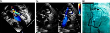
Echocardiography showed sinus rhythm with left bundle branch block, and the chest radiograph revealed cardiomegaly. Echocardiography demonstrated tetralogy of Fallot with a diffusely hypoplastic left pulmonary artery measuring 4 mm in diameter (z-score −3.5). The right pulmonary artery measured 9 mm (z-score 0.27). The papillary muscles of the mitral valve appeared echogenic with moderate mitral regurgitation from the coaptation point. Anomalous origin of the left coronary artery from the pulmonary artery was suspected with a dilated right coronary artery, multiple coronary collaterals, and flow from the left main coronary artery into the proximal pulmonary artery (Fig 1). The aortic arch was left sided. There was mild left ventricular systolic dysfunction (ejection fraction 48%).
Transthoracic echocardiogram. ( a ) Apical four-chamber view showing increased mitral papillary muscle echogenicity with mitral regurgitation. ( b ) Parasternal short-axis view demonstrating the left coronary artery arising from the pulmonary artery (*) with reversal of flow on colour Doppler imaging. Angiography. ( c ) Selective right coronary artery angiogram demonstrates anomalous left coronary artery from pulmonary artery with good collateral flow with subsequent filling of the left coronary artery (block arrow) and the main pulmonary artery. The confluent branch pulmonary arteries and small left pulmonary artery are also seen.

On cardiac catheter study, selective right coronary angiography demonstrated anomalous left coronary artery from the pulmonary artery (Fig 1) and a hypoplastic left pulmonary artery.
He underwent intracardiac repair with a right ventricle-to-pulmonary artery conduit (12-mm Contegra bovine jugular conduit, Medtronic Inc.) and translocation of the left coronary artery. Left atrial pressures were mildly elevated in the immediate postoperative period and he had multifocal atrial tachycardia once, which reverted to sinus rhythm with synchronised cardioversion. He was discharged on the 15th postoperative day.
Postoperative echocardiography demonstrated normal left ventricular function with good flow in the re-implanted left coronary artery with moderate mitral regurgitation and a diffusely small left pulmonary artery. At the 6-month follow-up, moderate mitral regurgitation persisted with normal left ventricular function (ejection fraction 71%).
Case 2
An 8-month-old boy was referred for evaluation of a murmur. The saturation was 70% with an ejection systolic murmur at the second left intercostal space. The chest radiograph showed a boot-shaped heart with mild pulmonary venous congestion. Echocardiography demonstrated sinus rhythm with right ventricular hypertrophy and early transition.
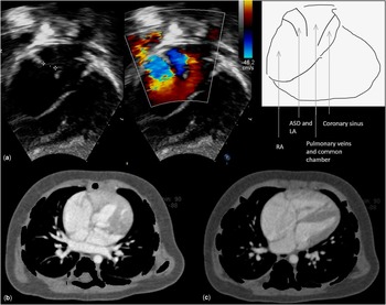
Echocardiography revealed tetralogy of Fallot with confluent branch pulmonary arteries with the right pulmonary artery being 6 mm and the left pulmonary artery being 8.7 mm in diameter. The pulmonary veins were draining into a common chamber above the left atrium and then directly into the right atrium, cardiac total anomalous pulmonary venous connection. A moderate secundum atrial septal defect was shunting right to left (Fig 2). Arterial relation was side by side with a rightward aorta. Multi-slice CT angiogram demonstrated the pulmonary venous confluence and drainage to the right atrium (Fig 2).
( a ) Transthoracic echocardiogram. Subcostal four-chamber view demonstrating the pulmonary veins draining via a common chamber to the right atrium. The secundum atrial septal defect with right-to-left shunt is also seen. ( b ) CT angiogram in the axial section demonstrating the pulmonary veins draining into a common chamber posterior to the atria. ( c ) The axial section demonstrates the common chamber draining to the right atrium.

He underwent pulmonary venous re-routing and intracardiac repair with a transannular patch and a bicuspid expanded polytetrafluoroethylene (0.1 mm) pulmonary valve. His immediate postoperative period was stormy with low cardiac output, requiring prolonged inotropic support. He was discharged from the hospital on the 11th postoperative day.
Follow-up echocardiography after 6 months revealed no residual ventricular septal defect and no right ventricular outflow tract obstruction with moderate pulmonary regurgitation. The pulmonary venous return to the left atrium was unobstructed.
Discussion
The common coronary abnormalities with tetralogy of Fallot include origin of a conal branch or left anterior descending coronary from the right coronary artery or right sinus and single coronary from the right sinus. Rarely, a left anterior descending from the pulmonary artery or intramural coronary artery has been described.Reference Perloff and Marelli 2 , Reference Bitar, Kveselis, Smith, Byrum and Quaegebeur 3 The incidence of coronary abnormalities is influenced by aortopulmonary rotation and is higher when the aortic root is anterior or side by side to the pulmonary artery.Reference Perloff and Marelli 2 Kurup et alReference Kurup, Daniel and Kumar 4 reported a 10-month-old male child with tetralogy of Fallot with normal left ventricular function where anomalous left coronary artery from pulmonary artery was incidentally detected on preoperative echocardiogram. The authors hypothesised that rapid development of collaterals from the right coronary artery may have preserved left ventricular function.
Physiologically, a patient with anomalous left coronary artery from pulmonary artery and tetralogy of Fallot would demonstrate features secondary to coronary steal and myocardial ischaemia earlier due to the low pulmonary arterial pressures and flow facilitating run-off from the left coronary artery into the pulmonary artery. The coronary flow into the pulmonary artery also constitutes a left-to-right shunt.
The origin of anomalous left coronary artery from pulmonary artery has various embryological explanations.As both defects involve the conotruncus, the coexistence of tetralogy of Fallot with anomalous left coronary artery from pulmonary artery, as seen in our patient, would give credence to theories related to the division of the embryological truncus arteriosus. Here, the coronary arteries are assumed to originate as two endothelial buds, before or simultaneously with the truncal division. At the time of enlargement of truncal cushions to form the truncal septum, displacement of the endothelial buds could assign either coronary artery to the future pulmonary artery. The proximity of the left aortic sinus to the truncal septum could explain the higher incidence of anomalous left coronary artery compared with anomalous right coronary artery from pulmonary artery.Reference Perloff and Marelli 2
The incidence of total anomalous pulmonary venous connection in patients with tetralogy of Fallot ranges from 0.25 to 0.34%.Reference Talwar, Choudhary and Shivaprasad 5 Talwar et alReference Talwar, Choudhary and Shivaprasad 5 reviewed 25 published cases of total anomalous pulmonary venous connection in patients with tetralogy, of which complete primary repair was carried out in 14 and only 11 patients survived. Patients who had palliative shunts, shunts with total anomalous pulmonary venous connection repair, and tetralogy intracardiac repair leaving the total anomalous pulmonary venous connection have not survived surgery, indicating that the only surgical option with this physiology is a single-stage primary repair.
Tetralogy of Fallot with total anomalous pulmonary venous connection causes chronic volume and pressure overload of the right ventricle and under-filling of the left atrium and left ventricle. The only source of left heart preload is the atrial septal defect.Reference Talwar, Choudhary and Shivaprasad 5 If the abnormal pulmonary venous drainage is not identified and corrected, there may be difficulty in weaning the patient off cardiopulmonary bypass and may even be fatal.Reference Talwar, Choudhary and Shivaprasad 5 As the left ventricle in this physiology is under-filled preoperatively, we expect an increased predisposition to low cardiac output state in the immediate postoperative period as was seen in our patient. Significant pulmonary regurgitation postoperatively with a transannular patch would further contribute to right ventricular failure, and was prevented in our patient with a bicuspid polytetrafluoroethylene pulmonary valve.Reference Quintessenza, Jacobs, Morell, Giroud and Boucek 6
In the classical embryological hypothesis, total anomalous pulmonary venous connection was believed to arise secondary to agenesis of the common pulmonary vein with persistence of primitive communications between the pulmonary venous and systemic venous beds.Reference Perloff and Marelli 2 Although anomalies of the venous pole were thought to be independent of conotruncal defects such as tetralogy of Fallot in the classical view, recent studies have postulated multiple embryological origins of conotruncal defects. The present view, corroborated by molecular biological techniques, is that the primary myocardium derived from the cardiac crescent is reinforced by other contributions of primary myocardium from the anterior, or secondary, heart field,Reference Anderson, Brown and Moorman 7 which is a source of cardiac progenitor cells in the pharyngeal mesoderm. This secondary heart field could give rise to both the arterial and venous poles of the heartReference Bajolle, Zaffran, Losay, Ou, Buckingham and Bonnet 8 , Reference Buckingham, Meilhac and Zaffran 9 and mutations affecting it would explain the association of tetralogy of Fallot with total anomalous pulmonary venous connection.
In conclusion, the rare associations of anomalous left coronary artery from the pulmonary artery with total anomalous pulmonary venous connection in patients with tetralogy of Fallot can be strongly suspected and diagnosed on transthoracic echocardiogram and have good immediate and mid-term surgical outcomes in our experience. Awareness of these rare associations will preclude missed diagnoses and consequent surgical surprises or catastrophes and they add to our understanding of the embryological origins of tetralogy of Fallot. Further cases with longer follow-up are required to study whether these associations alter the long-term outcomes of operated tetralogy of Fallot.
Acknowledgements
The authors thank Asavari Tawade, cardiac sonographer, Children’s Heart Centre, for help with non-invasive imaging of the patients.
Financial Support
This research received no specific grant from any funding agency, commercial, or not-for-profit sectors.
Conflicts of Interest
None.