Introduction
While most cerebrospinal fluid (CSF) leaks are caused by head trauma and surgical procedures, 6 to 24 per cent occur spontaneously. Known causes of spontaneous CSF leakage include empty sella syndrome, hydrocephalus and meningoencephalocoele. In some cases, the aetiology remains unclear.Reference Wolf, Greistorfer and Stammberger 1 , Reference Lindstrom, Toohill, Loehrl and Smith 2 The majority of spontaneous CSF leaks occur in the anterior skull base, and only a few cases of spontaneous leaks in the temporal bone have been described.Reference Leonetti, Marzo, Anderson, Origitano and Vukas 3 While patients with an anterior skull base CSF leak typically present with watery nasal discharge, leaks in the temporal bone may cause middle-ear effusion or rhinorrhoea via the eustachian tube.
We present a case of synchronous spontaneous CSF leaks in the anterior skull base and temporal bone, a previously unreported occurrence.
Case report
A 34-year-old woman presented with right-sided hearing loss and watery discharge from both nostrils. Her past medical history was unremarkable for previous otological or rhinological symptoms or significant head trauma.
Pure tone audiometry revealed a conductive hearing loss of 25 dB in the right ear. Impedance audiometry revealed a type B pattern on the right side and a normal pattern on the left.
Endoscopic examination of the nose showed clear secretion bilaterally, but was otherwise unremarkable.
Paracentesis of the right tympanic membrane revealed clear fluid in the middle ear.
Due to the watery quality of both the nasal and ear secretions, CSF leakage was included in the differential diagnosis. This was indeed the final diagnosis, as both the nasal and the ear secretions were proven to be CSF upon β trace protein testing (with a concentration of 31.3 mg/l).
Routine testing revealed the patient to be pregnant, at a gestational age of 13 weeks and 1 day. The clinicians and the patient discussed the risks of pre-delivery contrast-mediated imaging and surgery versus treatment after delivery. The patient requested that imaging and surgery be performed before delivery.
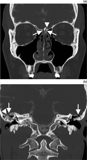
Computed tomography (CT) and cisternography demonstrated bilateral osseous defects in the ethmoid roof, with active CSF leakage (Figure 1a), as well as osseous defects in the mastoid roof on both sides, accompanied by a mastoid effusion on the right side (Figure 1b).
High resolution, coronal computed tomography scans with cisternography, showing: (a) osseous defects (arrows) in the cribiform plate on both sides, with active leakage (arrowhead) after intrathecal contrast application in the ethmoid roof; and (b) osseous defects in the right and left mastoid (arrows), with indication of active leakage in the right mastoid (arrowhead) accompanied by a mastoid effusion.

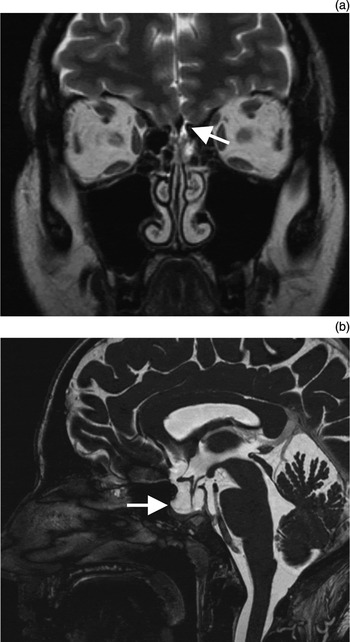
Magnetic resonance imaging (MRI) of the skull base showed a CSF-isointense signal in the intracranial vicinity of the osseous defects (Figure 2a) and an ‘empty sella’ sign (Figure 2b).
High resolution, T2-weighted magnetic resonance imaging scans. (a) Coronal section showing cerebrospinal fluid isointense signal in the intracranial vicinity of the osseous defects (arrow), without any bulging or herniated meningoencephalic sacs. (b) Axial section showing ‘empty sella’ sign (arrow) with typical thinned bone and a flattened pituitary gland.

Surgical procedure
Microsurgical exploration confirmed bilateral, 10 × 10 mm osseous defects at the transition from the ethmoid roof to the lamina cribrosa, with liquorrhoea from dural leaks of approximately 5 × 5 mm on both sides.
The CSF leaks were closed in three layers, working from inside to outside, using: (1) Tutopatch (bovine pericardium; Tutogen, Neunkirchen, Germany) in an intracranial-extradural position; (2) TachoSil (collagen sponge coated in fibrinogen and thrombin; Nycomed, Linz, Austria); and (3) mucosa from the inferior turbinate. All three layers were fixed with fibrin glue (Evicel; Ethicon, Norderstedt, Germany).
In the same surgical session, a subtotal mastoidectomy was performed on the right side, during which two osseous defects in the right mastoid roof (12 × 8 mm and 5 × 5 mm) were observed, both with exposed dura but no visible liquorrhoea at that time. A duraplasty was performed at the laterobasal aspect of each defect, in the same manner as in the nose.
A lumbar drain was placed, which was retained for 10 days post-operatively, in order to keep the patient's intracranial pressure (ICP) at 15 cmH2O; her pre-operative ICP had been found to be elevated to values of 25 cmH2O.
Six months after surgery, the patient delivered a healthy male infant via elective caesarean section.
Two years after surgery, the patient was free of CSF leakage, with an unremarkable neurological status.
Discussion
Spontaneous CSF leaks are often associated with an elevated ICP (with a mean value of 32.5 cmH2O, compared with a normal range of 5–15 cmH2O), as in the presented case.Reference Wise and Schlosser 4 So-called idiopathic intracranial hypertension is more prevalent in middle-aged, obese women than in the general population (with prevalences of 19.3/100 000 versus 0.9/100 000, respectively). Consistently, this subgroup develops spontaneous CSF leaks more often than the rest of the population.Reference Durcan, Corbett and Wall 5 – Reference Hannerz and Ericson 7 However, the incidence of idiopathic intracranial hypertension has not been shown to be generally higher during pregnancy.Reference Tang, Dorotheo, Schiffman and Bahrani 8 It remains a matter of speculation whether the pregnancy in our patient, who was not obese, contributed to her elevated ICP or not.
-
• This patient showed synchronous spontaneous cerebrospinal (CSF) leaks from the nose and ear
-
• Anterior skull base CSF leaks cause watery nasal discharge
-
• Temporal bone CSF leaks cause middle-ear effusion or pseudorhinorrhoea
-
• Spontaneous CSF leaks occur more often in obese women of child-bearing age
-
• Diagnosis involves β trace protein testing, computed tomography, cisternography and magnetic resonance imaging
-
• Treatment of limited CSF leaks is by multi-layer surgical closure
Identification of CSF is accomplished by testing for β trace protein; this test has high sensitivity and specificity. High resolution CT is accurate in delineating osseous defects, and when combined with MRI may reveal the contents of osteodural defects and dehiscences. In addition, CT cisternography is an important investigation for demonstrating actual CSF leakage.Reference Schlosser and Bolger 9 However, cisternography carries an additional – albeit low – radiation burden (comparable to a low-dose CT scan of the paranasal sinuses), and it is an invasive procedure with the potential risk of infection. Despite performance of this investigation, in the presented, especially complex case it was not entirely clear from which anatomical sites the liquorrhoea came. Therefore, our concern was that failure to close all the leaks in the one operation would result in the patient requiring multiple procedures. These risks and benefits of cisternography were weighed and discussed with the patient, who opted to undergo the procedure. In our patient, there was visible peri-operative CSF discharge at the frontobasal defects, but no active liquorrhoea at the two osseous temporal bone defects. Therefore, the possibility of retrograde CSF flow through the eustachian tube from the ethmoid roof to the middle ear had to be considered. Ayanoglu et al. demonstrated reflux of radionuclide-marked nasopharyngeal content into the middle ear via the eustachian tube in one of 38 non-selected patients.Reference Ayanoglu, Uneri, Turoglu and Dogan 10 However, in our patient the cisternographic findings, as well as the coincidence of osseous defects in the right mastoid roof and unilateral CSF middle-ear effusion on the same side, indicated a temporal bone CSF leak, which appeared to have spontaneously closed by the time of surgery.
Conclusion
A literature reviewed indicated that our patient is the first reported case of synchronous spontaneous CSF leaks in the nose and ear. The patient's pregnancy may have been a contributing factor to this unusual presentation.