Case A
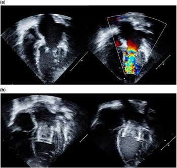
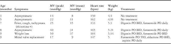
A newborn girl was prenatally diagnosed with left congenital diaphragmatic hernia. Her fetal echocardiogram demonstrated dextroposition, borderline hypoplastic left-sided structures, and diffuse mild hypoplasia of the aortic arch. At birth, she had features of short-limbed short stature. Her diaphragmatic hernia was primarily repaired at 2 days of life, and she was discharged 1 week after surgery. At 3 months of age, a follow-up echocardiogram showed a mildly dilated left atrium and mild mitral stenosis (mean gradient 8 mmHg, heart rate 150). Clinical whole-exome sequencing identified a de novo C1733G mutation in the FBN1 gene, confirming the diagnosis of geleophysic dysplasia. She had five follow-up visits (Table 1) because of progressive worsening in mitral valve gradients with thickening of the mitral leaflets (Fig 1a, Supplementary Figure 2). At 9 months of age, she underwent a transoesophageal echocardiogram (Supplementary Figure 3) and mitral valve replacement with an Edwards–Sapien 20-mm valve that was dilated to 16 mm (Fig 1b). The Edwards–Sapien valve is a transcatheter low-profile bioprosthetic valve, which was placed during a hybrid procedure by the surgeons and interventional cardiologists. At the time, her weight was 5.2 kg and her height was 59 cms (body surface area 0.29). It was felt that she was not a good candidate for mechanical mitral valve replacement. This particular valve was inserted at a diameter of 13–14 mm and expanded in place. Her postoperative course was uncomplicated, and she was discharged home on daily aspirin, spironolactone, and diuretics. At 15 months of age, she again developed signs of increasing mitral stenosis and pulmonary oedema. Her echocardiogram showed a severely dilated left atrium with anterior bowing of the atrial septum. Mean gradients across the mitral valve ranged from 16 to 22 mmHg, and her estimated right ventricular systolic pressure was 50–60 mmHg based on the tricuspid regurgitant jet; her systemic blood pressure at the time was 118/77 mmHg. Cardiac catheterisation showed elevated pulmonary artery pressures, a mean left atrial pressure of 24 mmHg, and a left ventricular end-diastolic pressure of 18 mmHg. The left ventricular outflow gradient was 10 mmHg, possibly from a combination of dynamic and mechanical obstruction by the prosthetic valve apparatus. Her prosthetic valve was balloon dilated from 16 to 18 mm (Fig 1b). Over the next 4 months, she developed increasing left ventricular outflow tract gradients (echocardiographic peak of 80 mmHg and mean of 49 mmHg) and increasing mitral valve gradients. Owing to recurrent severe mitral stenosis, she underwent a replacement of the Sapien valve with a 15-mm St. Jude Medical mechanical mitral valve at 19 months of age. She was placed under extra-corporeal life support in the operating room owing to poor respiratory function. She underwent successful decannulation in the paediatric ICU, but her respiratory function did not recover and she was removed from ventilatory support at 23 months of age.
( a ) Case A: thickened and restricted mitral valve with color Doppler showing flow acceleration. ( b ) Case B: the Edward–Sapien valve in the mitral position dilated to 16 and 18 mm.

Echocardiographic mitral valve gradients and clinical correlation.

BID=twice per day; bpm=beats per minute; PO TID=by mouth, three times per day
Case B
A 6-year-old girl with a presumptive clinical diagnosis of spondyloepiphyseal dysplasia-brachydactyly and distinctive speech, or Tattoo dysplasia, was transferred to our hospital for management of cardiorespiratory failure. She had a history of laryngotracheomalacia, subglottic stenosis, and restrictive lung disease for which she had laryngotracheal reconstruction. Her echocardiogram revealed a subaortic membrane with a peak gradient of 30 mmHg and mitral stenosis with a mean gradient of 18 mmHg (heart rate 137). She had evidence of pulmonary hypertension based on estimated systemic right ventricular pressure. She underwent aortic membrane resection and mitral valve replacement with a 16-mm CarboMedics valve (LivaNova PLC, London, United Kingdom). She had a difficult and prolonged recovery but was transferred to her home institution on postoperative day 35. At age 7, she underwent scheduled direct laryngoscopy with bronchoscopy. Upon induction of anaesthesia, she acutely decompensated requiring cardiopulmonary resuscitation with admission to the ICU on high-frequency oscillatory ventilation. A clinical genetics consultation was obtained, and she was recognised to have features characteristic of geleophysic dysplasia. Mutation analysis identified a p.Ala1728Glu mutation in the FBN1gene. She was subsequently transferred back to her home institution but did not have any meaningful recovery and died of respiratory complications at 8 years of age.
Case C
A 10-year-old boy was first hospitalised with pulmonary oedema in 1998. He had previously been diagnosed with a severe short-limb skeletal dysplasia, which is phenotypically similar to Tattoo dysplasia. He had been diagnosed with congenital mitral stenosis at the age of 4. At 6 years of age, a cardiac catheterisation showed a mean gradient across the mitral valve ranging between 30 and 35 mmHg, and he underwent a mitral valve commissurotomy. At the age of 10 years, a subsequent cardiac catheterisation was performed after hospitalisation for pulmonary oedema with respiratory infection. The mean gradient across the mitral valve was 20–22 mmHg. The valve was unable to be repaired, and he had a 23-mm CarboMedics mechanical valve implanted. After 4 months, he was noted to have a left ventricular outflow tract gradient of 26 mmHg that progressed to 50–70 mmHg 6 months later with increasing left ventricular hypertrophy. At the age of 14, he was hospitalised with a protracted respiratory illness with dyspnoea, desaturations, and pulmonary vascular congestion. His echocardiogram revealed a mean gradient of 15 mmHg across the mitral valve, a peak gradient of 95 mmHg across the left ventricular outflow tract, due to the growth of a subaortic membrane, and continued evidence of elevated pulmonary pressures, with an estimated right ventricular systolic pressure of 60 mmHg based on the tricuspid regurgitant jet. These findings were confirmed by cardiac catheterisation. His left ventricular end-diastolic pressure was markedly elevated at 26 mmHg, and his right ventricular pressure was systemic. He underwent subaortic membrane resection, but had a difficult postoperative course and expired during the first postoperative night. Unfortunately, molecular genetics evaluation was unavailable at the time of his presentation, but phenotypically he had acromelic dysplasia with progressive mitral valve disease and subaortic stenosis with pulmonary hypertension.
Discussion
Although phenotypically overlapping, the acromelic skeletal dysplasias group can be clinically distinguished on the basis of microspherophakia and ectopia lentis in Weill–Marchesani syndrome, deafness and intellectual disability in Myhre syndrome, and progressive cardiac valvular thickening, tracheal stenosis, and/or broncho-pulmonary insufficiency in geleophysic dysplasia.Reference Myhre, Ruvalcaba and Graham 1 , Reference Faivre, Le Merrer and Baumann 2 Geleophysic dysplasia is a genetically heterogeneous disorder caused by autosomal recessive mutations, either homozygous or compound heterozygous, in ADAMTSL2 (GPHYSD1) or autosomal dominant mutations in exons 41 and 42 of FBN1 (GPHYSD2).Reference Le Goff, Mahaut and Wang 3
The initial report in 1960s by Vanace et al described mitral stenosis in an atypical case of gargoylism. The patient died at 5 years of age of heart failure during a respiratory illness. Post-mortem evaluation revealed a markedly thickened and distorted mitral valve.Reference Vanace, Fried-man and Waagner 4
The first use of the term geleophysic dwarfism was by Spranger et alReference Spranger, Gilbert, Tuffli, Rossiter and Opitz 5 when they described the phenotypic features observed in three cases. Cardiomegaly was noted in two of the three patients. Multiple subsequent reports have associated the clinical diagnosis of geleophysic dysplasia with cardiac valve abnormalities.
Elhoury et alReference Elhoury, Faqeih, Almoukirish and Galal 6 described the cardiac findings of three siblings with geleophysic dysplasia, which affected one or more valves, particularly the pulmonary valve, in the form of thickening leading to stenosis. The patients did not show progression of disease within a 2-year follow-up period.
Scott et alReference Scott, Yeung, Dickinson, Karbani and Crow 7 reported on cardiac anomalies in 28 individuals described in previous case reports: 21 patients were reported to have cardiac involvement. In 10 of those cases, cardiac involvement was recognised in the first year of life.
Conclusion
The case studies of the three individuals in this report suggest that mitral valve disease in geleophysic dysplasia can be progressive and life threatening. In addition to progressive mitral stenosis, all of them had left ventricular outflow tract obstruction, diastolic dysfunction of the ventricles, and pulmonary hypertension due to multiple causes, including pulmonary pathology and left-sided heart disease. Case A and B had molecularly confirmed diagnoses of geleophysic dysplasia due to FBN1 mutations, whereas case C presented in the 1990s before the availability of advanced genetic testing, but with a similar phenotype. Patients with a diagnosis of acromelic dysplasia should undergo molecular diagnostic evaluation to identify the underlying gene locus, as the diagnosis of geleophysic dysplasia has important implications for clinical management. All affected individuals will benefit from a multidisciplinary approach with consultation from cardiology, otolaryngology, and genetics, among others. The diagnosis of geleophysic dysplasia is associated with thickening and dysplasia of all the cardiac valves. Rapidly progressive mitral valve disease, however, seems to markedly affect the overall outcome of these patients.
Acknowledgements
None.
Financial Support
This research received no specific grant from any funding agency, commercial, or not-for-profit sectors.
Conflicts of Interest
None.
Supplementary material
To view supplementary material for this article, please visit https://doi.org/10.1017/S1047951116002006