1. Introduction
Chilantaisaurus tashuikouensis is a colossal theropod represented by a partial postcranial skeleton from the Lower Cretaceous (Aptian–?Albian) Ulansuhai Formation of Inner Mongolia (Hu, Reference Hu1964). The dinosaur fauna of the Ulansuhai Formation also includes an ankylosaur (Vickaryous et al. Reference Vickaryous, Russell, Currie and Zhao2001), an iguanodontian (Kobayashi & Lü, Reference Kobayashi and Lü2003) and the ornithomimid Sinornithomimus (Kobayashi, Reference Kobayashi2001; Kobayashi & Lü, Reference Kobayashi and Lü2003).
The systematic position of Chilantaisaurus is interesting in regard to both biogeography and faunal composition, as the large theropod fauna of central Asia during the Early and ‘middle’ Cretaceous remains largely unknown compared to those of Africa (e.g. Sereno et al. Reference Sereno, Wilson, Larsson, Dutheil and Sues1994, Reference Sereno, Dutheil, Iarochene, Larsson, Lyon, Magwene, Sidor, Varricchio and Wilson1996, Reference Sereno, Beck, Dutheil, Gado, Larsson, Lyon, Marcot, Rauhut, Saleir, Sidor, Varicchio, Wilson and Wilson1998), Europe (e.g. Charig & Milner, Reference Charig and Milner1986; Hutt, Martill & Barker, Reference Hutt, Martill and Barker1996) and South America (e.g. Coria & Salgado, Reference Coria and Salgado1995; Kellner & Campos, Reference Kellner and Campos1996; Martill et al. Reference Martill, Cruikshank, Frey, Small and Clarke1996; Novas et al. Reference Novas, De Valais, Vickers-Rich and Rich2005; Coria & Currie, Reference Coria and Currie2006).
However, although the original description of C. tashuikouensis is well illustrated and moderately detailed, photographic images are not provided and the description primarily concerns general features found in a range of theropods rather than more detailed information that is useful in fine-grained species- or genus-level phylogenetic analysis. Consequently, although it has generally been recovered as a basal tetanuran in numerical phylogenetic analyses (Harris, Reference Harris1998; Rauhut, Reference Rauhut2003), the systematic position of C. tashuikouensis remains unresolved. It has been referred to both major groups of basal tetanurans: Allosauroidea (Harris, Reference Harris1998) and Spinosauroidea (Rauhut, Reference Rauhut2003), and Holtz, Molnar & Currie (Reference Holtz, Molnar, Currie, Weishampel, Dodson and Osmólska2004) assign Chilantaisaurus to Tetanurae incertae sedis. The aim of the present work is to provide detailed anatomical data and comparisons that will be useful in supporting a stable cladistic determination of the systematic position of Chilantaisaurus. The formulation of such a determination is currently problematic, as no published dataset includes a comprehensive taxon sample of both spinosauroids and allosauroids, and those that include the best sample of basal tetanuran taxa recover conflicting results (Harris, Reference Harris1998; Rauhut, Reference Rauhut2003). A large-scale analysis with thorough character and taxon sampling in this region of the theropod tree is beyond the scope of the present work but is currently in preparation (R. B. J. Benson, unpub. data).
2. Institutional abbreviations
Repositories for specimens and abbreviations used in the text are indicated by the following acronyms: BYU – Brigham Young University Museum of Earth Sciences, Provo, UT, USA; IVPP – Institute of Vertebrate Paleontology and Paleoanthropology, Beijing, China; MACN – Museo Argentino de Ciencias Naturales, Buenos Aires, Argentina; MCF – Museo Carmen Funes, Plaza Huincul, Argentina; MIWG – Dinosaur Isle, Isle of Wight Museum Services, Sandown, UK; ML – Museu da Lourinhã, Lourinhã, Portugal; MNN – Musée National du Niger, Niamey, Niger; MUCPv – Museo de Geología y Paleontología, Universidad Nacional del Comahue, Neuquén, Argentina; OMNH – The Sam Noble Oklahoma State Museum of Natural History, Norman, OK, USA; OUMNH – Oxford University Museum of Natural History, Oxford, UK; PVL – Instituto de Miguel Lillo, Tucumán, Argentina; UC – University of Chicago, Chicago, IL, USA; UMNH VP – University of Utah, Museum of Natural History, Salt Lake City, UT, USA.
3. Systematic palaeontology
DINOSAURIA Owen, Reference Owen1842
THEROPODA Marsh, Reference Marsh1881
Tetanurae Gauthier, Reference Gauthier and Padian1986
Genus Chilantaisaurus Hu, Reference Hu1964
Type species. Chilantaisaurus tashuikouensis.
Diagnosis. As for the type and only species.
Comments. Two species of Chilantaisaurus were originally proposed, based on material from separate localities (Hu, Reference Hu1964): C. tashuikouensis (p. 42 in Chinese; p. 56 in English), and C. maortuensis (p. 50 in Chinese; p. 59 in English) from the Early Cretaceous of Maortu, Inner Mongolian Autonomous Region, People's Republic of China. C. tashuikouensis was proposed earlier in the paper and is therefore considered to be the type species of Chilantaisaurus. Both species were referred to Chilantaisaurus on the basis of unspecified similarities between the vertebrae and teeth. ‘C’. maortuensis is based on cranial material and caudal vertebrae, and therefore cannot be compared with the holotype of C. tashuikouensis, which includes neither cranial nor axial material. Hu (Reference Hu1964) referred a tooth and two partial middle caudal vertebrae associated with the type material to C. tashuikouensis. However, none of the teeth referred to C. tashuikouensis or ‘C’. maortuensis are autapomorphic within Theropoda and the vertebrae referred to C. tashuikouensis are substantially different to those of ‘C’. maortuensis (Rauhut, Reference Rauhut2003). Furthermore, Chure (Reference Chure1998) lists numerous features uniting ‘C’. maortuensis with maniraptorans, and the phylogenetic analysis of Rauhut (Reference Rauhut2003) recovered ‘C’. maortuensis as a basal coelurosaur and C. tashuikouensis as a spinosauroid. On the basis of these observations it is clear that ‘C’. maortuensis cannot be referred to Chilantaisaurus.
‘Allosaurus’ sibiricus, based on a metatarsal from the Berriasian–Hauterivian of Russia described by Riabinin (Reference Riabinin1914), was considered a nomen dubium by Rauhut (Reference Rauhut2003). The species was referred to Chilantaisaurus by Holtz, Molnar & Currie (Reference Holtz, Molnar, Currie, Weishampel, Dodson and Osmólska2004, p. 73) to form the new combination ‘?C. sibiricus’. However, Holtz, Molnar & Currie (Reference Holtz, Molnar, Currie, Weishampel, Dodson and Osmólska2004) do not provide apomorphies that unite ‘A’. sibiricus and C. tashuikouensis. Furthermore, the whereabouts of the holotype of ‘A’. sibiricus is unknown and the specimen has not been figured, therefore ‘A’. sibiricus cannot be referred to Chilantaisaurus on the basis of current data.
A fourth species, Chilantaisaurus zheziangensis, was erected by Dong (Reference Dong1979), based on a fragmentary right tibia and complete foot from the Tangshang Formation (Upper Cretaceous) of Zhejiang Province, China (Weishampel et al. Reference Weishampel, Barrett, Coria, Le Loeuff, Xu, Zhao, Sahni, Gomani, Noto, Weishampel, Dodson and Osmólska2004). These remains have not been figured and were therefore not available for comparison in the present study. However, the material is currently considered to belong to a therizinosauroid (e.g. Barsbold & Maryanska, Reference Barsbold, Maryanska, Weishampel, Dodson and Osmólska1990; Clark, Maryanska & Barsbold, Reference Clark, Maryanska, Barsbold, Weishampel, Dodson and Osmólska2004), and it is therefore unlikely to belong to Chilantaisaurus. Clark, Maryanska & Barsbold (Reference Clark, Maryanska, Barsbold, Weishampel, Dodson and Osmólska2004) list C. zheziangensis as a nomen dubium.
Chilantaisaurus tashuikouensis Hu, Reference Hu1964
Figures 1–6; Hu, Reference Hu1964, figures 1–7
Right humerus of Chilantaisaurus tashuikouensis (IVPP V.2884.1): (a) anterior view; (b) medial view; (c) posterior view; (d) lateral view; (e) proximal view; (f) distal view. Abbreviations: dpc – deltopectoral crest; h – humeral head; rc – radial condyle; uc – ulnar condyle; uec – ulnar epicondyle. Scale bar = 200 mm.

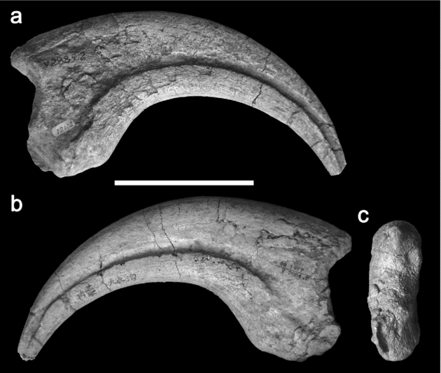
Manual ungual I of Chilantaisaurus tashuikouensis (IVPP V.2884.2): (a) lateral view; (b) medial view; (c) proximal view. Scale bar = 100 mm.

Left ilium of Chilantaisaurus tashuikouensis (IVPP V.2884.3): (a) lateral view; (b) medial view; (c) ventral view; (d) posteromedial view. Abbreviations: avp – anteroventral process; ms – medial shelf; pp – pubic peduncle; sac – supracetabular crest. Scale bar = 200 mm.

Femora of Chilantaisaurus tashuikouensis (IVPP V.2884.4): (a–d, f) left femur; (e, g) right femur; (a) anterior view; (b) medial view; (c, e) posterior view; (d) lateral view; (f) proximal view; (g) distal view. Abbreviations: h – femoral head; ft – fourth trochanter; gt – greater trochanter; lt – lesser trochanter. Scale bar = 200 mm.

Tibiae and fibula of Chilantaisaurus tashuikouensis (IVPP V.2884.5): (a–d) right tibia; (e–h) left tibia and fibula; (a, e) anterior view; (b, f) lateral view, (c, h) medial view; (d) distal view; (g) posterior view. Abbreviations: cnc – cnemial crest; fib – fibula; ff – fibular flange; mr – medial ridge. Scale bar = 200 mm.

Right metatarsus of Chilantaisaurus tashuikouensis (IVPP V.2884.7): (a–c) metatarsals II–III; (d–f) metatarsal II; (g–i) metatarsal III; (j–l) metatarsal IV; (a) anterior view; (b) proximal view; (c) distal view; (d, g, j) lateral view; (e, h, k) ventral view; (f, i, l) medial view. Scale bar = 200 mm.

Lectotype. IVPP V.2884.1, a right humerus.
Paralectotype series. Additional material probably from the same individual as IVPP V.2884.1: an ungual phalanx (IVPP V.2884.2); a fragment of left ilium (IVPP V.2884.3); left and right femora (IVPP V.2884.4); right and partial left tibiae (IVPP V.2884.5); a partial left fibula (IVPP V.2884.6); right metatarsals II–IV and left metatarsals III–IV (IVPP V.2884.7).
Revised diagnosis. Tetanuran theropod with the following autapomorphies of the humerus: subrectangular, anteromedially curving deltopectoral crest that protrudes almost as far anteriorly as it is long proximodistally and bears a pitted scar on its anterior surface (modified from Rauhut, Reference Rauhut2003), and an obliquely oriented ulnar condyle.
Locality and horizon. Ulansuhai Formation, People's Republic of China (60 km North of Chilantai, Nei Mongol Zizhiqu); ?Aptian–Albian or Upper Cretaceous. Hu (Reference Hu1964) suggested that Chilantaisaurus was more derived than Allosaurus (Antrodemus) and Acrocanthosaurus (and therefore later in geological age). This suggestion was mainly based on the possession of a long and robust humerus, a reduced fourth trochanter and short, robust metatarsals. This observation led to the conclusion that C. tashuikouensis, and therefore the Ulansuhai Formation, was Late Cretaceous in age. The Ulansuhai Formation was regarded as at least younger than 92 Ma (Late Cretaceous), based on K–Ar dating of basalts from the unconformably underlying Suhongtu Formation by Kobayashi & Lü (Reference Kobayashi and Lü2003). However, Weishampel et al. (Reference Weishampel, Barrett, Coria, Le Loeuff, Xu, Zhao, Sahni, Gomani, Noto, Weishampel, Dodson and Osmólska2004) list the age as Aptian–?Albian based on a personal communication from Dong, and in agreement with the age given by Rauhut (Reference Rauhut2003). In the future, further data may clarify uncertainty regarding the age of the Ulansuhai Formation.
Comments. Chilantaisaurus tashuikouensis has previously been diagnosed as follows: ‘Humerus massive and elongate. Ungual strongly curved. Fourth trochanter of femur less developed. Tibia shorter than femur. Three metatarsals, short and not compactly united’ (Hu, Reference Hu1964, p. 56); ‘Humerus massive and elongate, structure like that in allosaurid, but almost twice as long as that of Allosaurus. Ungual strongly curved, fourth trochanter of femur less developed, tibia shorter than femur. Three metatarsals, short, not compactly united’ (Sun et al. Reference Sun, Li, Ye, Dong and Hou1992, p. 132); ‘Humerus with distally placed, subrectangular, pterosaur-like deltopectoral crest’ (Rauhut, Reference Rauhut2003, p. 29). The characters used by Hu (Reference Hu1964) and Sun et al. (Reference Sun, Li, Ye, Dong and Hou1992) are not unique to C. tashuikouensis, as they are present in a variety of other tetanurans. The present study identified only two characters of the humerus as autapomorphies of C. tashuikouensis.
As reservations are expressed below over the status of some of the material referred to the taxon by Hu (Reference Hu1964), the autapomorphic humerus (IVPP V.2884.1) is designated as the lectotype of C. tashuikouensis. However, it is still considered likely that the original holotype, which now constitutes the lectotype and paralectotype series, belonged to a single individual.
An isolated tooth (IVPP V.2884.8) and two partial middle caudal vertebrae were found in association with the type material and are described by Hu (Reference Hu1964, p. 59) as ‘probably belonging to the same species’. He did not include this extra material in the holotype, so presumably it was not associated closely enough to be considered part of the type individual. As stated by Rauhut (Reference Rauhut2003), three vertebrae are stored with material bearing the specimen number IVPP V.2884. Of these, one possesses depressions ventral to the transverse processes similar to those of ‘C’. maortuensis. However, it is too small to belong to the holotype of C. tashuikouensis and is labelled IVPP V.2564.6, not IVPP V.2884 (contra Rauhut, Reference Rauhut2003). Therefore, this small vertebra does not belong to the type or referred material of C. tashuikouensis. The two other vertebrae are larger and bear the correct specimen number (IVPP V.2884). The larger of the two probably represents the anterior middle caudal vertebra mentioned by Hu (Reference Hu1964). It has a stout and only weakly constricted centrum with subrectangular articular surfaces, and the broken attachments of the neural arch show that the latter was narrow. These observations suggest that the centrum belongs to a sauropod and should be considered Sauropoda indet. (O. W. M. Rauhut, pers. comm.). As one of the vertebrae does not belong to a theropod, and the referral of both was based on a similar degree of association, neither can be referred to C. tashuikouensis with any confidence. The smaller of the two caudal vertebrae is represented by a centrum. It is elongate, similar in proportions to the distal caudal vertebrae of theropods but comparatively large in size. The articular surfaces are subequally biconcave and the ventral surface is flat at one end, developing into a broad longitudinal groove flanked by prominent ventral ridges towards the other end. The large size and lack of diagnostic features of this element mean that it should be considered Dinosauria indet. The tooth referred to C. tashuikouensis was not found during the course of this study. It was described by Hu (Reference Hu1964) as similar to the teeth of ‘C’ maortuensis, which are typically theropod-like, recurved, serrated and transversely compressed. These teeth do not possess any autapomorphic characters and therefore the tooth referred to C. tashuikouensis should be considered Theropoda indet.
4. Description
4.a. Humerus (Fig. 1)
The right humerus is 580 mm in length (Table 1), almost half the length of the femur (Table 2), a similar proportion to those of spinosauroids and other basal tetanurans (e.g. Piatnitzkysaurus, MACN CH 895). It is long in comparison with those of very large theropods in general, except for spinosaurids (Table 2). Chilantaisaurus has the largest known humerus of any non-coelurosaurian theropod, but the humerus is shorter than those of large maniraptorans with elongate forelimbs such as Deinocheirus (Osmólska & Roniewicz, Reference Osmólska and Roniewicz1970) and Gigantoraptor (Xu et al. Reference Xu, Tan, Wang, Zhao and Tan2007), which also have a higher ratio of humeral to femoral length (Table 2).
Measurements (in millimetres) of the humerus (IVPP V.2884.1) and right metatarsus (IVPP V.2884.7) of C. tashuikouensis

Abbreviations: Distal ap – anteroposterior length measured distally; Distal ml – mediolateral width measured distally; i – measurement incomplete; Proximal ap – anteroposterior length measured proximally; Proximal ml – mediolateral width measured distally. Length of manual ungual I measured in a straight line (not in brackets) and following the curve (in brackets).
Selected measurements (in millimetres) of the humerus, femur, and tibia of various colossal theropods

Measurement sources: Chilantaisaurus (IVPP V.2884), Suchomimus (MNN GDF 500), Acrocanthosaurus (Currie & Carpenter, Reference Currie and Carpenter2000), Mapusaurus (Coria & Currie, Reference Coria and Currie2006; humerus MCF-PVPH 108–45; femur MCF-PVPH 108–234; tibia MCF-PVPH 108–68), Deinocheirus (Osmólska & Roniewicz, Reference Osmólska and Roniewicz1970); Gigantoraptor (Xu et al. Reference Xu, Tan, Wang, Zhao and Tan2007). Abbreviations: e – estimated length.
The humeral shaft of Chilantaisaurus is straight in lateral view and the distal end is rotated 30–40° dextrally relative to the plane of the proximal expansion. In anterior view the outline of the head is continuous with those of the internal and external tuberosities, forming a smoothly convex arc similar to that of spinosauroids such as Afrovenator (UC OBA 1) and Eustreptospondylus (Sadleir, Barrett & Powell, Reference Sadleir, Barrett and Powell2008). By contrast, in allosauroids such as Acrocanthosaurus (Currie & Carpenter, Reference Currie and Carpenter2000) and Allosaurus (Madsen, Reference Madsen1976), the external tuberosity is higher proximally so that the outline of the proximal end of the humerus in anterior view is almost horizontal. The humeral head of Chilantaisaurus is anteroposteriorly thick relative to the tuberosities and bulges posteriorly, overhanging the posterior surface of the proximal expansion. The long axis of the head is oriented slightly obliquely to the plane of the proximal expansion as it is in Acrocanthosaurus (Currie & Carpenter, Reference Currie and Carpenter2000). However, in spinosauroids the long axis of the humeral head is collinear with the plane of the proximal expansion (Afrovenator, UC OBA 1; Baryonyx, Charig & Milner, Reference Charig and Milner1997; Eustreptospondylus OUMNH J.13558; Torvosaurus, Britt, Reference Britt1991).
The internal and external tuberosities are anteroposteriorly thick. The internal tuberosity curves slightly posteromedially and is small compared to spinosaurids such as Baryonyx (Charig & Milner, Reference Charig and Milner1997). A gentle longitudinal ridge located medially on the anterior surface of the humerus divides the anterior surface of the proximal expansion from the slightly anteromedially facing anterior surface of a robust buttress that supports the internal tuberosity distally. The external tuberosity is slightly longer mediolaterally than the internal tuberosity and forms a rounded, distolaterally curving, proximolateral shoulder to the outline of the humerus in anterior view.
The deltopectoral crest is a prominent, anteriorly protruding flange, with a sub-rectangular outline in lateral view. It originates distal to the external tuberosity (Fig. 1) and has a minimum proximodistal length of 97 mm around mid-height, and a maximum proximodistal length of 105 mm where it is expanded more anteriorly. It extends almost as far anteriorly as its proximodistal length and curves slightly anteromedially. The anterior surface of the crest is occupied by a crescentic and gently pitted muscle scar. This unusual morphology of the deltopectoral crest has been described as ‘pterosaur-like’ (Rauhut, Reference Rauhut2003, p. 29), and is substantially different from that of most other theropods, in which the deltopectoral crest is a low, proximodistally elongate protuberance with a subtriangular outline in lateral view that originates closer to the external tuberosity (Holtz, Molnar & Currie, Reference Holtz, Molnar, Currie, Weishampel, Dodson and Osmólska2004). The deltopectoral crest of Acrocanthosaurus (Currie & Carpenter, Reference Currie and Carpenter2000) is similar to that of Chilantaisaurus, as it takes the form of an anteromedially curving sub-rectangular flange; it differs from the deltopectoral crest of Chilantaisaurus in being markedly longer proximodistally than it is anteroposteriorly and lacking a pitted scar on its anterior surface. Therefore, the unique morphology of the deltopectoral crest is an autapomorphy of Chilantaisaurus.
The distal end of the humerus is transversely expanded relative to the shaft to accommodate the radial and ulnar condyles. The ulnar condyle has a sub-rectangular outline that is almost as broad anteroposteriorly as it is wide mediolaterally, and is oriented obliquely to the plane of the distal expansion (fig. 1e). Generally in basal tetanurans the ulnar condyle is also sub-rectangular, but is oriented parallel to the plane of the distal expansion (e.g. Acrocanthosaurus, Currie & Carpenter, Reference Currie and Carpenter2000; Baryonyx, Charig & Milner, Reference Charig and Milner1997; Eustreptospondylus, OUMNH J.13558; Piatnitzkysaurus, PVL 4073; Torvosaurus, Britt, Reference Britt1991), and an obliquely oriented ulnar condyle is therefore an autapomorphy of Chilantaisaurus. A prominent, anterolaterally directed epicondyle is present on the anterolateral surface of the humerus adjacent to the ulnar condyle; this continues a short distance proximally along the anterolateral surface of the humerus as a ridge. A prominent ulnar epicondyle is also present in Acrocanthosaurus (Currie & Carpenter, Reference Currie and Carpenter2000) and Allosaurus (e.g. UMNH VP 13832, UMNH VP 7794), but is absent in most basal tetanurans, including Mapusaurus (Coria & Currie, Reference Coria and Currie2006), and spinosauroids such as Baryonyx (Charig & Milner, Reference Charig and Milner1997), Eustreptospondylus (Sadleir, Barrett & Powell, Reference Sadleir, Barrett and Powell2008) and Torvosaurus (Britt, Reference Britt1991). The radial condyle is transversely wide and anteroposteriorly compressed and a low, mound-like tuber is present on the anteromedial surface of the humerus adjacent to this condyle. A broad longitudinal trough separates the condyles on the posterior surface of the humeral shaft, whereas the anterior surface of the distal end of the humerus is flat except in the region of the ulnar condyle, which projects anteriorly due to the oblique orientation of the condyle. In Piatnitzkysaurus (PVL 4073) and allosauroids (Acrocanthosaurus, Currie & Carpenter, Reference Currie and Carpenter2000; Allosaurus, Madsen, Reference Madsen1976; Mapusaurus, Coria & Currie, Reference Coria and Currie2006) there is a deep depression on the anterior surface of the distal humerus adjacent to the ulnar condyle. Contrastingly, in Ceratosaurus (Madsen & Welles, Reference Madsen and Welles2000) and spinosauroids (Baryonyx, Charig & Milner, Reference Charig and Milner1997; Eustreptospondylus, Sadleir, Barrett & Powell, Reference Sadleir, Barrett and Powell2008; Torvosaurus, BYU 2002), this surface is flat or only slightly concave as in Chilantaisaurus.
4.b. Ungual phalanx (Fig. 2)
A single, recurved, pointed manual ungual is preserved. It is large and elongate, more than three times as long as it is high at the proximal articular surface (Table 1). The first manual ungual of some spinosauroids is similarly enlarged and elongate relative to those of other non-coelurosaurian theropods, as is that of the basal coelurosaur Sinosauropteryx (Sereno et al. Reference Sereno, Beck, Dutheil, Gado, Larsson, Lyon, Marcot, Rauhut, Saleir, Sidor, Varicchio, Wilson and Wilson1998; Currie & Chen, Reference Currie and Chen2001; Rauhut, Reference Rauhut2003). A well-defined, vascular groove runs along both the lateral and medial surfaces just below mid-height of the phalanx. These grooves curve anteroventrally so that they are approximately parallel to the ventral margin of the bone in lateral view.
4.c. Ilium (Fig. 3)
A fragment of the left ilium is preserved in many pieces and comprises the anteroventral portion of the anterior blade, the base of the pubic peduncle and the region dorsal to the acetabulum. Several of the pieces were assembled during the course of this study (Fig. 3), but many other small fragments are preserved that could not be tessellated with the assembled portion of the bone. The blade is thin relative to the size of the bone, with fragments from the central portion measuring 6–7 mm in transverse thickness. Contrastingly, the ilia of basal tetanurans are relatively robust (e.g. Afrovenator, UC OBA 1; Allosaurus, Madsen, Reference Madsen1976; Giganotosaurus, MUCPv-Ch 1; Piatnitzkysaurus, PVL 4073). The ventral part of the anterior blade is slightly thickened and has an anteroventrally inclined ventral margin that develops into a weak anteroventral process, similar to those of compsognathids like Compsognathus (Ostrom, Reference Ostrom1978) and Sinosauropteryx (Currie & Chen, Reference Currie and Chen2001), but unlike the pronounced, hook-like, anteroventral processes of other basal tetanurans (Holtz, Molnar & Currie, Reference Holtz, Molnar, Currie, Weishampel, Dodson and Osmólska2004). A robust ridge on the medial surface of the ilium adjacent to the preacetabular notch forms the medial boundary of the preactabular fossa. It originates anteroventrally at the base of the pubic peduncle and curves anterodorsally onto the medial surface of the anterior blade. This prominent ridge is also present in tyrannosauroids (Holtz, Reference Holtz, Tanke and Carpenter2001) and Neovenator (BMNH R10001). In other allosauroids such as Allosaurus (UMNH VP 8240) and Giganotosaurus (MUCPv-Ch 1) a low ridge is present in this position. The preacetabular fossa and bounding ridge are absent in Piatnitzkysaurus (PVL 4073) and spinosauroids (Afrovenator, UC OBA 1; Eustreptospondylus, Sadleir, Barrett & Powell, Reference Sadleir, Barrett and Powell2008; Torvosaurus, Britt, Reference Britt1991). The pubic peduncle is incompletely preserved but is anteroventrally oriented, probably indicating a propubic pelvis. The prominent supracetabular shelf has been crushed and is now ventrally directed. It is likely that it was originally oriented ventromedially, overhanging the anterodorsal region of the acetabulum in lateral view as is usual for basal tetanurans.
4.d. Femur (Fig. 4)
Substantial portions of both femora are preserved (Fig. 4). The left femur is preserved in two pieces: a smaller proximal fragment comprising the head and the base of the lesser trochanter, and a larger distal fragment comprising the shaft and distal condyles; the proximal end is abraded and the medial portion of the distal end is broken away. The right femur is preserved in four pieces: one comprises the shaft and the remaining three are portions of the distal condyles; the head and lesser trochanter are broken off. The femoral shaft is gently curved posteriorly in medial view and the proximal portion of the shaft curves proximomedially in posterior view. The head is oriented strictly medially and is slightly proximally inclined as in other very large theropods such as Suchomimus (MNN GDF 500) and Tyrannosaurus (Brochu, Reference Brochu2003). The outline of the head in proximal view narrows from medial to lateral as is usual for non-coelurosaurian theropods (Rauhut, Reference Rauhut2003).
Only the base of the lesser trochanter is preserved. It is a broad flange that rises past the ventral level of the femoral head as in all tetanurans (Hutchinson, Reference Hutchinson2001). The portion of the anterior margin from which an accessory trochanter emerges in neotetanurans (Hutchinson, Reference Hutchinson2001) is not preserved, and so the presence of this structure in Chilantaisaurus cannot be determined.
The femoral fourth trochanter is reduced to a low ridge and is flanked medially by a broad suboval depression with a rugose surface (fig. 4b). In basal theropods the fourth trochanter is usually a prominent crest emerging from the medial margin of the posterior surface of the femoral shaft (Holtz, Molnar & Currie, Reference Holtz, Molnar, Currie, Weishampel, Dodson and Osmólska2004). It is absent or reduced to a weak ridge in some coelurosaurs (Rauhut, Reference Rauhut2003), but is also somewhat reduced in Suchomimus MNN GDF 500) and some allosauroids. This has been found as a synapomorphy of the Giganotosaurinae, a clade comprising Giganotosaurus, Mapusaurus and Tyrannotitan (Coria & Currie, Reference Coria and Currie2006). However, the degree of reduction of the fourth trochanter in Chilantaisaurus is greater than in any other basal tetanuran and is comparable to that in coelurosaurs.
The distal end of the femur is expanded transversely and anteroposteriorly. The condyles are sub-equal in transverse width and the crista tibiofibularis and posterior portion of the medial condyle are subequal in size. The anterior surface of the lateral condyle is gently rounded. The anterior surface of the medial condyle is poorly preserved but shows the proximal part of a depression, marked by coarse proximodistally oriented striae in the left femur. Only the proximal end of the medial distal crest is preserved, which forms a low longitudinal bar on the medial surface adjacent to this depression. The original morphology of these two structures when complete cannot be determined.
4.e. Tibia (Fig. 5)
Both tibiae are preserved (Fig. 5). The right tibia is complete and has slightly abraded proximal and distal ends. The left tibia is missing the proximal end and is preserved in articulation with the left fibula. The tibial shaft is anteroposteriorly compressed relative to its mediolateral width and the anterior surface is slightly flattened proximally. Most of the shaft curves medially in anterior view, although the proximal end curves laterally and the proximal surface is inclined to face slightly proximolaterally. The anterior and lateral portions of the cnemial crest are poorly preserved. The crest is prominent and rises proximally, substantially past the level of the tibial condyles, which are also poorly preserved. The outline of the fibular condyle is separated from that of the cnemial crest in proximal view by a gentle embayment, the incisura tibialis, as is usual for tetanurans (Rauhut, Reference Rauhut2003). A low mound-like eminence is present on the medial surface of the tibia at the base of the cnemial crest.
The most prominent portion of the fibular flange measures 160 mm proximodistally and is a robust, laterally projecting lamina. Proximally it becomes a low, anteroposteriorly expanding ridge that is inclined anteroproximally. This is similar to the situation in Mapusaurus (Coria & Currie, Reference Coria and Currie2006), but unlike that in Suchomimus (MNN GDF 500), Allosaurus (Madsen, Reference Madsen1976) and Neovenator (MIWG 6348), in which the proximal termination of the fibular flange is abrupt.
The distal end of the tibia is broad relative to its anteroposterior thickness. The distal end of the right tibia appears to have a distally projecting lateral malleolus. However, both malleoli are broken and this appearance is a consequence of the breakage of a greater portion of the medial malleolus. The astragalar facet is poorly defined but is bound medially by a robust longitudinal ridge along the medial margin of the anterior surface of the distal tibia. Such a ridge is also present in Suchomimus (MNN GDF 500) and is different to the proximolaterally oriented suprastragalar buttress of most basal tetanurans (Rauhut, Reference Rauhut2003). In derived allosauroids the astragalar buttress is sometimes reduced so that the astragalar facet takes the form of a depression on the anterior surface of the tibia (e.g. Acrocanthosaurus, OMNH 10147; Giganotosaurus, MUCPv-Ch 1), but this is also unlike the morphology in Chilantaisaurus. The morphology of the astragalar facet suggests an ascending process of the astragalus with a maximum height of 95 mm (which is the proximodistal length of the medial ridge; this is probably an overestimate, as the depression on the anterior surface to accommodate the ascending process is slightly lower). The maximum value of the ratio of the height of the ascending process to the length of the tibia is therefore 0.1. The ascending process of the astragalus is less than one-sixth of tibial length in spinosauroids (Eustreptospondylus, Torvosaurus) and approximately equal to one-sixth tibial height in allosauroids (Allosaurus, Giganotosaurus) (Harris, Reference Harris1998).
4.f. Fibula (Fig. 5)
The proximal half of the left fibula is preserved (Fig. 5). The proximal expansion is missing. The shaft is anteroposteriorly broad and transversely narrow, tapering slightly distally. It has a semi-oval cross-section, with a flat medial surface that becomes anteroposteriorly concave due to the presence of a gentle longitudinal trough distal to the anterolateral tubercle. The tubercle takes the form of a dorsoventrally elongate swelling on the anteromedial surface of the shaft around the level of the distal end of the fibular flange of the tibia, similar to those of other basal tetanurans such as Allosaurus (Madsen, Reference Madsen1976), Piatnitzkysaurus (PVL 4073) and Suchomimus (MNN GDF 500).
4.g. Metatarsus (Fig. 6)
Left metatarsals II–IV are preserved and only the distal end of the fourth metatarsal is damaged (Fig. 6). The second right metatarsal and a partial third right metatarsal, the distolateral portion of which has been abraded, are preserved.
The cross-section of the shaft of the second metatarsal is semi-circular with a flattened lateral surface. An extensive, proximodistally elongate swelling, bearing a sub-oval depression centrally, is present on the ventral surface of the shaft. It originates around mid-length and extends distally. The proximal end of the metatarsal is expanded, primarily dorsally, relative to the shaft. The proximal surface is slightly depressed and has a squared outline in proximal view with rounded dorso- and ventromedial angles and a stout, ventrally projecting ventrolateral lobe. The ventrolateral region of this surface is expanded proximally.
The distal articular surface of the second metatarsal is convex, forming a single condyle that is emarginated ventrally to form low, ventrally projecting, medial and lateral lobes. The medial lobe is slightly larger and projects further ventrally than the lateral lobe. Both sides of the distal end bear deep collateral ligament fossae; that of the lateral side is broader and is set into an extensive depression.
The shaft of the third metatarsal has a trapezoidal cross-section with a broad, flattened dorsal surface and a narrower, flattened ventral surface; this morphology was considered a synapomorphy of Neotetanurae by Holtz, Molnar & Currie (Reference Holtz, Molnar, Currie, Weishampel, Dodson and Osmólska2004). There are no muscle scars or significant irregularities on the shaft. The proximal end of the third metatarsal is dorsoventrally expanded and medially inclined. It is broader dorsally than ventrally. The medial surface is gently concave and the ventral portion of the lateral surface is emarginated to accommodate the proximomedial process of the fourth metatarsal. As a consequence, the outline of the third metatarsal in proximal view is constricted around mid-height so that it has an hourglass shape. The distal end of the metatarsal forms a single, transversely broad, dorsoventrally convex articular surface that has a sub-rectangular outline in distal view with a gently concave ventral margin.
The shaft of the fourth metatarsal curves distolaterally and has a semi-oval cross-section that is dorsoventrally low with a flat ventral surface. A broad, flat-topped longitudinal ridge originates just distal to mid-length on the ventral surface of the shaft. The ridge is as broad as the ventral surface proximally but tapers distally, curving distolaterally and reaching the lateral margin of the shaft around fourth-fifths of shaft length. The proximal articular surface of the metatarsal is gently concave and has a semi-oval outline that is embayed dorsomedially to receive the dorsolaterally expanded proximal portion of the third metatarsal.
5. The systematic position of Chilantaisaurus
Chilantaisaurus was originally referred to Megalosauridae by Hu (Reference Hu1964, p. 58), on the basis of the ‘unreduced shafts of the metatarsals’ (lack of the ‘arctometatarsalian’ condition of Holtz, Reference Holtz1994), a massive and elongate humerus and a tibia that is short relative to the femur, characters which exclude the material from referral to Tyrannosauridae. It is clear that Hu (Reference Hu1964) considered Megalosauridae to comprise a range of basal tetanurans, including taxa currently considered to be allosauroids.
Subsequent authors have all stated that C. tashuikouensis is a basal tetanuran outside of Coelurosauria (Harris, Reference Harris1998; Rauhut, Reference Rauhut2003; Holtz, Molnar & Currie, Reference Holtz, Molnar, Currie, Weishampel, Dodson and Osmólska2004), but there is uncertainty over its exact systematic position. The phylogenetic analysis of Harris (Reference Harris1998) recovered Chilantaisaurus as a derived allosauroid, whereas that of Rauhut (Reference Rauhut2003) recovered it as the sister taxon of Spinosauridae within Spinosauroidea.
5.a. Evidence for an allosauroid or neotetanuran affiliation of Chilantaisaurus
Harris (Reference Harris1998) found that Chilantiasaurus was an allosauroid related to carcharodontosaurids, Acrocanthosaurus and Neovenator. However, Harris (Reference Harris1998) scored characters of the braincase and axis for his Chilantaisaurus, presumably based on ‘Chilantaisaurus’ maortuensis. He also made a number of scoring errors, considering Chilantaisaurus to possess a sigmoidal humerus, a hook-like anteroventral process of the ilium, a robust fourth trochanter and a proximally inclined femoral head. Chilantaisaurus was erroneously united with carcharodontosaurids on the basis of its proximally inclined femoral head (Harris, Reference Harris1998, fig. 40A), but is not otherwise united with allosauroid taxa on the basis of synapomorphies pertaining to elements known for C. tashuikouensis.
Strong proximal inclination of the femoral head was found to be a synapomorphy of Carcharodontosauridae (excluding Acrocanthosaurus) in the phylogenetic analysis of Coria & Currie (Reference Coria and Currie2006). However, it is also present in Acrocanthosaurus (OMNH 10147) and Neovenator (MIWG 6348). Although the femoral head of C. tashuikouensis is inclined slightly proximally, those of derived allosauroids are much more strongly inclined (approximately 65° in Acrocanthosaurus, based on OMNH 10147). Consequently, there is little support for an allosauroid affiliation of Chilantaisaurus based on characters used by Harris (Reference Harris1998). None the less, Chilantaisaurus does possess several anatomical features which suggest a neotetanuran, if not allosauroid, affiliation.
A well-defined preacetabular fossa bounded by a prominent medial shelf is present on the ilium of Chilantaisaurus. This fossa is absent in some basal tetanurans, such as Piatnitzkysaurus (MACN CH 895) and spinosauroids (e.g. Eustreptospondylus, OUMNH J.13558; Suchomimus, MNN GDF 500), and has been recovered as a synapomorphy of Neotetanurae (Holtz, Molnar & Currie, Reference Holtz, Molnar, Currie, Weishampel, Dodson and Osmólska2004). This determination is consistent with its presence in only allosauroids among non-coelurosaurian tetanurans (e.g. Allosaurus, Madsen, Reference Madsen1976; Giganotosaurus, MUCPv-Ch 1). Therefore, the presence of a preacetabular fossa in Chilantaisaurus suggests that it is a neotetanuran. The prominent medial shelf bounding the preacetabular fossa has been listed as a tyrannosauroid feature by Holtz (Reference Holtz, Tanke and Carpenter2001), but it is also present in the allosauroid Neovenator (BMNH R10001) and is absent in all other basal tetanurans. As Chilantaisaurus lacks all other tyrannosauroid features, such as those enumerated by Holtz (Reference Holtz, Tanke and Carpenter2001), the presence of this shelf may suggest an affinity with Neovenator.
The dorsal surface of the third metatarsal of Chilantaisaurus is transversely broad relative to the ventral surface, giving the bone a trapezoidal or ‘wedge-shaped’ cross-section. This was originally considered to be a synapomorphy of Tetanurae (Gauthier, Reference Gauthier and Padian1986). However, although this morphology is present in allosauroids such as Acrocanthosaurus (OMNH 10147), Allosaurus (UMNH VP 10380) and Mapusaurus (MCF-PVPH 108-32), it is absent in spinosauroids such as Eustreptospondylus (Sadleir, Barrett & Powell, Reference Sadleir, Barrett and Powell2008) and Torvosaurus (BYU 5277). Holtz, Molnar & Currie (Reference Holtz, Molnar, Currie, Weishampel, Dodson and Osmólska2004) recovered this feature as a possible neotetanuran synapomorphy, and its presence in Chilantaisaurus may suggest that it is a member of this clade.
The humeral head is oriented obliquely to the plane of the proximal humerus in Chilantaisaurus. This has never been used as a character in phylogenetic analysis but may be of systematic utility, because in Ceratosaurus (UMNH VP 5278) and spinosauroids (Afrovenator, UC OBA 1; Baryonyx, Charig & Milner, Reference Charig and Milner1997; Eustreptospondylus OUMNH J.13558; Suchomimus, MNN GDF 500; Torvosaurus, Britt, Reference Britt1991) the long axis of the head is collinear with the plane of the proximal end of the humerus, whereas in Dilophosaurus (UCMP 37302), basal tetanurans such as Piatnitzkysaurus (PVL 4073), allosauroids (Allosaurus, UMNH VP 10779; Acrocanthosaurus, Currie & Carpenter, Reference Currie and Carpenter2000) and basal coelurosaurs (e.g. Tanycolagreus, Carpenter, Miles & Cloward, Reference Carpenter, Miles, Cloward and Carpenter2005), the situation is similar to that in Chilantaisaurus.
A prominent ulnar epicondyle is present in Chilantaisaurus. This structure has never been scored for use in phylogenetic analysis but is variable in basal theropods; it is absent in most basal tetanurans but is present in the allosauroids Acrocanthosaurus (Currie & Carpenter, Reference Currie and Carpenter2000) and Allosaurus (UMNH VP 10779). The presence of a prominent ulnar epicondyle in Chilantaisaurus may suggest a phylogenetic relationship with these taxa.
5.b. Evidence for a spinosauroid or non-allosauroid affiliation of Chilantaisaurus
Rauhut (Reference Rauhut2003) found a sister taxon relationship between Chilantaisaurus and Baryonychidae (Spinosauridae of Holtz, Molnar & Currie, Reference Holtz, Molnar, Currie, Weishampel, Dodson and Osmólska2004), which are united within Spinosauroidea on the basis of a bluntly rounded vertical ridge on the anteromedial side of the distal end of the tibia. A more inclusive clade comprising Torvosaurus, Chilantaisaurus and Baryonychidae was supported by a humerus that is straight, rather than sigmoid, in lateral view, as well as characters of the skull, forearm and pubis that are unknown in Chilantaisaurus. A straight humerus is also present in the allosauroids Acrocanthosaurus (Currie & Carpenter, Reference Currie and Carpenter2000) and Mapusaurus (Coria & Currie, Reference Coria and Currie2006). Although Rauhut (Reference Rauhut2003) included Acrocanthosaurus in the supraspecific taxon Carcharodontosauridae, he scored the shape of the humerus as uncertain in carcharodontosaurids. The character ‘manual ungual I more than half the length of the radius’ (Rauhut, Reference Rauhut2003, p. 146) is only uncertainly present in Chilantaisaurus as, although the radius is not known, the first manual ungual is elongated. Sereno et al. (Reference Sereno, Beck, Dutheil, Gado, Larsson, Lyon, Marcot, Rauhut, Saleir, Sidor, Varicchio, Wilson and Wilson1998, p. 1302) describe a character for elongation of the first manual ungual without reference to the length of the radius as ‘manual digit I-ungual, length: 2.5 times (0) or 3 (1) times the depth of the proximal end’, and this may be a more convenient way to describe the character, as it allows taxa for which the radius is not known to be scored positively for this character.
In the analysis of Rauhut (Reference Rauhut2003), Spinosauroidea as a whole (Eustreptospondylus, Torvosaurus, Chilantaisaurus and Baryonychidae) was supported by cranial characters that are not known for Chilantaisaurus. Therefore, the only character that unambiguously supports a spinosauroid affiliation of Chilantaisaurus in Rauhut's (Reference Rauhut2003) analysis is the vertical ridge on the anterior surface of the distal end of the tibia, which is also present in the spinosaurid Suchomimus (MNN GDF 500). However, although Rauhut (Reference Rauhut2003) does not score this ridge as present in any other theropods, it is present in the basal coelurosaur Coelurus agilis (Galton & Molnar, Reference Galton, Molnar and Carpenter2005) and may therefore have a wider distribution within Theropoda.
The presence of an enlarged first manual ungual may also support the spinosauroid affinities of Chilantaisaurus. However, this feature is also present in the basal coelurosaur Sinosauropteryx (Currie & Chen, Reference Currie and Chen2001). Therefore, neither of the characters suggesting a spinosauroid affiliation of Chilantaisaurus is unique to Spinosauroidea, in contrast with the presence of a preacetabular fossa and ‘wedge’-shaped third metatarsal, which are unique synapomorphies of Neotetanurae.
However, Chilantaisaurus does possess several other characters that are not present in allosauroids but are primitively present in basal tetanurans, including spinosauroids.
The facet on the tibia for the ascending process of the astragalus is low in Chilantaisaurus. The maximum value of the ratio of the height of the ascending process to the length of the tibia is 0.1. The ascending process of the astragalus is less than one-sixth of tibial length in spinosauroids (Eustreptospondylus, Torvosaurus) and approximately equal to one-sixth tibial height in neotetanurans such as the allosauroids Allosaurus and Giganotosaurus (Harris, Reference Harris1998). As such, the relatively low tibial astragalar facet of Chilantaisaurus suggests a non-neotetanuran, and therefore possibly spinosauroid, affiliation.
The humerus is approximately half the length of the femur, and is therefore long relative to those of allosauroids and the majority of large theropods, such as tyrannosaurids and abelisaurids, in which the ratio is less than 0.4 (Rauhut, Reference Rauhut2003). Contrastingly, spinosaurids and basal theropods such as Ceratosaurus and Piatnitzkysaurus have a ratio more similar to that in Chilantaisaurus (Table 2; Rauhut, Reference Rauhut2003).
The anterior surface of the humerus adjacent to the distal condyles is almost flat, and therefore similar to those of basal theropods such as Ceratosaurus (Madsen & Welles, Reference Madsen and Welles2000), Dilophosaurus (UCMP, 37302) and spinosauroids (Baryonyx, Charig & Milner, Reference Charig and Milner1997; Eustreptospondylus, Sadleir, Barrett & Powell, Reference Sadleir, Barrett and Powell2008; Torvosaurus, BYU 2002). In allosauroids (Acrocanthosaurus, Currie & Carpenter, Reference Currie and Carpenter2000; Allosaurus, Madsen, Reference Madsen1976; Mapusaurus, Coria & Currie, Reference Coria and Currie2006) and Piatnitzkysaurus (PVL 4073), however, there is a marked triangular depression in this region, adjacent to the ulnar condyle, and the absence of this depression suggests that Chilantaisaurus is not an allosauroid.
6. Conclusions
Chilantaisaurus possesses an unusual combination of anatomical features that do not unequivocally support either an allosauroid or spinosauroid affiliation based on comparison alone. However, the presence of a preacetabular fossa of the ilium and of a ‘wedge’-shaped cross-section to the third metatarsal strongly suggest that it is a neotetanuran, rendering a spinosauroid affiliation unlikely.
Although Chilantaisaurus is probably a neotetanuran, it lacks several features that are present in all allosauroids, a shortened humerus and a marked depression on the anterior surface of the humerus adjacent to the ulnar condyle. Consequently, Chilantaisaurus may not be an allosauroid. It is possible that Chilantaisaurus represents a lineage of neotetanuran theropods outside of Allosauroidea that continued into the Cretaceous from the diversification of basal neotetanurans in the Middle Jurassic, producing extremely large-bodied taxa independent of the allosauroid carcharodontosaurids. This hypothesis may explain the unusual combination of characters seen in Chilantaisaurus. For instance, Chilantaisaurus possesses a reduced femoral fourth trochanter, and lacks a prominent anteroventral process of the ilium, features it shares with basal coelurosaurs such as Compsognathus (Ostrom, Reference Ostrom1978; Rauhut, Reference Rauhut2003). Although this hypothesis has not been recovered by previous phylogenetic analyses, these analyses have included only a fraction of the relevant characters, and none of them include a comprehensive sample of basal tetanuran theropods. Consequently, confirmation of the relationships of Chilantaisaurus relative to Allosauroidea, Spinosauroidea and Coelurosauria awaits thorough phylogenetic re-examination.
Acknowledgements
The authors would like to thank S. Chapman, R. Coria, M. Getty, P. A. Jeffery, A. Kramarz, M. Munt, B. Masek, O. Mateus, J. Person, J. Powell and R. Scheetz for access to specimens. They thank P. J. Currie and O. W. Rauhut for constructive reviews. RBJB thanks P. M. Barrett and D. B. Norman for supervision, S. L. Brusatte for comments on a draft manuscript, and the Department of Earth Sciences, University of Cambridge and Department of Palaeontology, BMNH for support. RBJB is supported by NERC studentship NER/S/A/2005/13488. X.X.'s work is supported by the Chinese Academy of Sciences.