Acute pulmonary embolism is a potentially life-threatening condition and requires prompt diagnosis and timely management. Acute pulmonary embolism can be fatal with the reported mortality rate of 21–39% in children.Reference Biss, Brandao, Kahr, Chan and Williams1 The incidence of acute pulmonary embolism in children is low at <1 per 100,000.Reference Stein, Kayali and Olson2 There is a paucity of data on the management of acute pulmonary embolism in children. In contrast, acute pulmonary embolism frequently occurs in adults with significant improvement in the care of patients with acute pulmonary embolism. Recently, pulmonary embolism response team has been formed in adult centres to provide immediate and appropriate care to patients with suspected pulmonary embolism.Reference Jaber, Fong and Weisz3
Catheter-directed therapy has been reported to be an effective modality in adults with acute sub-massive and massive pulmonary embolism.Reference Kuo, Banerjee and Kim4 Among the various catheter-directed therapy options, EkoSonic ultrasound-accelerated thrombolysis system (EkoS: EKOS Corp., Bothell, Washington, United States of America) is an innovative tool to facilitate thrombolysis and improve right ventricular strain with fewer bleeding complications in adults with massive and sub-massive pulmonary embolism.Reference Piazza, Hohlfelder and Jaff5 In adults, EkoS is now recognised as a choice of therapy for sub-massive and massive pulmonary embolism. However, the use of EkoS is yet to be well described in children.Reference Bavare, Naik and Lin6 In our study, we describe our experience of catheter-directed therapy for acute pulmonary embolism in children.
Methods
This is a retrospective study to evaluate the safety and efficacy of catheter-directed therapy in children at a single tertiary care academic institution. This study was approved by the Institutional Review Board. Patients were identified through the cardiac catheterisation database. Inclusion criteria were children aged ⩽20 years with a structurally normal heart who underwent catheter-directed therapy for acute pulmonary embolism at Detroit Medical Center. Exclusion criteria were those with underlying CHD except patent foramen ovale. The study period was for 12 years from January 2005 to July 2017. Definitive diagnosis of acute pulmonary embolism was made based on chest CT angiography in all the included cases.
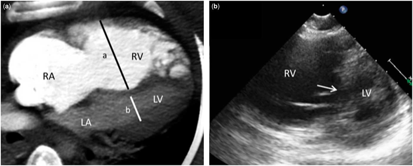
Demographic characteristics, clinical presentation, laboratory data, haemodynamic data of cardiac catheterisation, hospitalisation course, outcome, adverse events associated with catheter-directed therapy, and follow-up data were collected. Obesity was defined as body mass index above 95th percentile based on age and gender. Elevated cardiac biomarkers were defined as increased levels of troponin I, B-type natriuretic peptide, or N-terminal-pro B-type natriuretic peptide above the reference value. Chest CT angiography, echocardiography, and cardiac catheterisation data were reviewed by two investigators (JA and DK). Right ventricle to left ventricle cavity ratio was measured at the four-chamber view on chest CT angiography imaging (Fig. 1a), and a ratio of >0.9 was defined as abnormal dilation.Reference Schoepf, Kucher, Kipfmueller, Quiroz, Costello and Goldhaber7 Pulmonary embolism was classified into massive and sub-massive: massive pulmonary embolism was defined by the presence of hypotension, whereas sub-massive pulmonary embolism was defined by right ventricular strain by echocardiography, chest CT angiography, and/or elevated cardiac biomarkers in normotensive patients.Reference Jaff, McMurtry and Archer8 Right ventricular strain was defined by right ventricular dilation and/or systolic dysfunction detected by echocardiography and/or chest CT angiography. Severity risk of pulmonary embolism was stratified into four levelsReference Konstantinides, Torbicki and Agnelli9 of low risk – no haemodynamic compromise or right ventricular strain; intermediate to low risk – right ventricular strain without haemodynamic compromise or elevation of cardiac biomarkers; intermediate to high risk – right ventricular strain and elevation of cardiac biomarkers without haemodynamic compromise; and high risk – haemodynamic compromise.
RV strain on imaging studies. (a) Chest CT angiography showed dilated RV and RA. RV:LV ratio measures 2.3 (a = 51.5 mm, b = 22.0 mm). (b) Echocardiography shows dilated RV with D-shaped LV on the parasternal short-axis view. Interventricular septum (arrow) is bowed to the LV. LA = left atrium; LV = left ventricular; RA = right atrium; RV = right ventricular;

EkoSonic ultrasound-accelerated thrombolysis
EkoSonic endovascular system consists of ultrasound-accelerated thrombolysis with local delivery of thrombolytic agents through the ultrasonic core within a 5.4-Fr EkoSonic infusion catheter (Fig. 2a) and an EkoSonic control unit (Fig. 2b). First, right heart cardiac catheterisation was performed to evaluate the haemodynamic data in the cardiac catheterisation laboratory. After pulmonary artery angiography was performed, the EkoSonic infusion catheters were inserted to unilateral or bilateral branch pulmonary arteries (Fig. 2c). The EkoSonic infusion catheter (Fig. 2d) is a multi-lumen catheter and is 106 or 135 cm in length with 6–50 cm treatment zones. It was positioned via the femoral, internal jugular, or brachial vein approach using a 6-Fr sheath. The ultrasonic core was inserted into the infusion catheter. Then the patients were monitored in the ICU, while the EkoSonic endovascular system delivered the ultrasound-accelerated thrombolysis. Tissue plasminogen activator was administered as an initial bolus dose of 2 mg followed by an infusion at the rate of 1 mg/hour through the infusion catheter. During the ultrasound-accelerated fibrinolysis, normal saline was given to cool the EkoSonic ultrasonic core at the rate of 35 ml/hour through each EkoS infusion catheter. Concomitant low-dose intravenous heparin was given at 500 units/hour during EkoS therapy. EkoS catheter was removed either at the bedside or in the cardiac catheterisation laboratory.
(a) EkoSonic infusion catheter (5.4-Fr, 106 and 135 cm working length) and ultrasonic core (6,12, 18, 24, 30, 40, and 50 cm treatment zones); (b) EkoSonic control unit; (c) EkoSonic infusion catheters are placed in the left and right pulmonary arteries; (d) The infusion catheter shaft consists of a main coolant lumen with three surrounding small drug lumens. The guidewire or the ultrasonic core is inserted into the coolant lumen. In addition, normal saline is continuously infused through the coolant lumen to cool the ultrasonic core during use. Tissue plasminogen activator is infused through the drug lumens. Images: Courtesy of EKOS Corporation.

Results
Patient characteristics
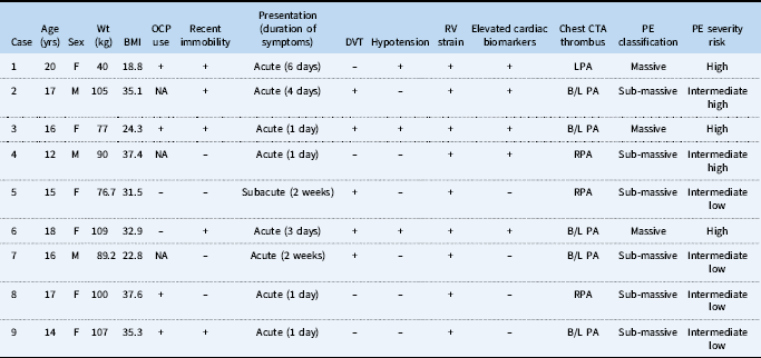
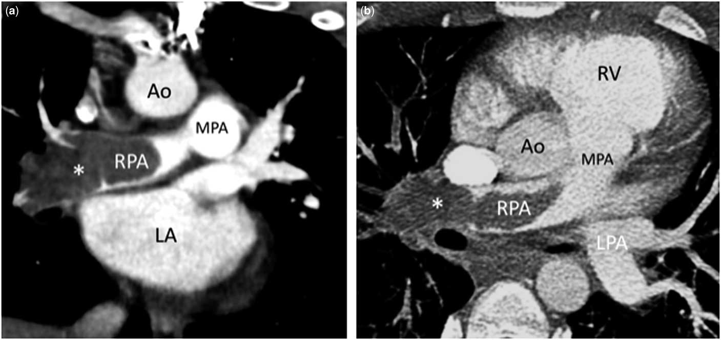
Our study cohort consisted of nine patients, of which six (66.67%) were females (Table 1). The median age of the patients was 16 years, with the range from 12 to 20 years. The median weight was 89.2 kg with a range from 40 to 109 kg. Among nine patients, six patients (67%) were obese with body mass index >30. The predisposing risk factors of pulmonary embolism include recent immobility in 5/9 patients (56%), oral contraceptive use in 4/6 female patients (67%), underlying haematological diseases in 3/9 patients (33%), and systemic lupus erythematosus in 2/9 patients (23%). All the patients complained of dyspnoea with/without chest pain or haemoptysis. Majority of the patients (7/9) had an acute presentation (<1 week) except two who had a subacute presentation (>1 week). Concurrent deep venous thrombosis was found in five patients (56%); three patients (33%) were hypotensive requiring inotropic support. Moreover, five (56%) had elevated cardiac biomarkers such as Trop I or NT-pro brain natriuretic peptide. All the patients were hypoxic requiring intubation in three patients, oxygen supplementation via mask, or nasal cannula in six patients. Inhaled nitric oxide was used in two patients because of severe hypoxia. All the patients had right ventricular strain (Fig. 1). A total of three patients with massive pulmonary embolism had severely dilated right ventricular with right ventricule to left ventricule ratio >2 on chest CT angiography (Fig. 1a). Extensive embolus in both pulmonary arteries was seen in five patients, whereas thrombus in one branch pulmonary artery was seen in the other four patients (Fig. 3). Pulmonary infarct was present in five patients.
Patient characteristics, presentation, and severity risk of pulmonary embolism.

B/L PA = bilateral pulmonary artery; BMI = body mass index; CTA = computed tomography angiography; DVT = deep venous thrombosis; LPA = left pulmonary artery; NA = not applicable; OCP = oral contraceptives; PE = pulmonary embolism; RPA = right pulmonary artery; RV = right ventricle; wks = weeks; Wt = weight; yrs = years
Chest CT angiography showed a large occlusive thrombus in the proximal RPA. (a) Coronal view. Thrombus (*) is occlusive in RPA. (b) Axial view. Ao, aorta; LA = left atrium; LPA = left pulmonary artery; MPA = main pulmonary artery; RPA = right pulmonary artery; RV = right ventricle.

Catheter-directed therapy
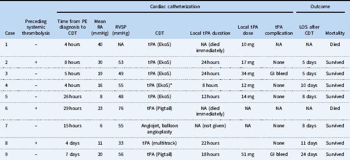
Three patients had preceding systemic thrombolytic therapy. The time from pulmonary embolism diagnosis to catheter-directed therapy was variable, with a median duration of 8 hours, varying from 4 hours to 7 days. In our cohort five patients were urgently taken to the catheterisation laboratory within 8 hours of pulmonary embolism diagnosis. The median right atrial pressure was 19 mmHg, with the range from 6 to 40 mmHg. The median right ventricular systolic pressure was 55 mmHg, with the range from 33 to 76 mmHg. All the patients received catheter-directed thrombolysis therapy, except one patient received mechanical thrombectomy using Angioget® thrombectomy system (Boston Scientific, Marlborough, MA, United States of America) and balloon angioplasty because of concern for risk of bleeding associated with thrombolysis (Table 2). EkoS was used recently in five patients.
Catheter-directed therapy and outcome.

CDT = catheter-directed therapy; EkoS = EkoSonic catheter; GI = gastrointestinal; LOS = length of stay; NA = not applicable; PE = pulmonary embolism; RA = right atrium; RVSP = right ventricular systolic pressure; tPA = tissue plasminogen activator
* Patient 4 required second CDT using an infusion catheter and received 25 mg tPA locally over 24 hours
EkoSonic ultrasound-accelerated thrombolysis
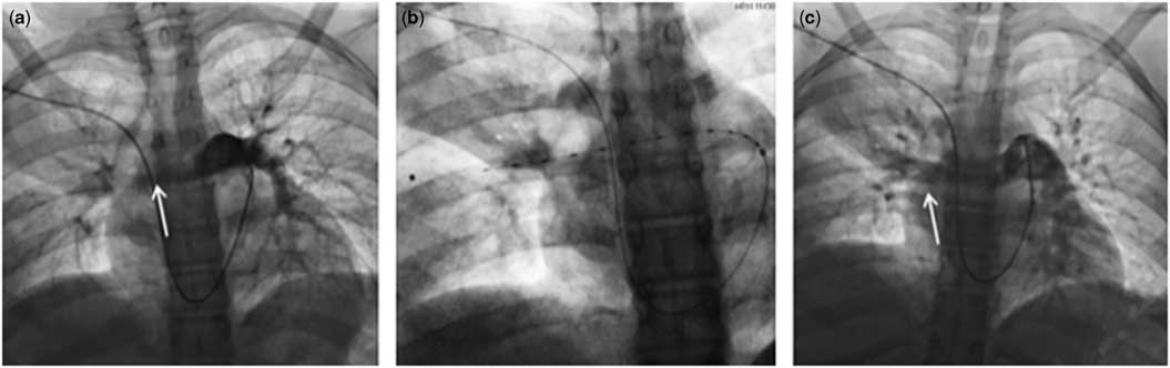
A total of five patients received accelerated thrombolytic therapy using EkoSonic endovascular system (Fig. 4). EkoS system was placed in bilateral pulmonary artery branches in one patient and in unilateral pulmonary artery branch in four patients through the femoral venous access (four patients) and right subclavian vein (one patient), based on thrombus burden in pulmonary artery system. Placement of EkoS was technically straightforward in all patients. Duration of tissue plasminogen activator infusion through the EkoS ranged from 8 to 24 hours. In three patients oxygenation improved within 2 hours of therapy with EkoS system, whereas Case 1 died of extremis pulmonary embolism immediately after EkoS placement and Case 4 required second local tissue plasminogen activator infusion 3 days after the initial EkoS therapy. In fact, this patient received only 8 hours of tissue plasminogen activator infusion through the EkoS.
EkoSonic ultrasound-accelerated thrombolysis system for RPA occlusive thrombus in a 15-year-old girl with sub-massive pulmonary embolism (Case 5). (a) Baseline pulmonary artery angiography using a 5-Fr pigtail catheter showed a large occlusive thrombus (arrow) with no distal flow in the proximal RPA. (b) EkoSonic catheter is positioned in the RPA. (c) 12 hours post-EkoSonic therapy, there was a significant improvement in flow (arrow) to the distal RPA branches with reduction of thrombus. RPA= right pulmonary artery.

Outcome
A total of seven patients responded to catheter-directed therapy and concomitant anticoagulation therapy using intravenous heparin infusion. The median length of hospital stay after catheter-directed therapy was 8 days with the range from 5 to 24 days. Of seven patients, six patients were discharged on enoxaparin and one patient on apixaban. In a median follow-up of 11 months with the range from 3 to 96 months, these patients remained alive and had normal right ventricular size and systolic function on the follow-up echocardiography. Two patients with systemic lupus erythematosus had recurrent pulmonary embolism while being on enoxaparin. Two patients had minor gastrointestinal bleeding that was medically managed without the need for blood transfusion.
There were two deaths (22%). Case 1 had delayed diagnosis of pulmonary embolism. This patient developed massive pulmonary embolism after a surgical revision of ventriculo-atrial shunt. The diagnosis of pulmonary embolism was made 3 days after the onset of hypoxia. She was emergently taken to the catheterisation laboratory but became haemodynamically unstable quickly. The mean right atrium pressure was extremely elevated at 40 mmHg. Even though EkoS catheter was successfully placed, she died in 1 hour after the procedure. Case 6 had a poor response to preceding systemic thrombolysis. However, she was taken to the catheterisation laboratory 29 hours later due to clinical decompensation. The mean right atrial pressure was severely elevated at 23 mmHg and the right ventricular systolic pressure was moderately/severely elevated at 76 mmHg. The local tissue plasminogen activator infusion was initiated through the Pigtail catheter in the pulmonary artery, but she died immediately following the catheterisation.
Discussion
Acute pulmonary embolism in children is rare but can lead to rapid cardiovascular decompensation with a high risk of mortality. In our cohort, two patients (22%) died just after the initiation of catheter-directed therapy. They became critically ill before being taken to the catheterisation laboratory because one patient had delayed diagnosis and the other had an initial poor response to systemic tissue plasminogen activator administration. Hence prompt diagnosis and timely intervention of pulmonary embolism are critical, and it is important to have a high index of suspicion in children with unexplained dyspnoea, hypoxia, with or without risk factors of pulmonary embolism. A previous study of 14 children with pulmonary embolism showed that the average time to diagnosis was 7 days, highlighting the difficulty in making a timely diagnosis in children.Reference Rajpurkar, Warrier and Chitlur10 The common risk factors for pulmonary embolism in our cohort included oral contraceptive use, immobility along with recent surgery, obesity, and underlying haematological and rheumatological diseases. This finding was consistent with previous reports.Reference Rajpurkar, Warrier and Chitlur10, Reference Wang, Ignjatovic and Francis11
Pharmacological thrombolysis is the mainstream therapy for massive and sub-massive pulmonary embolism. Thrombolytic agents are enzymes that convert plasminogen to plasmin which degrades the fibrin to dissolve the thrombus. Active thrombolytic therapy aims to promote a faster improvement of right ventricular strain by reduction of thrombus burden in the pulmonary arterial circulation. Systemic administration of thrombolytic agents is an effective therapy but carries a higher risk of major bleeding, including intracranial haemorrhage, which could be life-threatening or cause permanent neurological deficits.Reference Marti, John and Konstantinides12 It can also cause minor haemorrhage, which could increase the morbidity and prolong the hospital stay.Reference Vedantham, Piazza, Sista and Goldenberg13 More recently, catheter-directed thrombectomy and/or thrombolytic therapy has emerged as an alternative option because of its potential effectiveness and less bleeding risk.Reference Konstantinides, Barco, Lankeit and Meyer14 There is a paradigm shift in the acute care of pulmonary embolism in adults. Pulmonary embolism response team has been formed in many centres to provide timely diagnosis and optimal treatment plan for patients with acute pulmonary embolism. Pulmonary embolism response team typically consists of multiple specialists, including pulmonary critical care, emergency medicine, interventional cardiology/radiology, haematology, surgery, etc.Reference Jaber, Fong and Weisz3 In paediatric centres, pulmonary embolism response team has not been well described. For children with pulmonary embolism, we need to develop a standardised approach for diagnosis and provide a timely and optimal therapy.
Catheter-directed therapy consists of various techniques, including clot fragmentation, aspiration, low-dose thrombolytic administration with/without an ultrasound-accelerated thrombolysis system. The benefit of catheter-directed therapy is the reduction of thrombolysis agent doses and lesser bleeding complications. Among various catheter-directed therapy options, the EkoSonic catheter is the only Food and Drug Administration approved catheter for the use of thrombolytic therapy in pulmonary embolism.15 There are a series of randomised clinical trials showing the efficacy and safety of EkoS in adults with acute pulmonary embolism.Reference Piazza, Hohlfelder and Jaff5, Reference Kucher, Boekstegers and Muller16 The landmark study was the SEATTLE II Trial.Reference Piazza, Hohlfelder and Jaff5 This was a prospective multi-centred study of 150 patients to evaluate the efficacy and safety of EkoS endovascular system on massive and sub-massive pulmonary embolism. EkoS effectively treated right ventricular strain, pulmonary artery obstruction, and pulmonary hypertension. Most strikingly, there was no intracranial haemorrhage in 150 patients. Although EkoS appears to be a very attractive modality, the use of EkoS in paediatric pulmonary embolism cases has not been well described. The largest experience of EkoS use on acute pulmonary embolism in children was reported by Texas Children’s Hospital in 2014.Reference Bavare, Naik and Lin6 In four children aged 11–17 years who were treated with EkoS, all had improvement in clinical and echocardiographic parameters following the treatment, without any mortality or bleeding complications. In our study, we describe a total of nine children who received catheter-directed therapy. Among them, five children received EkoSonic ultrasound-accelerated thrombolysis as our preferred choice of catheter-directed therapy. All the children were of adult size, and there was no technical difficulty in placing the EkoS catheter in the branch pulmonary artery. The duration of local tissue plasminogen activator administration through the EkoS catheter was variable (8–24 hours) and on the discretion of the practitioner. Except for two patients who died just after the EkoS placement, the length of stay was relatively short at a median duration of 8 days with the range from 5 to 24 days.
The EkoSonic ultrasound-accelerated thrombolysis is our first choice of therapy for patients with intermediate and high risk acute pulmonary embolism, when there is no contraindication for the use of thrombolytic agents. Although the body size may be a limiting factor for the use of the EkoSonic endovascular system because of a concern for fluid load, a recent report showed the successful use of the EkoSonic system in an infant weighing as low as 6.8 kg.Reference Khan, Gowda, Parekh and Qureshi17 The dosage of tissue plasminogen activator needs to be weight adjusted in small children. When there is a significant concern for a bleeding risk associated with the use of thrombolytic agents and/or non-acute pulmonary embolism, other adjunctive mechanical thrombectomy techniques including Angiojet thrombectomy may be considered. Angiojet may provide effective thrombectomy but potentially causes significant haemodynamic compromise in children.Reference Qureshi, Petit, Crystal, Liou, Khan and Justino18
Limitation
Our study had a small sample size, which limits the generalisability of our results. The decision to perform catheter-directed therapy was at the discretion of the individual practitioners, and there was no standardised approach for children with acute pulmonary embolism during the study period. The follow-up period was limited in some patients, but the focus of our study was to evaluate the acute result of the catheter-directed therapy.
Conclusion
Catheter-directed therapy with/without EkoSonic ultrasound thrombolysis system was effective and safe for sub-massive and massive pulmonary embolism in children when instituted in a timely fashion. Delay in diagnosis and delay in therapy resulted in a poor outcome. A standardised and team-based approach would be important to facilitate the prompt diagnosis and treatment in children.
Acknowledgement
None.
Financial support
This research received no specific grant from any funding agency, commercial, or not-for-profit sectors.
Conflicts of interest
None.
Ethical standards
The authors assert that all procedures contributing to this work comply with the ethical standards of the relevant national guidelines on human experimentation and with the Helsinki Declaration of 1975, as revised in 2008, and has been approved by the Institutional Review Board.