Introduction
After fertilization, a mouse zygote begins the series of mitotic cell divisions that eventually gives rise to the 8-cell stage embryo at embryonic day (E)2.5. At this stage, the boundaries between individual blastomeres become indistinct due to junctional complex formation; the process is termed compaction. The resultant morula develops into the blastocyst, where embryonic and extraembryonic tissues first differentiate at E3.5. The following few days are the peri-implantation period when the blastocyst hatches and implants, and feto-maternal communication is established (reviewed in Kaufman (Reference Kaufman1992)). Intercellular communication via gap junctions first appears in the early compacted embryo at the 8-cell stage (Lo & Gilula, Reference Lo and Gilula1979b; Ducibella & Anderson, Reference Ducibella and Anderson1975). The intercellular channels equilibrate the status of adjacent cells, thereby regulating many aspects of development such as epithelial-mesenchymal transformation in the endocardium, glucose uptake in the placenta, and ocular morphogenesis (Nishii et al., Reference Nishii, Shibata and Kobayashi2014). Nine connexin (Cx) mRNAs are expressed (Cx30, Cx30.3, Cx31, Cx31.1, Cx36, Cx40, Cx43, Cx45, and Cx57) before implantation (Davies et al., Reference Davies, Barr, Jones, Zhu and Kidder1996; Houghton et al., Reference Houghton, Barr, Walter, Gabriel, Grümmer, Traub, Leese, Winterhager and Kidder2002). However, a systematic search of Cxs expressed in mouse embryonic stem cells revealed mRNAs for Cx26, Cx30.3, Cx31, Cx32, Cx37, Cx43, and Cx45, but only Cx31, Cx43, and Cx45 are present as proteins (Wörsdörfer et al., Reference Wörsdörfer, Maxeiner, Markopoulos, Kirfel, Wulf, Auth, Urschel, von Maltzahn and Willecke2008). Embryonic stem cells are derived from the inner cell mass, and therefore gap junctions during peri-implantation development are likely composed primarily of these three Cxs. Indeed, there have been extensive studies on how Cx31, Cx43, and Cx45 are expressed during peri-implantation development. Cx31-, Cx43-, and Cx45-positive gap junctional plaques are detected as early as the compacted 8-cell stage (De Sousa et al., Reference De Sousa, Valdimarsson, Nicholson and Kidder1993, Reference De Sousa, Juneja, Caveney, Houghton, Davies, Reaume, Rossant and Kidder1997; Reuss et al., Reference Reuss, Hellmann, Traub, Butterweck and Winterhager1997). In blastocysts at E3.5, they are still ubiquitously expressed, especially in the trophectoderm and inner cell mass (De Sousa et al., Reference De Sousa, Valdimarsson, Nicholson and Kidder1993, Reference De Sousa, Juneja, Caveney, Houghton, Davies, Reaume, Rossant and Kidder1997; Dahl et al., Reference Dahl, Winterhager, Reuss, Traub, Butterweck and Willecke1996; Houghton et al., Reference Houghton, Barr, Walter, Gabriel, Grümmer, Traub, Leese, Winterhager and Kidder2002; Grümmer et al., Reference Grümmer, Reuss and Winterhager1996; Reuss et al., Reference Reuss, Hellmann, Traub, Butterweck and Winterhager1997). Shortly after implantation, however, cells expressing Cx31 show striking segregation from those expressing Cx43: extraembryonic tissues express Cx31, while embryonic tissues express Cx43 (Dahl et al., Reference Dahl, Winterhager, Reuss, Traub, Butterweck and Willecke1996; Reuss et al., Reference Reuss, Hellmann, Traub, Butterweck and Winterhager1997). This change is strongly correlated with compartmentalisation of communication, wherein extraembryonic tissues become less communicative with embryonic tissues after implantation (Reuss et al., Reference Reuss, Hellmann, Traub, Butterweck and Winterhager1997; Dahl et al., Reference Dahl, Winterhager, Reuss, Traub, Butterweck and Willecke1996; Lo & Gilula, Reference Lo and Gilula1979a). Expression of Cx45 seems to follow that of Cx43: knocked-in lacZ expression is confined to the embryonic tissues but absent in the extraembryonic tissues (Krüger et al., Reference Krüger, Plum, Kim, Winterhager, Maxeiner, Hallas, Kirchhoff, Traub, Lamers and Willecke2000; Kumai et al., Reference Kumai, Nishii, Nakamura, Takeda, Suzuki and Shibata2000). Although both Cx43 and Cx45 are widely expressed in early embryonic tissues, the primitive heart is an exceptional organ where precedent expression of Cx45 is required for initial peristaltic contractions (Alcoléa et al., Reference Alcoléa, Théveniau-Ruissy, Jarry-Guichard, Marics, Tzouanacou, Chauvin, Briand, Moorman, Lamers and Gros1999; Kumai et al., Reference Kumai, Nishii, Nakamura, Takeda, Suzuki and Shibata2000; Seki et al., Reference Seki, Nishii and Hagiwara2015). Due to cardiac insufficiency, Cx45 −/− lethality occurs around E9.5 (Nishii et al., Reference Nishii, Kumai and Shibata2001, Reference Nishii, Kumai, Egashira, Miwa, Hashizume, Miyano and Shibata2003). In contrast, Cx43 −/− embryos show normal development until a defect in gonadal development appears at E11.5 (Juneja et al., Reference Juneja, Barr, Enders and Kidder1999; Reaume et al., Reference Reaume, de Sousa, Kulkarni, Langille, Zhu, Davies, Juneja, Kidder and Rossant1995). In the Cx43 −/− preimplantation embryos, active gap junctions exhibit permeability characteristics typical of Cx45 channels (De Sousa et al., Reference De Sousa, Juneja, Caveney, Houghton, Davies, Reaume, Rossant and Kidder1997). These reports suggest complementarity between Cx43 and Cx45 may be important for early development of the embryo proper, while Cx31 alone is required for early placental development (Plum et al., Reference Plum, Winterhager, Pesch, Lautermann, Hallas, Rosentreter, Traub, Herberhold and Willecke2001).
The functional complementarity of two Cxs can be studied in mice lacking the genes for both; this double knockout approach has been applied to Cx30.2/Cx40, Cx31/Cx43, Cx32/Cx43, Cx37/Cx40, Cx37/Cx43, Cx40/Cx43, and Cx46/Cx50 (Schrickel et al., Reference Schrickel, Kreuzberg, Ghanem, Kim, Linhart, Andrié, Tiemann, Nickenig, Lewalter and Willecke2009; Simon et al., Reference Simon, McWhorter, Dones, Jackson and Chen2004; Houghton et al., Reference Houghton, Thönnissen, Kidder, Naus, Willecke and Winterhager1999; Kanady et al., Reference Kanady, Dellinger, Munger, Witte and Simon2011; Simon & McWhorter, Reference Simon and McWhorter2002; Xia et al., Reference Xia, Cheng, Huang, Cheung, Li, Dunia, Benedetti, Horwitz and Gong2006; Kibschull et al., Reference Kibschull, Magin, Traub and Winterhager2005). Mice doubly deficient in Cx31 and Cx43 in different communication compartments during peri-implantation development show no additive phenotypes in comparison with single knockout strains (Kibschull et al., Reference Kibschull, Magin, Traub and Winterhager2005). Here we report an unexplored mouse strain doubly deficient in Cx43 and Cx45. Although they constitute major gap junctional proteins in the same communication compartment, the Cx43 −/− Cx45 −/− embryos showed normal peri-implantation development, as did the single knockout strains.
Materials and methods
Mutant mice and genotyping
Null Cx43 and Cx45 alleles were previously described (Kumai et al., Reference Kumai, Nishii, Nakamura, Takeda, Suzuki and Shibata2000; Reaume et al., Reference Reaume, de Sousa, Kulkarni, Langille, Zhu, Davies, Juneja, Kidder and Rossant1995). For genotyping of the Cx43 locus, three primers (5′-AAACTGCCTTATTTTGACTGACC-3′, 5′-GGGCACAGACACGAATATGAT-3′, and 5′-CCGGATCAAGCGTATGC-3′) were used to distinguish the 0.52-kb Cx43 null and 0.39-kb wild-type (WT) alleles. Cx45 genotyping was performed as described (Kumai et al., Reference Kumai, Nishii, Nakamura, Takeda, Suzuki and Shibata2000). This study followed institutional guidelines for animal experiments.
Histological examination
Samples were fixed and processed as described (Kumai et al., Reference Kumai, Nishii, Nakamura, Takeda, Suzuki and Shibata2000).
Blastocyst culture
Blastocysts were isolated, cultured in embryonic stem cell culture medium without leukaemia inhibitory factor, photographed, and genotyped as described (Nishii et al., Reference Nishii, Tsuzuki, Kumai, Takeda, Koga, Aizawa, Nishimoto and Shibata1999).
Statistical analysis
Quantitative data are represented as mean ± standard deviation. Statistical significance was evaluated using one-way analysis of variance (ANOVA).
Results
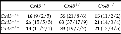
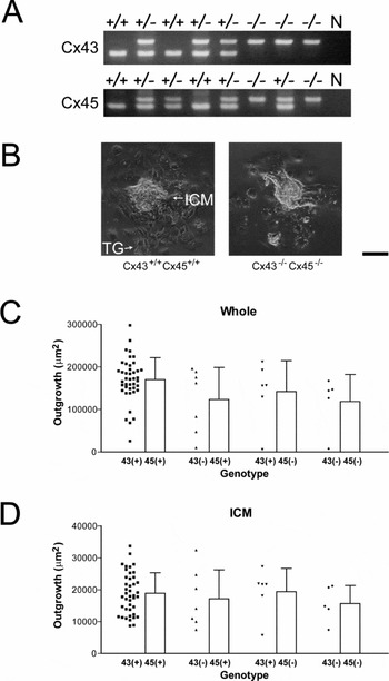
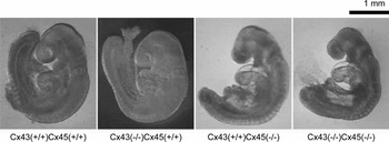
Cx43 −/− and Cx45 −/− are lethal strains (Kumai et al., Reference Kumai, Nishii, Nakamura, Takeda, Suzuki and Shibata2000; Reaume et al., Reference Reaume, de Sousa, Kulkarni, Langille, Zhu, Davies, Juneja, Kidder and Rossant1995). Cx43 −/− Cx45 −/− mice were generated from Cx43 +/− Cx45 +/− intercrosses in a C57BL/6 background. The genotype distribution conformed to the expected ratio (Fig. 1 A, Table 1; χ2-test). Given that gap junctions of preimplantation embryos mostly consist of Cx43 and Cx45 (De Sousa et al., Reference De Sousa, Juneja, Caveney, Houghton, Davies, Reaume, Rossant and Kidder1997), blastocysts were cultured and examined for peri-implantation defects (Fig. 1). Cx43 −/− Cx45 −/− blastocysts grew as efficiently as those of other genotypes, and some developed to E9.5 embryos (Fig. 2). The Cx43 −/− Cx45 −/− embryos showed cardiovascular defects nearly identical to those of the Cx45 −/− embryos (precise evaluation of cardiovascular functions will be described elsewhere). Contrary to prior suggestions that Cx43 and Cx45 are required for left-right patterning in vertebrate development (Levin, Reference Levin2007; Hatler et al., Reference Hatler, Essner and Johnson2009; Vandenberg & Levin, Reference Vandenberg and Levin2013), Cx43 −/− Cx45 −/− and single knockout hearts all showed dextro-looping. These results indicated that early development was not significantly affected by the loss of Cx43 or Cx45, or both.
Genotype distribution of embryos from Cx43 +/− Cx45 +/− intercrosses

Total numbers are shown in bold; numbers of E9.5 and E8.5 embryos and blastocyst cultures are shown in parentheses.
Normal growth of Cx43 −/− Cx45 −/− blastocysts. (A) PCR genotyping of mouse strains. N, negative control. (B) Representative micrographs of cultured day 3 blastocysts. ICM, inner cell mass; TG, trophoblast giant cells. Scale bar = 100 μm. (C, D) Surface area of whole blastocyst outgrowth (ICM + TG region) (C) and ICM only (D) was determined from micrographs of day 3 blastocysts. Data from +/+ and +/− genotypes were pooled as +, while −/− is represented as −. Raw data are plotted on the left side of each bar (mean ± SD). Differences between groups were not statistically significant.

External appearances of E9.5 embryos of WT (Cx43 +/+ Cx45 +/+), Cx43 −/− (Cx43 −/− Cx45 +/+), Cx45 −/− (Cx43 +/+ Cx45 −/−), and Cx43 −/− Cx45 −/−. The Cx43 −/− embryo looked similar to the WT embryo, while developmental arrest in the Cx45 −/− embryo also occurred in the Cx43 −/− Cx45 −/− embryos. Scale bar = 1 mm.

Cx43 and Cx45 are co-expressed in many tissues, and can be assembled into the same gap junction channel (Martinez et al., Reference Martinez, Hayrapetyan, Moreno and Beyer2002). In the E9.5 embryos, Cx43 and Cx45 were highly expressed in somite mesenchyme and neuroepithelial cells, but were absent in the Cx43 −/− Cx45 −/− embryos (Fig. 3 A, B). Cx40, reported in pre- and early post-implantation embryos, was weakly detected in tissues where Cx43 was expressed (Fig. 3 C), suggesting gap junctional intercellular communication would be marginally maintained through other Cxs including Cx40 in Cx43 −/− Cx45 −/− embryos.
Immunohistochemical detection of Cx40, Cx43, and Cx45 at E9.5. Centre panels are the overlay of images to the left and right. Signals were slightly enhanced for −/− to increase the background signal. (A, B) Cx43 and Cx45 in somites (A) and neuroepithelial cells (B). Cx43 and Cx45 are highly expressed in somite mesenchyme and neuroepithelial cells. Cx43 gap junctional plaques are typically larger than and frequently co-localises with Cx45 plaques. No immunoreactivity was observed in Cx43 −/− Cx45 −/−. (C) Cx43 and Cx40 in the trunk mesenchymal region. Weak Cx40 expression was detected in both Cx43 +/− Cx45 +/− and Cx43 −/− Cx45 −/−. Scale bar = 50 μm.

Discussion
Although Cx43 and Cx45 are the major gap junctional proteins, embryos lacking both of these proteins nevertheless completed peri-implantation development indistinguishable from that of WT, Cx43 −/−, and Cx45 −/−. Cx-knockout strains exhibit developmental and electrophysiological abnormalities that are closely related to their expression patterns and channel properties (Nishii et al., Reference Nishii, Shibata and Kobayashi2014). However, in this study, the Cx43 −/− Cx45 −/− embryos seemed to perform the developmental programme through compensatory mechanisms such as expression of Cx40.
Transcripts of Cx31 and Cx43 are found in zygotes; they disappear before compaction, but are expressed again from the compacted 8-cell stage onward (Reuss et al., Reference Reuss, Hellmann, Traub, Butterweck and Winterhager1997). The pattern of Cx45 mRNA expression in preimplantation development is similar to that of Cx31 and Cx43 (Davies et al., Reference Davies, Barr, Jones, Zhu and Kidder1996). The absence of Cx43 was shown in the Cx43 −/− morula (early post-compaction) stage embryos (De Sousa et al., Reference De Sousa, Juneja, Caveney, Houghton, Davies, Reaume, Rossant and Kidder1997). In addition, the lack of Cx45 protein and mRNA has been shown in the Cx45 −/− early post-implantation embryos (Kumai et al., Reference Kumai, Nishii, Nakamura, Takeda, Suzuki and Shibata2000). The half-lives of Cx43 and Cx45 are very short: 2 and 3 h, respectively (Darrow et al., Reference Darrow, Laing, Lampe, Saffitz and Beyer1995). Thus, functional gap junctions in the peri-implantation Cx43 −/− Cx45 −/− embryos, if they exist, would be comprised of other minor Cxs rather than from zygote-derived (i.e. maternal), residual Cx43 and Cx45. The candidate Cxs include Cx31, Cx31.1, and Cx40, which are expressed in blastocysts (De Sousa et al., Reference De Sousa, Valdimarsson, Nicholson and Kidder1993, Reference De Sousa, Juneja, Caveney, Houghton, Davies, Reaume, Rossant and Kidder1997; Dahl et al., Reference Dahl, Winterhager, Reuss, Traub, Butterweck and Willecke1996; Davies et al., Reference Davies, Barr, Jones, Zhu and Kidder1996; Grümmer et al., Reference Grümmer, Reuss and Winterhager1996; Houghton et al., Reference Houghton, Barr, Walter, Gabriel, Grümmer, Traub, Leese, Winterhager and Kidder2002). Cx31 and Cx31.1 are confined to the extraembryonic tissues after implantation (Dahl et al., Reference Dahl, Winterhager, Reuss, Traub, Butterweck and Willecke1996; Reuss et al., Reference Reuss, Hellmann, Traub, Butterweck and Winterhager1997). We showed weak expression of Cx40 in the trunk mesenchyme region (Fig. 3). However, Cx40 does not contribute to gap junctions in preimplantation embryos, because it is localised in the cytoplasm (Davies et al., Reference Davies, Barr, Jones, Zhu and Kidder1996; Houghton et al., Reference Houghton, Barr, Walter, Gabriel, Grümmer, Traub, Leese, Winterhager and Kidder2002). Whether Cx40 can serve as a functional gap junctional protein within the peri-implantation Cx43 −/− Cx45 −/− embryos merits further investigation.
Some reports have described essential roles of gap junctions for preimplantation development. A demethylating teratogenic agent, 5-AZA-2′-deoxycytidine, downregulates Cx31, Cx43, Cx45, E-cadherin, and β-catenin; as a result, preimplantation development is limited (Yu et al., Reference Yu, Xue, Wang, Lin, Liu, Lu and Liu2009). ‘Decompaction’ is induced after injecting antisense RNA or antibodies against Cxs (Lee et al., Reference Lee, Gilula and Warner1987; Bevilacqua et al., Reference Bevilacqua, Loch-Caruso and Erickson1989; Becker et al., Reference Becker, Evans, Green and Warner1995). Interestingly, after blastocyst injections, embryonic stem cells lacking Cx43 can integrate into chimaeric tissues, but those lacking Cx45 are extruded from the recipient cells (Egashira et al., Reference Egashira, Nishii, Nakamura, Kumai, Morimoto and Shibata2004; Gutstein et al., Reference Gutstein, Morley, Vaidya, Liu, Chen, Stuhlmann and Fishman2001b).
In contrast, the recent leading hypothesis suggests gap junctions are not important for preimplantation development (reviewed in Houghton (Reference Houghton2005)). Treatment with a gap junctional intercellular communication inhibitor, α-glycyrrhetinic acid, eliminates dye coupling in preimplantation embryos, but their growth into blastocysts is unaffected (Vance & Wiley, Reference Vance and Wiley1999; Houghton et al., Reference Houghton, Barr, Walter, Gabriel, Grümmer, Traub, Leese, Winterhager and Kidder2002). No abnormality is reported in Cx43 −/− embryonic stem cells whose expression of Cx31 and Cx45 is suppressed by RNA interference (Wörsdörfer et al., Reference Wörsdörfer, Maxeiner, Markopoulos, Kirfel, Wulf, Auth, Urschel, von Maltzahn and Willecke2008). Showing normal peri-implantation development of the Cx43 −/− Cx45 −/− embryos, our result also seems to confirm the hypothesis. The ubiquity of gap junctions in the pre- and peri-implantation period may prepare blastocysts for differentiation and morphogenesis in later stages. Cx expression patterns during pre- and peri-implantation development and those within the placenta vary between mammalian species (Houghton, Reference Houghton2005; Malassiné & Cronier, Reference Malassiné and Cronier2005). This fact, together with the previous and current studies, suggests that the role of intercellular communication in pre- and peri-implantation development is probably minor.
The Cx43 −/− Cx45 −/− embryos were obtained at a frequency as low as 1/16, and these deletions were lethal in early embryogenesis. Although these limitations make further molecular analyses difficult, it is a basic and interesting question of whether zygotes without gap junctions can develop into post-implantation stage embryos. Our findings suggest that it would be difficult to create a mouse strain with multiple null mutations in Cx factors relevant to pre- and peri-implantation development by natural breeding. Recent advancements in induced pluripotent stem cell (iPSC) technology may provide an improved, reproducible platform for the analysis of early stage embryos lacking multiple Cxs (Nishii et al., Reference Nishii, Shibata and Kobayashi2014). iPSCs derived from adult somatic cells have a differentiation potential similar to that of embryonic stem cells; they differentiate into various kinds of tissues and can recapitulate initial embryogenesis through formation of embryoid bodies (Yamanaka, Reference Yamanaka2012). There are several healthy ‘flox’ Cx mutant strains, including those for Cx26, Cx43, and Cx45 in which expression is reported in pre- and peri-implantation development (Gutstein et al., Reference Gutstein, Morley, Tamaddon, Vaidya, Schneider, Chen, Chien, Stuhlmann and Fishman2001a; Liao et al., Reference Liao, Day, Damon and Duling2001; Theis et al., Reference Theis, de Wit, Schlaeger, Eckardt, Krüger, Döring, Risau, Deutsch, Pohl and Willecke2001; Cohen-Salmon et al., Reference Cohen-Salmon, Ott, Michel, Hardelin, Perfettini, Eybalin, Wu, Marcus, Wangemann, Willecke and Petit2002; Nishii et al., Reference Nishii, Kumai, Egashira, Miwa, Hashizume, Miyano and Shibata2003; Maxeiner et al., Reference Maxeiner, Dedek, Janssen-Bienhold, Ammermüller, Brune, Kirsch, Pieper, Degen, Krüger, Willecke and Weiler2005). They can likely be combined to create mouse strains with multiple Cx flox alleles. Using iPSCs from such strains, the floxed Cx coding regions can be simultaneously deleted after expression of Cre recombinase in vitro. Embryonic stem cells show normal growth performance even if expression of Cx31, Cx43, and Cx45 is significantly reduced (Wörsdörfer et al., Reference Wörsdörfer, Maxeiner, Markopoulos, Kirfel, Wulf, Auth, Urschel, von Maltzahn and Willecke2008). Thus, iPSCs with multiple Cx-deletions are probably viable and retain their original differentiation potential. It will be interesting to see how these iPSCs differentiate into specific tissues or form embryoid bodies.
Acknowledgements
The authors thank Y. Miyano, K. Hashizume, M. Kamizono, and T. Omura for their care of experimental animals and assistance in experiments.
Financial support
This work was supported by Grants-in-Aid for Scientific Research from the Japan Society for the Promotion of Science (K.N., grant number 22590197).
Conflict of interest
None.