Introduction
DNA methylation has been implicated in a number of specialized biochemical functions, including mammalian development (Rogers et al., Reference Rogers, Francis, Sulik, Alles, Massaro, Zucker, Elstein, Rosen and Chernoff1994; Okano et al., Reference Okano, Bell, Haber and Li1999; Walsh and Bestor, Reference Walsh and Bestor1999; Jackson-Grusby et al., Reference Jackson-Grusby, Beard, Possemato, Tudor, Fambrough, Csankovszki, Dausman, Lee, Wilson, Lander and Jaenisch2001), gene expression (Reik & Dean, Reference Reik and Dean2001), cell differentiation (Jost et al., Reference Jost, Oakeley, Zhu, Benjamin, Thiry, Siegmann and Jost2001), genomic imprinting (Reik et al., Reference Reik, Dean and Walter2001a), X chromosome inactivation (Heard et al., Reference Heard, Clerc and Avner1997), chromatin modification (Okano et al., Reference Okano, Bell, Haber and Li1999), cancer (Baylin, Reference Baylin1997) and aging (Cooney, Reference Cooney1993; Issa, Reference Issa2000).
5-AZA-2′-deoxycytidine (5-AZA-CdR) was synthesized by Piskala & Sorm in Reference Piskala and Sorm1964. As a DNA methylation inhibitor, 5-AZA-CdR was widely used to study mammalian development (Branch et al., Reference Branch, Francis, Brownie and Chernoff1996; Cisneros et al., Reference Cisneros and Branch2003), cell differentiation (Gattei et al., Reference Gattei, Aldinucci, Petti, Da Ponte, Zagonel and Pinto1993; Jüttermann et al., Reference Jüttermann, Li and Jaenisch1994; Jackson-Grusby et al., 1997) and cancer therapy (Issa et al., Reference Issa, Gharibyan, Cortes, Jelinek, Morris, Verstovsek, Talpaz, Garcia-Manero and Kantarjian2005; Ikeda et al., Reference Ikeda, Iyama, Ishikawa, Egami, Nakao, Sado, Ninomiya and Baba2006). What's more, 5-AZA-CdR has been shown to induce developmental defects in mouse and rat embryos (Branch et al., Reference Branch, Francis, Brownie and Chernoff1996, Reference Branch, Chernoff, Brownie and Francis1999); however, the effects of 5-AZA-CdR have only been shown for implantation embryos and not for preimplantation embryos. In our previous study, the preimplantation mouse embryos treated with 5-AZA-CdR (0.2 and 1.0 μM) were unable to develop fully, especially for 8-cell embryos, morulae and blastulae (submitted). In detail, the pronuclear embryos only developed to 5–7-cell embryos not 8-cell embryos, when 5-AZA-CdR was added from pronuclear stage. In fact, 2-cell embryos could develop to 8-cell, but the 8-cell embryos could not compact as the control, when treated with 5-AZA-CdR from the 2-cell stage. When 4-cell embryos were exposed to 5-AZA-CdR, the rate of morulae in 1.0 μM 5-AZA-CdR was significantly lower than that in the control (p < 0.05). From the embryonic morphology, 5-AZA-CdR affected the compaction and blastocoel cavitation. We also detected genome DNA methylation by immunofluorescence and found that 5-AZA-CdR led to the decrease of DNA methylation in 8-cell embryos and early morulae (submitted), which can tell us that DNA methylation is very important for the normal development of 8-cell embryos, morulae and blastulae.
5-AZA-CdR-substituted DNA forms covalent adducts with cellular DNA methyltransferase 1 (Dnmt1), thereby depleting the cells of enzyme activity and causing demethylation of genomic DNA (Creusot et al., Reference Creusot, Acs and Christman1982; Michalowsky & Jones, Reference Michalowsky and Jones1987; Christman, Reference Christman2002). Dnmt1o protein is synthesized from an oocyte-specific transcript, which is translated into a truncated version of Dnmt1 that is missing its N-terminal 118 amino acids (Mertineit et al., Reference Mertineit, Yoder, Taketo, Laird, Trasler and Bestor1998). It has been proved that Dnmt1o contributes to maintaining the right DNA methylation in 8-cell embryos (Howell et al., Reference Howell, Bestor, Ding, Latham, Mertineit, Trasler and Chaillet2001). So the aims of this study were to determine: (1) if Dnmt1o mediated the decrease of DNA methylation or not; and (2) how sequential gene expression changed in 8-cell embryos and early morulae after treatment with 5-AZA-CdR, especially the genes that have key roles for compaction and blastocoel cavitation, such as gap junction family and tight junction genes.
Materials and Methods
In vitro fertilization and embryo culture
Female 21–28-day-old Kun-ming mice were superovulated with 5 IU of pregnant mares' serum gonadotropin (PMSG) and then treated 48 h later with 5 IU of human chorionic gonadotropin (hCG). Unfertilized metaphase II-arrested oocytes were collected 14–15 h later in Whitten's medium (Whitten, Reference Whitten1971) from the ampullae of the oviducts. Sperm were collected from the caudal epididymides of adult Kun-ming males and preincubated in Whitten's medium for 2 h in an atmosphere of 5% CO2 and 95% air at 37.5°C. The oocytes were then inseminated with the capacitated sperm in Whitten's medium. Six hours after insemination, the fertilized oocytes were washed with CZB medium (Chatot et al., Reference Chatot, Ziomek, Bavister, Lewis and Torres1989) and cultured in a humidified atmosphere of 5% CO2 and 95% air at 37.5°C.
Treatment with 5-AZA-CdR
5-AZA-CdR was purchased from Sigma. Pronuclear, 2-cell and 4-cell embryos were incubated with 5-AZA-CdR (diluted in CZB) at 1.0 μM continuously and allowed to develop in vitro. The medium was replaced with CZB containing fresh 5-AZA-CdR every 24 h.
Immunochemical analysis
For the detection of Dnmt1o, the embryos were washed in PBS containing 3 mg/ml polyvinylpyrrolidone (PBS/PVP), fixed for 1 h in 3.7% paraformaldehyde in PBS and permeabilized with 0.5% Triton X-100 in PBS for 20 min at room temperature. The fixed cells were blocked for at least 1 h in blocking buffer (3% BSA in PBS) and then incubated with anti-Dnmt1o antibodies (Santa Cruz Biotechnology, Inc.), followed by incubation with a Cy3-conjugated anti-goat IgG secondary antibody (Sigma). The DNA was stained by treatment with 3 mg/ml DAPI for 20 min and the cells were mounted on a glass slide in Vectashield. Immunofluorescence was visualized by using a confocal microscope.
Real-time quantitative PCR
We detected the transcripts of the five genes – Cx31, Cx43, Cx45 (connexin family members), Cdh1 (E-cadherin) and ctnnb1 (β-catenin), which are crucial for the development of preimplantation mouse embryos. As an external standard for quantification, a predetermined amount of rabbit globin mRNA (0.05 pg/embryo) was added to each embryo batch before lysis (Manejwala et al., Reference Manejwala, Logan and Schultz1991) to control for variation in the RNA recovery, reverse transcription and PCR steps. So changing levels of the five genes mRNA on a per embryo basis could be seen as changes in the ratio between the five genes and globin mRNA amplicons. Total RNA was extracted using TRIzol (Invitrogen) from about 30–50 embryos according to the manufacturer's procedures and was treated with RNase-free DNase (Promega) to remove the genomic DNA.
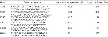
cDNA was synthesized from 1–2 mg RNA using 0.5 μg/μl oligo-dT primer (Promega), 10 mM dNTPs, 0.1 M DTT, 40 U of Maloney murine leukemia virus reverse transcriptase (Promega) and 20 U of RNasin, in a total volume of 25 μl. The resulting cDNA was diluted 1:50 to use in a quantitative PCR reaction (Q-PCR). Q-PCR was performed using SYBRGreen (Invitrogen) for the detection of fluorescence during amplification. Each amplification reaction contained 0.1 μg of forward and reverse primers, 6.25 μl of SYBRGreen mix and 5.25 μl of diluted cDNA in a total volume of 12.5 μl. All primers were designed with annealing temperatures of 56–61°C. PCR conditions were 95°C for 1 min, followed by 40–45 cycles at 94°C for 10 s, 56–61°C for 20 s and 72°C for 20 s. After each PCR run, melting-curve analysis was performed for each sample to verify that a single specific product was generated. Amplicon size was confirmed by 8% polyacrylamide gel electrophoresis (PAGE). Negative controls, composed of the PCR mix without nucleic acid, were also run with each group of samples. The relative amount of each sample was determined using a standard curve obtained by amplifying a 10-fold dilution series of cDNA prepared by PCR. Details of the primer sequences and annealing temperatures for the genes tested are shown in Table 1.
Primers used for real-time PCR.

Data analysis
Each experiment included controls and three trials were conducted for each treatment. The data were analysed by one-way analysis of variance (ANOVA) after being transformed via LSD (least-significant difference) using SPSS software (SPSS Inc.; p < 0.05 was considered significant).
Results
Effects of 5-AZA-CdR on Dnmt1o in preimplantation mouse embryos
Pronuclear embryos, 2-cell embryos and 4-cell embryos were treated with CZB containing 1.0 μM 5-AZA-CdR continuously and they were allowed to develop fully in vitro. Then Dnmt1o was detected by immunofluorescence assay before and after treatment with 5-AZA-CdR.
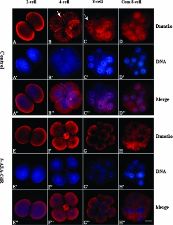
We found that Dnmt1o was concentrated in peripheral cytoplasm and nuclei did not contain detectable Dnmt1o in 2-cell and 4-cell embryos obtained from pronuclear and 2-cell embryos exposed to 5-AZA-CdR (Fig. 1e, f), which was almost the same with that in control (Fig. 1a, b). From the figures, we also noticed that most of Dnmt1o was distributed around the membranes of 2-cell embryos in a dispersal morpha. However, in 4-cell embryos, some were still in a dispersal morpha, but others were changed to cytoplasmic granules.
Dnmt1o immunofluorescence. Cy3 (red) represents the expression of Dnmt1o, DAPI (blue) shows DNA of nucleus and the first two figures were merged to one photo. The above photographs were gotten from the control groups and the below photos shows the experimental groups, which were treated with 1.0 μM 5-AZA-CdR. Com 8-cell is referred to as compacted 8-cell, arrow in B, cytoplasmic granules; arrow in C, dispersal Dnmt1o in the nucleus. Scale bar, 20 μm.

For 8-cell embryos and early morulae (compacted 8-cell), there was great difference between the experimental group and the control group. In the control, Dnmt1o was not only in the cytoplasm but also was present in large amounts in nuclei except for nucleoli (Fig. 1c, d). In the 8-cell embryos and early morulae exposed to 5-AZA-CdR, their nuclei did not contain detectable Dnmt1o (Fig. 1g, h), although their cytoplasmic Dnmt1o was no difference with that in the controls. Dnmt1o accumulated into granules in cytoplasm at this developmental stage both in experimental group and control group.
5-AZA-CdR led to down-regulation of gene transcript
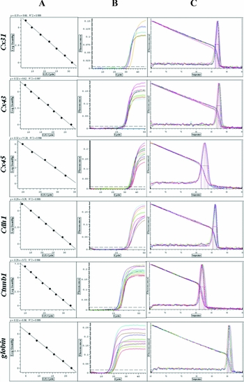
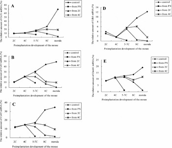
Real-time quantitative PCR was used to determine the changing of Cx31, Cx43, Cx45, Cdh1 and Ctnnb1 mRNA after preimplantation mouse embryos were treated with 1.0 μM 5-AZA-CdR. Figure 2 showed the standard, amplification and melting curves of Cx31, Cx43, Cx45, Cdh1 Ctnnb1 and rabbit globin. And the relative ratios between Cx31, Cx43, Cx45, Cdh1, Ctnnb1 and rabbit globin were shown in Fig. 3a–e, respectively. Cx31, Cx43, Cx45, Cdh1 and Ctnnb1 were all expressed at the 2-cell stage and accumulated with the development of early mouse embryos.
The standard, amplification and melting curves of Cx31, Cx43, Cx45, Cdh1, Ctnnb1 and rabbit globin in real-time PCR. A, B and C represent the standard, amplification and melting curves, respectively.

The relative amount of mRNA for Cx31, Cx43, Cx45, Cdh1 and Ctnnb1. PN, pronuclear embryo; 2C, 2-cell embryo; 4C, 4-cell embryo; 5–7C, 5–7-cell embryo; 8C, 8-cell embryo. Note: the morulae in ‘from 2C’ group were referred to the arrested 8-cell embryos.

As shown in Fig. 3a, the relative amount of Cx31 mRNA did not change in 2-cell and 4-cell embryos, compared to that in the control. For 5–7-cell embryos, the amount of ‘from PN’ was significantly lower than that in the control (p < 0.05), while the left two groups (‘from 2C’ and ‘from 4C’) were the same with the control. The transcript amount in 8-cell embryos obtained from 2C and 4C decreased significantly than that in the control (p < 0.05). However, the arrested 8-cell obtained from 2-cell almost decreased to zero. When 4-cell embryos were treated with 5-AZA-CdR, the embryos could develop to early morulae, but their transcription level of Cx31 was much lower than that in the control (p < 0.05). The whole changing trends of the other four genes were very similar with Cx31. The only difference was the extent of decrease in contrast with their respective control. In detail, the decrease extent of Cx43 and Cx45 (Fig. 3b, c) was higher than that of Cx31, Cdh1 and Ctnnb1 in 8-cell embryos both from 2C and from 4C. In morulae, the relative amount of Cx31, 43 and 45 decreased much more than Cdh1 and Ctnnb1 (Fig. 3d, e), compared to their respective control.
Discussion
5-AZA-CdR and Dnmt1o
Maintenance of genomic methylation patterns in mammalian somatic cells depends on DNA methyltransferase-1 (Dnmt1). Mouse oocytes and preimplantation embryos lack Dnmt1 but express a variant of this protein called Dnmt1o. Dnmt1o is synthesized and stored in the oocyte cytoplasm and in the cytoplasm of all preimplantation cleavage stages, but only trafficks to the 8-cell nucleus during preimplantation development (Carlson et al., Reference Carlson, Page and Bestor1992; Mertineit et al., Reference Mertineit, Yoder, Taketo, Laird, Trasler and Bestor1998; Cardoso & Leonhardt, Reference Cardoso and Leonhardt1999; Howell et al., Reference Howell, Bestor, Ding, Latham, Mertineit, Trasler and Chaillet2001). In our experiment, we also saw that Dnmt1o was in the cytoplasm of 2-cell and 4-cell embryos, while Dnmt1o was found in the nuclei of 8-cell embryos and early morulae (compacted 8-cell embryos) in the control without 5-AZA-CdR. It has been proved that 5-AZA-CdR can induce the rapid degradation of Dnmt1 by the proteasomal pathway in cancer cells (Ghoshal et al., Reference Ghoshal, Datta, Majumder, Bai, Kutay, Motiwala and Jacob2005). Our data indicate that 5-AZA-CdR may also make Dnmt1o degradation by some pathway in 8-cell embryos of mouse.
Studies in mice indicate that heterozygous mutant embryos derived from homozygous mutant oocytes showed a loss of methylation at certain imprinted loci during their preimplantation development (Howell et al., Reference Howell, Bestor, Ding, Latham, Mertineit, Trasler and Chaillet2001), most likely because the loss of nuclear Dnmt1o protein at the 8-cell stage leads to a loss of maintenance methylation in 8-cell blastomeres (Ratnam et al., Reference Ratnam, Mertineit, Ding, Howell, Clarke, Bestor, Chaillet and Trasler2002). Howell et al. (Reference Howell, Bestor, Ding, Latham, Mertineit, Trasler and Chaillet2001) also thought that transient nuclear localization of Dnmt1o in 8-cell embryos provided maintenance methyltransferase activity specifically at imprinted loci during the fourth embryonic s phase. Studies on cloned preimplantation mouse embryos showed that defects in the regulation of Dnmt1o expression and cytoplasmic–nuclear trafficking may prevent clones from completing essential early developmental events (Chung et al., Reference Chung, Ratnam, Chaillet and Latham2003). Therefore, aberrant loss of Dnmt1o in 8-cell embryos induced by 5-AZA-CdR may result in the decrease of DNA methylation, which may lead to the developmental arrest of 8-cell embryos and early morulae.
5-AZA-CdR only decreased Dnmt1o in 8-cell embryos and morulae (compacted 8-cell), but not 2- or 4-cell embryos. Most of Dnmt1o was concentrated in the peripheral cytoplasm and the nuclei in 2- and 4-cell embryos do not contain detectable Dnmt1o (Carlson et al., Reference Carlson, Page and Bestor1992; Mertineit et al., Reference Mertineit, Yoder, Taketo, Laird, Trasler and Bestor1998; Cardoso & Leonhardt, Reference Cardoso and Leonhardt1999; Howell et al., Reference Howell, Bestor, Ding, Latham, Mertineit, Trasler and Chaillet2001). And the 5-AZA-CdR-induced degradation of Dnmt1 is nuclear (i.e. 5-AZA-CdR does not degrade Dnmt1 in the cytoplasm; Ghoshal et al., Reference Ghoshal, Datta, Majumder, Bai, Kutay, Motiwala and Jacob2005). Therefore, Dnmt1o in the 5-AZA-CdR-treated 2- and 4-cell embryos was unchanged. In contrast, large amounts of Dnmt1o transiently enter the nuclei in 8-cell embryos. Thus, the observed decrease of Dnmt1o in the 8-cell embryos is logical.
As for the pathway of Dnmt1o cytoplasmic–nuclear traffic, Dnmt1o does not colocalize with either mitochondria or endoplasmic reticulum, it does colocalize with annexin V (Doherty et al., Reference Doherty, Bartolomei and Schultz2002). So the decrease of Dnmt1o induced by 5-AZA-CdR may be caused by the blockage of Dnmt1o cytoplasmic–nuclear traffic.
5-AZA-CdR and developmental gene expression
During the development of mouse preimplantation embryos, there are two morphological changes, compaction and cavitation, which happen at 8-cell stage and blastocyst stage, respectively. Our results showed that 5-AZA-CdR mainly affected the above two changes (submitted) when preimplantation mouse embryos were exposed to 5-AZA-CdR. 5-AZA-CdR as a demethylating agent can lead to up-regulation and down-regulation of genes in cancer cells (Gius et al., Reference Gius, Cui, Bradbury, Cook, Smart, Zhao, Young, Brandenburg, Hu, Bisht, Ho, Mattson, Sun, Munson, Chuang, Mitchell and Feinberg2004; Arai et al., Reference Arai, Yokosuka, Hirasawa, Fukai, Chiba, Imazeki, Kanda, Yatomi, Takiguchi, Seki, Saisho and Ochiai2006). It is interesting that there are nearly as many genes were downregulated as upregulated after demethylation caused by 5-AZA-CdR (Gius et al., Reference Gius, Cui, Bradbury, Cook, Smart, Zhao, Young, Brandenburg, Hu, Bisht, Ho, Mattson, Sun, Munson, Chuang, Mitchell and Feinberg2004). The transcription of the five genes (Cx31, Cx43, Cx45, Cdh1 and Ctnnb1) was downregulated after treated with 5-AZA-CdR in our research.
Gap junctions and tight junctions are crucial for compaction, the formation of blastocysts and cell differentiation during preimplantation development (Chen et al., Reference Chen, Zhang and Zhang2002). Gap junctional communication plays a central role in the maintenance of cellular homeostasis by allowing the passage of small molecules between adjacent cells (Wei et al., Reference Wei, Xu and Lo2004; Houghton et al., Reference Houghton2005). Gap junctions are composed of a family of proteins termed connexins and Davies' findings indicate that at least four connexins (Cx31, 31.1, 43 and 45) contribute to gap junctions in preimplantation development. The 8-cell embryo obtained from treatment with 5-AZA-CdR showed the decrease of Cx31, 43 and 45 mRNA in our experiment. Also in rat studies, the uncompacted 8-cell stage lacks the transcripts of Cx31 and Cx43 and re-expression of Cx31 and 43 mRNA is found from the compacted 8-cell stage onward (Reuss et al., Reference Reuss, Hellmann, Traub, Butterweck and Winterhager1997). Early experiments involved attempts to disrupt gap junctional coupling in preimplantation mouse embryos by means of connexin antibodies (Lee et al., Reference Lee, Gilula and Warner1987; Becker et al., Reference Becker, Evans, Green and Warner1995) or antisense RNA (Bevilacqua et al., Reference Bevilacqua, Erickson and Hieber1988). In each of these three studies, abnormal gap junctional coupling affected blastomeres underwent ‘decompaction’. Thus it was concluded that the maintenance of compaction and, as a consequence, blastocyst development depend on gap junctional coupling. However, when intercellular communication was severely reduced by Cx43 knockout, there was no apparent effect on either compaction or blastocyst formation (De Sousa et al., Reference De Sousa, Juneja, Caveney, Houghton, Davies, Reaume, Rossant and Kidder1997). These discrepancies remain unresolved.
Epithelial cadherin (E-cadherin), catennin and zonula occludents are involved in the establishment of tight junctions and there are two functions served by the tight junction, the movement of water and solutes between adjacent cells and maintenance of cell polarity (Chen et al., Reference Chen, Zhang and Zhang2002). β-Catenin binds to the cytoplasmic domain of E-cadherin (Nagafuchi & Takeichi, Reference Nagafuchi and Takeichi1989; Ozawa et al., Reference Ozawa, Baribault and Kemler1989) and β-catenin is one of several intracellular mediators necessary for the maintenance and function of E-cadherin in cell–cell interactions (De Vries et al., Reference De Vries, Evsikov, Haac1, Fancher, Holbrook, Kemler, Solter and Knowles2004). E-cadherin mediates compaction of the individual blastomere in the 8-cell-stage embryo and this adhesion triggers the development of the trophectoderm and other epithelial cell layers at later developmental stages (Gumbiner, Reference Gumbiner1996). E-cadherin null embryos fail to form an intact trophectoderm cell layer (Larue et al., Reference Larue, Ohsugi, Hirchenhain and Kemler1994). Lack of β-catenin results in the early gastrulation lethality, with no mesoderm formation and a block in anterior–posterior axis formation and head development (Haegel et al., Reference Haegel, Larue, Ohsugi, Fedorov, Herrenknecht and Kemler1995; Huelsken et al., Reference Huelsken, Vogel, Brinkmann, Erdmann, Birchmeier and Birchmeier2000). Another study shows that absence of wild-type β-catenin in oocytes does statistically compromise developmental success rates and the blastomeres of embryos can not adhere as normal (De Vries et al., Reference De Vries, Evsikov, Haac1, Fancher, Holbrook, Kemler, Solter and Knowles2004).
Collectively, the decrease of Cx31, Cx43, Cx45, Cdh1 and Ctnnb1 mRNA may affect gap junctions and tight junctions, which contribute to the developmental arrest, decompaction and failure to blastulae caused by 5-AZA-CdR.
As for the 5–7-cell embryos, although there was no change in the expression of Dnmt1o, a sharp decrease took place in gene expression just like arrested 8-cell embryos. 5-AZA-CdR is a deoxycytidine analogue that incorporates into DNA, which may block the synthesis of DNA (Christman, Reference Christman2002), then may have effects on the early embryos development. On the other hand, it has been proved that cells exposed to low doses of 5-AZA-CdR exhibit not only altered gene expression but also reduced proliferation and/or increased apoptosis (Issa et al., Reference Issa, Garcia-Manero, Giles, Mannari, Thomas, Faderl, Bayar, Lyons, Rosenfeld, Cortes and Kantarjian2004). Further research is needed to find the real reasons for the arrest of 5–7 cell embryos induced by 5-AZA-CdR.
In conclusion, the developmental arrest induced by 5-AZA-CdR may be caused by the disappearance of Dnmt1o, the transcriptional down-regulation of Cx31, Cx43, Cx45, Cdh1 and Ctnnb1 and other unknown reasons. Dnmt1o traffic to 8-cell nuclei and right genes expression are essential for normal compaction and blastocoel cavitation in mouse preimplantation embryos.
Acknowledgements
The authors thank Miao Deqiang Ph.D and Sui Hongshu Ph.D, College of Animal Science and Veterinary Medicine, Shandong Agriculture University, for their helpful suggestions. This research was supported by grants from the 973 Programme of the Ministry of Science and Technology of the People's Republic of China (No. 2007CB947403).