INTRODUCTION
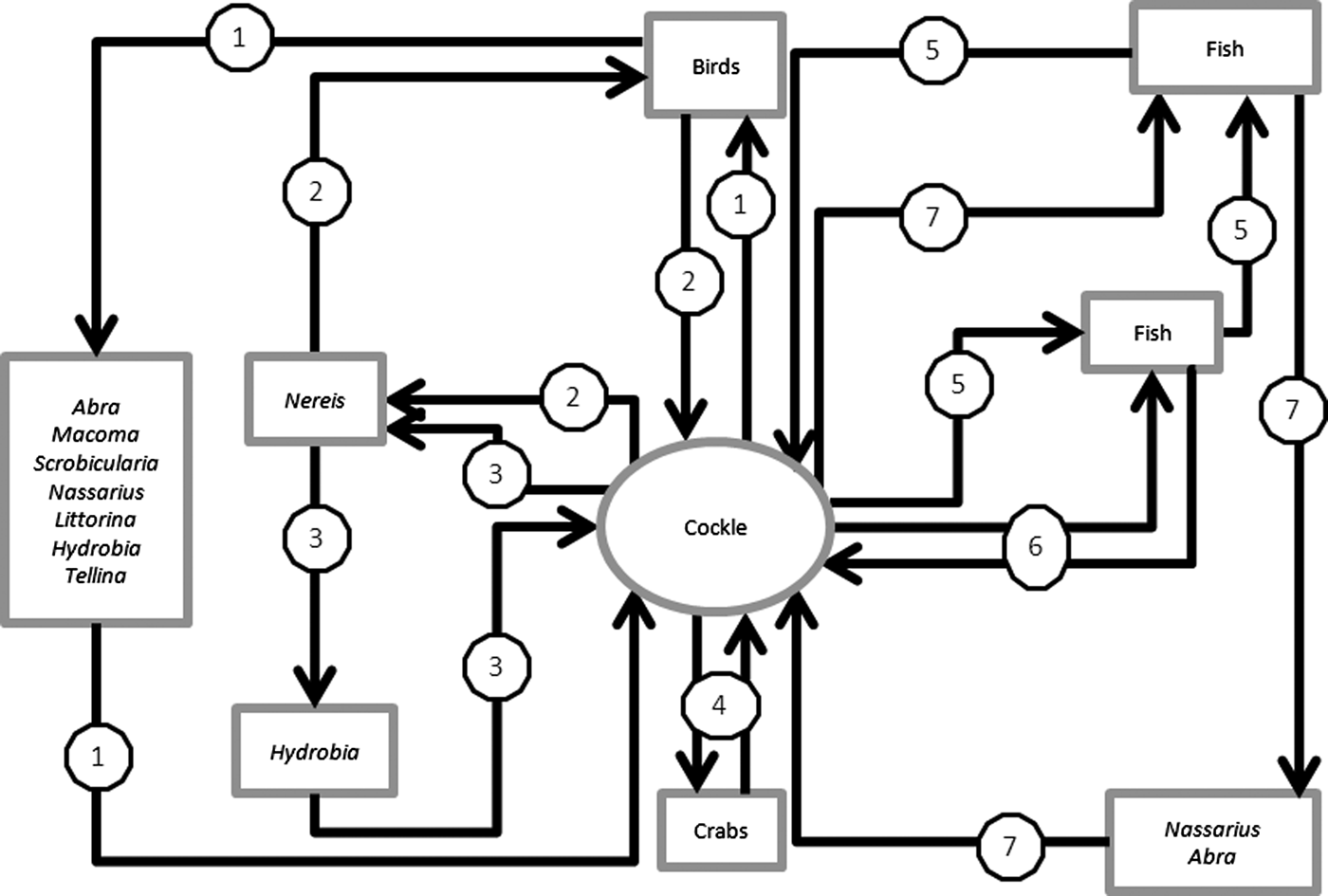
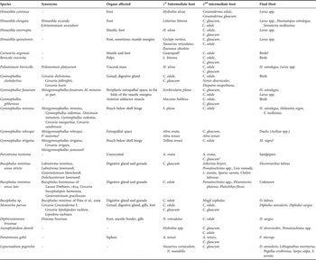
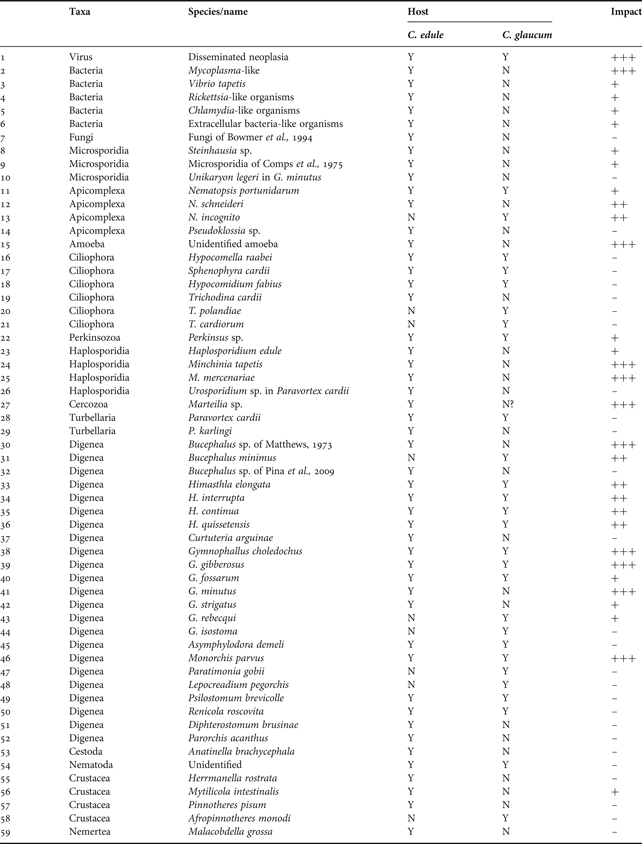
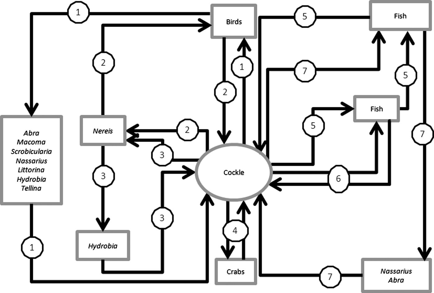
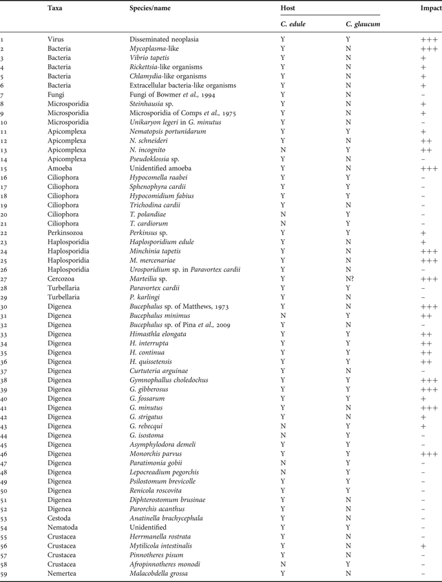
Cockles are a small bivalve occurring intertidally and subtidally around the coastline of Europe. Under normal conditions they remain buried in the sediment and feed via a siphon. Cerastoderma glaucum (Bruguière, 1789), the lagoon cockle, tends to occur in non-tidal areas such as lagoons and salt marshes whilst C. edule (L. 1758), the edible cockle, is more likely to be found on open coasts and estuaries (Reise, Reference Reise2003). They occur from Norway to Morocco and through the Baltic, Mediterranean and Black Sea. Mortalities of cockles and catastrophic declines in cockle populations have been reported for many years with predation, disease, climactic events, pollution, failed recruitment and over-fishing being suggested as important factors (Ducrotoy et al., Reference Ducrotoy, Rybarczyk, Souprayen, Bachelet, Beukema, Desprez, Dörjes, Essink, Guillou, Michaelis, Sylvand, Wilson, Elkaïm and Ibanez1989). Furthermore, it is recognized that cockles are hosts to a wide variety of parasites and diseases. Many of these are normally relatively innocuous but can become problematic particularly if prevailing environmental conditions are poor and have been implicated in mass mortality events leading to declines in cockle populations. Mortalities of cockles were reported in the Burry Inlet (Loughor Estuary, Wales) in the 1950s and 1960s, which were attributed to environmental conditions and predation by man and oystercatchers (Hancock, Reference Hancock, den Boer and Gradwell1970). However, whilst Hancock (Reference Hancock, den Boer and Gradwell1970) acknowledged that there were a number of parasites in cockles from the Burry Inlet that had been previously implicated in mass mortalities, that study only considered that they might have been problematic during ‘times of stress, such as cold winters and hot summers’. Lauckner (Reference Lauckner1984) in examining cockles from the German North Sea coast concluded that digeneans were an extremely important component of the biota and, in agreement with Hancock & Urquhart (Reference Hancock and Urquhart1965) thought infected cockles were likely to be less resistant to thermal stress as a result of infections with Digenea. To date almost 60 parasites and disease conditions have been described or reported from C. edule and C. glaucum (see Table 1). A schematic of the life cycles of most of the cockle parasites is shown in Figure 1. This systematic review covers all the groups of pathogens reported for both the lagoon cockle and the edible cockle throughout its range (Table 2).
Life cycle patterns of cockle parasites that involve more than one host. It does not include infections transmitted between cockles such as viruses and bacteria. Where line 1 includes Himasthla continua, H. elongata, H. interrupta, H. quissetensis, Renicola roscovita, Psilostomum brevicolle, Gymnophallus fossarum, G. gibberosus, G. minutus, G. rebecqui and G. strigatus; line 2 includes G. choleodochus; line 3 includes Asymphylodora demeli; line 4 includes Nematopsis sp.; line 5 includes Bucephalus spp.; line 6 includes Monorchis parvus and line 7 includes Diphterostomum brusinae, Paratimonia gobii and Lepocreadium pegorchis.
Digenea reported from cockles (Cerastoderma edule and C. glaucum) along with details of the organs affected, other hosts directly involved in the life cycle and appropriate references.

1Reported as C. edule by Dolgikh (Reference Dolgikh1968). However, C. edule is absent from the Black Sea. It is therefore assumed that the report is of C. glaucum.
Summary of parasites/diseases/commensals reported in Cerastoderma edule and C. glaucum. Where: – No effect recorded or negligible impact; + Localised pathology, limited effect on survival; ++ Individual mortality or impact on growth, metabolism; +++ Population level effect, mass mortalities reported.
VIRUSES AND VIRUS-LIKE PARTICLES
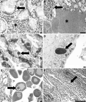
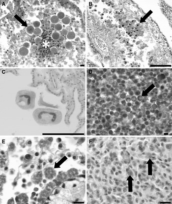
Disseminated neoplasias, also called haematopoietic, haemic, haemocytic neoplasia or disseminated sarcoma (Figure 2A) have been widely reported in Cerastoderma edule and to a lesser extent C. glaucum in Ireland, Spain and France (Poder & Auffret, Reference Poder and Auffret1986; Twomey & Mulcahy, Reference Twomey and Mulcahy1988; Carballal et al., Reference Carballal, Iglesias, Santamarina, Ferro-Soto and Villalba2001; Le Grand et al., Reference Le Grand, Kraffe, de Montaudouin, Villalba, Marty and Soudant2010). Spatio-temporal variations in prevalence have been reported and mortalities associated with the condition are known to occur (Twomey & Mulcahy, Reference Twomey and Mulcahy1988; Carballal et al., Reference Carballal, Iglesias, Santamarina, Ferro-Soto and Villalba2001; Villalba et al., Reference Villalba, Carballal and López2001; Ordás & Figueras, Reference Ordás and Figueras2005; Le Grand et al., Reference Le Grand, Kraffe, de Montaudouin, Villalba, Marty and Soudant2010). Prevalence of infection is significantly higher in surfaced cockles compared with buried animals and the condition is considered a mortality driver (Villalba et al., Reference Villalba, Carballal and López2001; Le Grand et al., Reference Le Grand, Kraffe, de Montaudouin, Villalba, Marty and Soudant2010). The aetiological agent for disseminated neoplasias in cockles and other bivalve species has been the subject of numerous studies with suggestions that they may be due to contaminants or biotoxins (Twomey & Mulcahy, Reference Twomey and Mulcahy1988; Landsberg, Reference Landsberg1996; Barber, Reference Barber2004). Transmissibility of the condition between cockles suggested an infectious agent; subsequent studies have shown evidence of a retrovirus (Twomey & Mulcahy, 1998; Collins & Mulcahy, Reference Collins and Mulcahy2003). Furthermore, Romalde et al. (Reference Romalde, Vilariño, Beaz, Rodriguez, Diaz, Villalba and Carballal2007) provided ultrastructural evidence of virus-like particles in the cytoplasm of neoplastic cells which they considered ‘could resemble a retrovirus-like agent’ and utilized an assay to demonstrate the presence of retroviruses in disseminated neoplasias-affected cockles. Increases in levels of the heat shock proteins Hsp 70 and Hsp 90 were noted in cockles affected by disseminated neoplasias (Díaz et al., Reference Díaz, Cao, Villalba and Carballal2010). In the same study, the authors reported an increase in a 53 kDa protein analogous to mutant p53 which they considered was related to an alteration of the cell cycle.
Histopathology of infections of Cerastoderma spp.: (A) disseminated neoplasias infection showing affected cells (*) in the connective tissue surrounding digestive gland tubules (arrow); (B) large foci of heavy haemocyte infiltration (*) in connective tissue. Note presence of ovaries (arrow); (C) Chlamydia-like organisms (CLOs) (arrow) in the digestive gland tubule epithelial cells. Note the lack of host response to the infection; (D) large extracellular bacterial-like organism (arrow) of the mantle; (E) Steinhausia sp. (arrow) infection of the ovocyte with no obvious associated pathology; (F) Nematopsis sp. (arrow) infection in the gills of cockle. Scale bars for A, C, D and E = 10 µm; B and F = 100 µm. Histological sections stained with haematoxylin and eosin (H&E).

Recently, Carrasco et al. (Reference Carrasco, Roque, Andree, Rodgers, Lacuesta and Furones2011) reported a virosis of the epithelial cells of the digestive gland tubules. However, they were unable to unequivocally link the presence of the virosis with a mortality event in cockles from the Mediterranean coast of Spain. The lesions were noted in the digestive glands of all cockles examined by histology.
Large foci of heavy haemocyte infiltration (Figure 2B) have been linked with the presence of a putative Picornaviridae virus in C. edule (Carballal et al., Reference Carballal, Villalba, Iglesias and Hine2003). However, the authors point out that some haemocytes contain up to 4 spherical to elongate cells measuring 2–5 µm in diameter within the areas of infiltration and provide ultrastructural evidence of phagocytosed cells within host cells. It is probable that these cells are in fact developmental stages of a haplosporidian and thus the causative agent of the foci of heavy haemocyte infiltration rather than the virus. Further work is required to confirm this hypothesis.
BACTERIA AND BACTERIAL-LIKE ORGANISMS
Excluding bacteria of concern for human consumption that are acquired through feeding, a number of bacteria have been reported in cockles. Azevedo (Reference Azevedo1993) reported the presence of a Mycoplasma-like organism in the gills of Cerastoderma edule from central Portugal. Prevalence in gaping cockles was around 65 to 70% and was found ‘less frequently in living cockles’. Mortalities were attributed to a combination of the Mycoplasma-like organisms and high water temperatures (Azevedo, Reference Azevedo1993). The cytoplasm of infected cells showed evidence of lysis and contained pyknotic nuclei, few mitochondria, no ribosomes and numerous vacuoles. The infection has not been reported since.
Brown ring disease (BRD) caused by Vibrio tapetis Borrego, Castro, Luque, Paillard, Maes, Garcia & Ventossa, 1996 is a shell disease occurring predominately in clams (Ruditapes spp.) (Paillard et al., Reference Paillard, Maes and Oubella1994). However, despite the bacteria being isolated from several species of bivalves, mortalities in the wild are only recorded in the Manila clam (Paillard, Reference Paillard2004). Under experimental conditions, mortality of cockles was 100% (Paul-Pont et al., Reference Paul-Pont, Gonzalez, Baudrimont, Jude, Raymond, Bourrasseau, Le Goïc, Haynes, Legeay, Paillard and de Montaudouin2010); in the wild the bacteria does not appear to induce mortalities (Paillard et al., Reference Paillard, Maes and Oubella1994; Lassalle et al., Reference Lassalle, de Montaudouin, Soudant and Paillard2007). Despite high concentration of bacteria in cockles, their role in transmitting the bacteria to naïve clams and the subsequent manifestation of BRD is unknown.
Symbiotic bacteria occurring in the epithelial cells of a number of hosts have been reported either as Rickettsia-like organisms (RLOs) or Chlamydia-like organisms (CLOs) (Cajaraville & Angulo, Reference Cajaraville and Angulo1991) and have occasionally been reported as causing mortality in a range of hosts (Sun & Wu, Reference Sun and Wu2004; Nowak & LaPatra, Reference Nowak and LaPatra2006; Horn, Reference Horn2008). RLOs in the gills and CLOs in the digestive gland epithelial cells (Figure 2C) of C. edule from Spain have been described but with limited pathological changes noted (Carballal et al., Reference Carballal, Iglesias, Santamarina, Ferro-Soto and Villalba2001; Ordás & Figueras, Reference Ordás and Figueras2005). An additional, unclassified extracellular bacteria-like organism was reported in the gills and mantle (Figure 2D) of around 40% of edible cockles from Galicia (Carballal et al., Reference Carballal, Iglesias, Santamarina, Ferro-Soto and Villalba2001). The cysts, which altered gill architecture, were generally larger than those reported for RLOs and CLOs in the same animals (Carballal et al., Reference Carballal, Iglesias, Santamarina, Ferro-Soto and Villalba2001). Similar extracellular cysts have been reported in oysters with limited pathology and mortality (Azevedo & Villalba, Reference Azevedo and Villalba1991).
Changes in burrowing behaviour or survival of cockles during low tide have been attributed to a number of factors including the presence of bacteria and it has been suggested that anaerobic pathogenic bacteria intimately associated with cockles may be responsible for some of the mortalities observed (de Zwaan et al., Reference de Zwaan, Babarro, Monari and Cattani2002). Indeed, the addition of broad-spectrum antibiotics that target gram-positive bacteria can increase cockle survival time two-fold under anoxic conditions (Babarro & de Zwaan, Reference Babarro and de Zwaan2001, Reference Babarro and de Zwaan2008). Blanchet et al. (Reference Blanchet, Raymond, de Montaudouin, Capdepuy and Bachelet2003) considered that the marine bacteria Pseudomonas fluorescens (Flügge, 1886) triggered the emergence of cockle from the sediment but that such action did not lead directly to the death of the affected animals.
FUNGI
Fungal infections of cockles have been rarely reported. Bowmer et al. (Reference Bowmer, Jenner, Foekema and Van der Meer1994) reported the presence of branched fungal hyphae in the epithelial cells of the digestive gland of Cerastoderma edule leading to oedema and subsequent degeneration of the digestive gland. No further details were provided and its relationship to Leptolegniella (=Leptolegnia) marina (Atkins, Reference Atkins1954) reported from a solitary Acanthocardia echinata (Linnaeus, 1758) is unknown (Atkins, Reference Atkins1954).
Microsporidia, previously considered to be protistans, are now considered to be fungi (Hirt et al., Reference Hirt, Logsdon, Healy, Dorey, Doolittle and Embley1999; Fischer & Palmer, Reference Fischer and Palmer2005; Gill & Fast, Reference Gill and Fast2006). A brief report of an unidentified microsporidian in the digestive gland epithelium of C. edule in France was provided by Comps et al. (Reference Comps, Grizel, Tige and Duthoit1975). Spores measured 2.5 × 1.5 µm and had 4 turns to the polar filament; the absence of other stages in the host precluded a full description. Comps et al. (Reference Comps, Grizel, Tige and Duthoit1975) suggested that the infection was unlikely to be a major contributory factor in cockle mortalities as it occurred at low prevalence (10%) with limited pathology associated with infection.
A Steinhausia sp. (Microsporidia) was noted in the ovocytes (Figure 2E) of 2 C. edule collected from 2 sites in Galicia, north-west Spain with no significant pathology recorded (Carballal et al., Reference Carballal, Iglesias, Santamarina, Ferro-Soto and Villalba2001). Cysts measured about 18 µm in length and contained spores measuring approximately 1.5 µm in diameter. A second, slightly larger Steinhausia sp. measuring 2–3 µm in diameter has been reported from cockles from Baie des Veys, France (Comtet et al., Reference Comtet, Garcia, Le Coguic and Joly2003). Intensity of infection was generally low and unlike some species of Steinhausia, pathological changes associated with the parasite were limited (Anderson et al., Reference Anderson, Hine and Lester1995). Although prevalence was significantly higher in surfaced cockles compared with buried cockles (11% versus 20%), the unequivocal role of the parasite in mortalities was unclear (Comtet et al., Reference Comtet, Garcia, Le Coguic and Joly2003). These parasites require further characterization and discrimination from related Steinhausia spp.
Unikaryon (=Nosema) legeri (Dollfus, 1912) (Microsporidia) (Figure 5B), is a hyperparasite of the tissues of the digenean Gymnophallus (=Meiogymnophallus) minutus (Cobbold, 1859). Infection levels of U. legeri in G. minutus increase with increasing size and age of cockles, peaking in G. minutus in 2–3 year old cockles (Goater, Reference Goater1993; Fermer et al., Reference Fermer, Culloty, Kelly and O'Riordan2009). Subsequent loss of U. legeri infections from G. minutus of older cockles is attributed to hyperparasite-induced death of the digenean host with U. legeri being considered to be a major mortality driver for G. minutus (Goater, Reference Goater1993; Fermer et al., Reference Fermer, Culloty, Kelly and O'Riordan2009). Whilst the hyperparasite can be detected throughout the year in its digenean host, transmission is restricted to spring and summer and is strongly correlated to water temperature (Fermer et al., Reference Fermer, Culloty, Kelly and O'Riordan2010). The complete life cycle of U. legeri has not yet been fully elucidated although it appears to be restricted to the metacercarial stages of G. minutus in cockles (Fermer et al., Reference Fermer, Culloty, Kelly and O'Riordan2009). Following inhalation of spores by the cockle, host transmission between metacercariae leads to the development of paired spores occurring within a sporophorous vesicle (Canning & Nicholas, Reference Canning and Nicholas1974; Azevedo & Canning, Reference Azevedo and Canning1987). The hyperparasite has no direct impact on the cockle.
PROTISTA—PHYLUM APICOMPLEXA
Gregarines of the genus Nematopsis undergo sporogony in lamellibranch molluscs and have a vegetative and reproductive phase in marine decapod crustaceans (Clopton, Reference Clopton, Lee, Leedale, Patterson and Bradbury2002) although the life cycles of Nematopsis spp. occurring in cockles have not been fully elucidated (Figure 2F). Whilst pathogenicity associated with members of the genus are inconclusive, Azevedo & Cachola (Reference Azevedo and Cachola1992) considered that a mortality of Portuguese Cerastoderma edule was possibly due to a Nematopsis sp. The parasite caused lysis of the tissues of cockles leading to ‘complete destruction of the gill cells in the immediate area in contact with the oocyst’. Prevalence is generally higher in the summer months and more intense in larger specimens (Azevedo & Cachola, Reference Azevedo and Cachola1992). At least 30 Nematopsis species have been described from bivalves worldwide (Belofastova, Reference Belofastova1996), including N. portunidarum (Frensel, 1885) and N. schneideri Léger, 1903 in C. edule and N. incognito Belofastova, Reference Belofastova1996 and N. portunidarum in C. glaucum (van Banning, Reference van Banning1979; Azevedo & Cachola, Reference Azevedo and Cachola1992; Soto et al., Reference Soto, Pascual, Rodriguez, Gestal, Abollo, Arias and Estévez1996; Belofastova & Dmitrieva, Reference Belofastova and Dmitrieva1999; Ordás & Figueras, Reference Ordás and Figueras2005).
Low level infections by an unidentified coccidian were noted in cockles from Portugal with light to moderate lesions in the kidney tubules (Carballal et al., Reference Carballal, Iglesias, Santamarina, Ferro-Soto and Villalba2001). All developmental stages were noted in affected hosts suggesting that life cycle was direct. Hypertrophy of the renal cells coupled with a localized haemocytic response is typical with subsequent loss and disruption of infected cells in heavy infections. Carballal et al. (Reference Carballal, Iglesias, Santamarina, Ferro-Soto and Villalba2001) considered that these heavy infections had the potential to cause renal dysfunction. To date no adequate description of the kidney-infecting coccidian of cockles has been provided (Ghimire, Reference Ghimire2010) although it is likely to be a Pseudoklossia sp. (Figure 3A).
Histopathology of infections in Cerastoderma spp.: (A) Pseudoklossia sp. infection of the kidney (arrow). Note loss of infected cells and haemocyte infiltration (*) associated with presence of gamont and oocyst stages; (B) aggregation of Hypocomella raabei (arrow) in the gills of Cerastoderma sp. with typical dark stained nucleus apparent in most individuals; (C) cross-section through three individual Trichodina sp. on the gills; (D) trophozoites of Perkinsus sp. (arrow); (E) plasmodia containing developing spores of Haplosporidium edule (arrow) in the connective tissue of edible cockle; (F) high power view of large foci of heavy haemocyte infiltration in Figure 2B. Several haemocytes containing numerous single celled organisms are apparent (arrows). Scale bars for A, D, E and F = 10 µm; B and C = 100 µm. Histological sections stained with haematoxylin and eosin (H&E).

PROTISTA—FAMILY AMOEBIDAE
An unidentified amoeba, measuring 18–20 µm in diameter, was described from the sub-epithelial gill tissues of Cerastoderma edule from Portugal (Azevedo, Reference Azevedo1997). The amoeba was associated with haemocytic infiltration and necrosis of host cells. Affected cockles were found gaping at the surface and the infection was considered to be responsible for a mass mortality event of cockles (Azevedo, Reference Azevedo1997). It has not been reported since.
PROTISTA—PHYLUM CILIOPHORA
In general, ciliates from cockles have been poorly described with few recent studies reporting below the level of genus or even family (Carballal et al., Reference Carballal, Iglesias, Santamarina, Ferro-Soto and Villalba2001; Ordás & Figueras, Reference Ordás and Figueras2005). Typically ciliates have not been associated with mortalities in cockles. They generally occur on the gills and mantle surfaces of cockles and detailed descriptions of ciliates in cockles provided by Fenchel (Reference Fenchel1965). These include Hypocomella raabei (=cardii) Chatton & Lwoff, 1950 (Figure 3B), Sphenophyra cardii Chatton & Lwoff, 1950, Hypocomidium fabius Raabe, 1938 on the gills of Cerastoderma glaucum and C. edule. In addition, trichodinids, which typically inhabit external surfaces of aquatic animals have been reported on cockles (Figure 3C) including Trichodina cardii from C. edule and Trichodina polandiae ( = T. domergui f. cardii) Fenchel, Reference Fenchel1965 and T. cardiorum Raabe & Raabe, 1959 from C. glaucum. Fenchel (Reference Fenchel1965) also reported the presence of unidentified species of Trichodina and thus it is likely that several species exist.
PROTISTA—PHYLUM PERKINSOZOA
Perkinsus sp. hypnospores (Figure 3D), measuring between 16 and 45 µm in diameter, were reported at prevalences of up to 28% in Cerastoderma glaucum from St Gilla Lagoon, Sardinia (Culurgioni et al., Reference Culurgioni, D'Amico, De Murtas, Trotti and Figus2006; Figus et al., Reference Figus, Culurgioni, De Murtas and Trotti2006). As hypnospores were obtained through culturing of mantle and gill pieces in thioglycollate solution, no information was provided on pathological responses in the host. The parasite is apparently absent from cockles from Galicia (Villalba et al., Reference Villalba, Carballal and López2001) but has been reported in C. edule from the French Atlantic coast at an average prevalence of 33% (range 3 to 67%). Density was estimated at around 36 cells g−1 tissue, although maximum observed was 103 cells g−1 tissue in cockles from Oléron (Lassalle et al., Reference Lassalle, de Montaudouin, Soudant and Paillard2007). Whilst mortalities have been associated with Perkinsus spp. in other bivalve hosts, to date, no information has been provided on the impact of the parasite on cockle populations. The parasite in cockles remains to be properly described and adequately discriminated from pathogenic species.
PROTISTA—PHYLUM HAPLOSPORIDIA
Different stages of the haplosporidian Haplosporidium edule Azevedo, Conchas & Montes, Reference Azevedo, Conchas and Montes2003 (Figure 3E), have been described from various tissues of Cerastoderma edule from Spain by Azevedo et al. (Reference Azevedo, Conchas and Montes2003). Spores, measuring approximately 3 × 2 µm, have bifurcated projections and a large number of folds to the wall. Plasmodia, sporonts and sporocysts occur in the connective tissue of the gills, mantle and gonad, and predominately in the digestive gland. A strong haemocytic response is noted, particularly towards plasmodial stages and sporogony is asynchronous (Carballal et al., Reference Carballal, Iglesias, Santamarina, Ferro-Soto and Villalba2001). No data exist on the impact of this haplosporidian on cockle survival although given the strong inflammatory response throughout the tissues it is likely to be detrimental.
Large foci of heavy haemocyte infiltration have been reported in C. edule from Spain (Carballal et al., Reference Carballal, Iglesias, Santamarina, Ferro-Soto and Villalba2001; Villalba et al., Reference Villalba, Carballal and López2001). Prevalence was up to 88% and led to loss of normal architecture and destruction of organs and tissues in infected animals (Villalba et al., Reference Villalba, Carballal and López2001). Carballal et al. (Reference Carballal, Villalba, Iglesias and Hine2003) considered that a member of the picornaviridae was involved in the production of the condition based on ultrastructural studies. Virus-like particles were detected in paracrystalline arrays in the cytoplasm of infected cells. However, in all reports of the condition, the authors also report the presence of up to 4 unidentified cells measuring 2–5 µm in diameter within haemocytes in these foci (Figure 3F) (Carballal et al., Reference Carballal, Iglesias, Santamarina, Ferro-Soto and Villalba2001, Reference Carballal, Villalba, Iglesias and Hine2003; Villalba et al., Reference Villalba, Carballal and López2001). Preliminary data from The Netherlands and the UK strongly implicate Minchinia tapetis (Vilela, 1951) and M. mercenariae Ford, Stokes, Burreson, Scarpa, Carnegie, Kraeuter & Bushek, Reference Ford, Stokes, Burreson, Scarpa, Carnegie, Kraeuter and Bushek2009 in the formation of these lesions (Engelsma et al., Reference Engelsma, Roozenburg, Voorbergen-Laarman, Van den Brink, Troost, Ysebaert, Bateman and Longshaw2011; Longshaw & Engelsma, unpublished data) originally described from their respective natural hosts, Ruditapes decussatus (Linnaeus, 1758) and Mercenaria mercenaria (Linnaeus, 1758) by Navas et al. (Reference Navas, Castillo, Vera and Ruiz-Rico1992), Burreson & Ford (Reference Burreson and Ford2004) and Ford et al. (Reference Ford, Stokes, Burreson, Scarpa, Carnegie, Kraeuter and Bushek2009). The host specificity of M. tapetis is questionable as it has also been reported in Ruditapes philippinarum (Adams & Reeve, 1850) and Venerupis aurea (Gmelin, 1791) by Navas et al. (Reference Navas, Castillo, Vera and Ruiz-Rico1992). Further characterization, such as molecular methods, of cells involved in the lesions in cockles is required to confirm the role of these haplosporidians in the condition.
Carballal et al. (Reference Carballal, Diaz and Villalba2005) have reported the presence of a Urosporidium sp. in the connective tissue of a single Paravortex cardii Hallez, 1908 from the lumens of C. edule. The authors were unable to discriminate the hyperparasite from others within the genus and suggested that further characterization was required. Its impact on P. cardii and its ability to infect cockles is unknown although it does not appear to be directly harmful to cockles.
PROTISTA—PHYLUM CERCOZOA
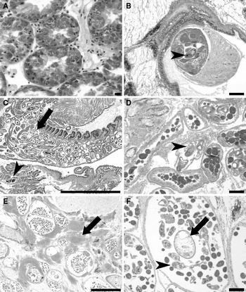
Marteilia species have been associated with mortality in a number of bivalve hosts and the discrimination of species requires the use of both molecular and ultrastructural data (Novoa et al., Reference Novoa, Posada and Figueras2005; Thébault et al., Reference Thébault, Bergman, Pouillot, Le Roux and Berthe2005; López-Flores et al., Reference López-Flores, Garrido-Ramos, de la Herran, Ruiz-Rejón, Ruiz-Rejón and Navas2008; Feist et al., Reference Feist, Hine, Bateman, Stentiford and Longshaw2009). Comps et al. (Reference Comps, Grizel, Tige and Duthoit1975) provided a brief description of a Marteilia sp. in the digestive gland of Cerastoderma edule from the Brittany region of France which they were unable to discriminate from M. refringens Grizel, Comps, Bonami, Cosserans, Duthoit & Le Pennec, 1974; prevalence was approximately 10% (Figure 4A). A second report of the parasite in three populations of cockles from the same region was made by Auffret & Poder (Reference Auffret and Poder1987) although no description was provided and prevalence was reported as ‘low’.
Histopathology of infections in Cerastoderma spp.: (A) histological section through digestive gland showing the presence of Marteilia sp. in the epithelial cells. Note large primary cell containing numerous secondary and tertiary cells; (B) Paravortex karlingi in the intestine of Cerastoderma sp. with numerous developmental stages, including one with obvious eyespots (arrowhead); (C) low power view of edible cockle with heavy infection with Bucephalus minimus sensu lato. Note presence of parasite sporocysts both within the gill (arrowhead) and mantle and gonad (arrow); (D) high power view of B. minimus sensu lato sporocysts with containing developing cercaria with obvious furcae (arrowhead). Note asynchronous development of other sporocysts nearby containing early germ cells only; (E) low power view of Gymnophallus choleodochus infection with marked inflammatory response to sporocysts (arrow); (F) sporocysts of G. choleodochus containing both developing cercaria (arrowhead) and metacercaria (arrow). Scale bars for A = 10 µm; B, D and F = 100 µm; C and E = 1 mm. Histological sections stained with haematoxylin and eosin (H&E).

Carrasco et al. (Reference Carrasco, Roque, Andree, Rodgers, Lacuesta and Furones2011) reported on a mass mortality event of edible cockles on the Mediterranean coast of Spain, with a Marteilia sp. being noted in around 40% of the animals examined during 2008. The authors acknowledged that they could not unequivocally link the presence of the parasite to the mortality event in light of the comparatively low prevalence of infection. However, they did argue that the relatively moderate to high intensity of infection coupled with the presence of other stressors may have contributed to the mortality event. Subsequently Carrasco et al. (Reference Carrasco, Andree, Lacuesta, Roque, Rodgers and Furones2012) revisited the site of the 2008 mortality event and collected samples for molecular studies. Prevalence of infection was much lower (23% in May 2010 and 1.7% in June 2010) compared with previous years. Molecular characterization of the cockle parasite demonstrated that the parasite showed around 85% homology of the internal transcribed spacer 1 (ITS-1) and intergenic spacer (IGS) with M. refringens from oysters and mussels and the authors consider that the parasite is a new genotype. Ultrastructural studies are required to further discriminate the parasite in cockles from M. refringens reported in other European bivalves. Furthermore, studies on the life cycle of the parasite, similar to those completed for M. refringens would need to be conducted to further understand its potential to spread and affect other cockle beds (Audemard et al., Reference Audemard, Le Roux, Barnaud, Collins, Sautour, Sauriau, de Montaudouin, Coustau, Combes and Berthe2002; Carrasco et al., Reference Carrasco, Arzul, Chollet, Robert, Joly, Furones and Berthe2008; López-Flores et al., Reference López-Flores, Garrido-Ramos, de la Herran, Ruiz-Rejón, Ruiz-Rejón and Navas2008). In addition, confirmation of the species of cockle infected would need to be completed as C. edule rarely occurs in the Mediterranean Sea and thus it is possible that the cockles examined were in fact C. glaucum.
PHYLUM PLATYHELMINTHES—CLASS TURBELLARIA
Turbellaria are generally considered to be commensals of a number of molluscs, being able to freely move from the mantle cavity of the mollusc to the alimentary canal and do not generally cause a problem for the host. Paravortex cardii occurs in both Cerastoderma edule and C. glaucum with C. edule also being host to P. karlingi Pike & Burt, Reference Pike and Burt1981 (Figure 4B). Brusa et al. (Reference Brusa, Ponce de Leon and Damborenea2006) lists C. edule as a host for P. gemellipara (Linton, 1910) citing the record of Leigh-Sharpe (Reference Leigh-Sharpe1933) from cockles in Plymouth, UK. However, P. gemellipara is an endocommensal of bivalves from the eastern Atlantic Ocean and thus the report is considered dubious at best. Furthermore, Atkins (Reference Atkins1934), also working in Plymouth retracted his earlier record of P. gemellipara in cockles (Atkins, Reference Atkins1933) stating that his form in Plymouth and that of Leigh-Sharpe (Reference Leigh-Sharpe1933) was clearly P. cardii. The record of a ‘ciliated sporocyst’ in C. edule by Nicoll (Reference Nicoll1906) is likely to be P. cardii. Paravortex cardii is approximately twice the size of P. karlingi and has three times as many brood capsules as P. karlingi. Paravortex cardii is restricted to the digestive gland tubules, whilst P. karlingi occurs in the intestine of its host. Pike & Burt (Reference Pike and Burt1981), in describing P. karlingi, showed that both species were widespread throughout the UK. Paravortex cardii has been recorded in edible and lagoon cockles in the Baltic Sea, Wadden Sea, Black Sea, Mediterranean Sea, France, Spain and Morocco (Belofastova & Dmitrieva, Reference Belofastova and Dmitrieva1999; de Montaudouin et al., Reference de Montaudouin, Kisielewski, Bachelet and Desclaux2000; Carballal et al., Reference Carballal, Iglesias, Santamarina, Ferro-Soto and Villalba2001; Zander & Reimer, Reference Zander and Reimer2002; Culurgioni et al., Reference Culurgioni, D'Amico, De Murtas, Trotti and Figus2006; Thieltges et al., Reference Thieltges, Krakau, Andresen, Fottner and Reise2006; Gam et al., Reference Gam, Bazaïri, Jensen and de Montaudouin2008).
PHYLUM PLATYHELMINTHES—CLASS TREMATODA
Cockles are hosts to a wide range of digeneans (trematodes), some of which have been implicated in mortality events or in reducing growth or fecundity. Identification keys to trematodes occurring in Cerastoderma edule are provided by de Montaudouin et al. (Reference de Montaudouin, Thieltges, Gam, Krakau, Pina, Bazairi, Dabouineau, Russell-Pinto and Jensen2009). Typically digenean life cycles are complex with eggs released from adult stages producing free-swimming ciliated miracidia that infect the first intermediate host, normally a mollusc. This develops into a mother sporocyst within the first intermediate host with further development into either daughter sporocysts or redia, depending on species. A further asexual phase takes place with the production of cercaria which is infective to the second intermediate host. Cockles can act as either first or second intermediate host, depending on the species of digenean. Once in the second intermediate host, cercaria develops into metacercaria before transmission to the final host where they undergo sexual reproduction followed by release of eggs into the environment.
One of the main Digenea reported as causing castration and death of cockles occurs in the genus Bucephalus (family Bucephalidae). The taxonomic history of bucephalids in cockles (and other bivalves) is somewhat confused, leading to some erroneous records and complications with the nomenclature. A detailed historical account of the family is provided by Kniskern (Reference Kniskern1952) and Overstreet & Curran (Reference Overstreet, Curran, Gibson, Jones and Bray2002). Bucephalus haimeanus (Lacaze-Duthiers, Reference Lacaze-Duthiers1854) was described from Ostrea edulis (Linnaeus, 1758) and Acanthocardia tuberculata (Linnaeus, 1758) (originally reported as Cardium rusticum) collected at Mahon on the Balearic Islands and Sète on the French coast (Mediterranean Sea) respectively (Lacaze-Duthiers, Reference Lacaze-Duthiers1854). Due to misinterpretations by various authors, cercaria in a variety of bivalve hosts have been referred to as B. haimeanus, including those occurring in European Cerastoderma spp. as well as the cercaria of B. cuculus McGrady, 1873 from Crassostrea gigas (Thunberg, 1793) collected in the United States of America (Tennent, Reference Tennent1906; Hopkins, Reference Hopkins1954; Cheng & Burton, Reference Cheng and Burton1965). Bucephalus haimeanus sensu stricto is a parasite of O. edulis (Príncep & Durfort, Reference Príncep and Durfort1993; Príncep et al., Reference Príncep, Bigas and Durfort1996); the digenean reported as B. haimeanus in A. tuberculata by Lacaze-Duthiers (Reference Lacaze-Duthiers1854) requires redescription and reports of the parasite in other European bivalves are incorrect.
Subsequently, Stossich (Reference Stossich1887) described B. minimus Stossich, Reference Stossich1887 from the intestine of Dicentrarchus labrax (Linnaeus, 1758) caught off Trieste in the Adriatic Sea as Gasterostomum minimum. Several nomenclatural changes followed culminating in the description of Labratrema lamirandi (Maillard, 1975) as a new species by Maillard (Reference Maillard1975b) from the intestine of D. labrax. However, Maillard & Saad-Fares (Reference Maillard and Saad-Fares1981) later listed B. lamirandi as a junior synonym of B. minimus. Overstreet & Curran (Reference Overstreet, Curran, Gibson, Jones and Bray2002) did not refer to this work and, whilst correctly proposing that Labratrema should be a junior synonym of Bucephalus, they considered that B. lamirandi was a species inquirendae. Maillard (Reference Maillard1975b), in completing the life cycle of B. minimus, showed experimentally that it cycled from C. glaucum as a first host, through several fish species as second intermediate hosts and used D. labrax as the final host. The proposed life cycle for the C. glaucum bucephalid has subsequently been confirmed by other workers (Faliex & Morand, Reference Faliex and Morand1994; Failex et al., Reference Faliex, Pagès, Combes, Combes and Jourdane2003).
Cercaria in C. glaucum have a flame cell arrangement of 2 [(6 + 6 + 6) + (6 + 6 + 6)] = 72 and measure 200–350 µm × 80–100 µm with the furcae measuring approximately 350 µm. The parasite has been reported in C. glaucum at various locations within the Mediterranean Sea (Faliex & Morand, Reference Faliex and Morand1994; Faliex et al., Reference Faliex, Pagès, Combes, Combes and Jourdane2003; Culurgioni et al., Reference Culurgioni, D'Amico, De Murtas, Trotti and Figus2006; Bartoli & Gibson, Reference Bartoli and Gibson2007). Based on the life cycle data, morphological characteristics and its geographical location (Mediterranean Sea, close to the type locality of B. minimus from sea bass), it is proposed that the bucephalid occurring in C. glaucum be referred to as Bucephalus minimus sensu stricto (Stossich, Reference Stossich1887).
A second bucephalid parasite has been reported from C. edule in the UK (Lebour, Reference Lebour1912; Cole, Reference Cole and Graham1956; James et al., Reference James, Bowers and Richards1966; Bowers, Reference Bowers1969; Matthews, Reference Matthews1973), The Netherlands (Thieltges & Reise, Reference Thieltges and Reise2006; Thieltges et al., Reference Thieltges, Krakau, Andresen, Fottner and Reise2006; Reference Thieltges, Hussell, Hermann, Jensen, Krakau, Taraschewski and Reise2008; Engelsma et al., Reference Engelsma, Roozenburg, Voorbergen-Laarman, Van den Brink, Troost, Ysebaert, Bateman and Longshaw2011), France (Huet, Reference Huet1888; Pelseneer, Reference Pelseneer1906; Deltreil & His, Reference Deltreil and His1970; de Montaudouin et al., Reference de Montaudouin, Kisielewski, Bachelet and Desclaux2000; Blanchet et al., Reference Blanchet, Raymond, de Montaudouin, Capdepuy and Bachelet2003), Spain (Carballal et al., Reference Carballal, Iglesias, Santamarina, Ferro-Soto and Villalba2001; Ordás & Figueras, Reference Ordás and Figueras2005), and Morocco (Gam et al., Reference Gam, Bazaïri, Jensen and de Montaudouin2008). To date only part of the life cycle has been experimentally completed with the parasite being transmitted from naturally infected C. edule to Pomatoschistus microps (Krøyer, 1838) by Matthews (Reference Matthews1973). Matthews (Reference Matthews1973), referring to the parasite as Cercaria bucephalopsis haimeana of Lacaze-Duthiers (Reference Lacaze-Duthiers1854), suggested that Pleuronectes platessa Linnaeus, 1758 were accidental hosts as only juvenile plaice could be infected and that the majority of the parasites did not survive beyond 2 weeks in the flatfish host. It was suggested that the final host was D. labrax based on ‘comparative morphology and ecology of the host’. Matthews (Reference Matthews1973) described the cercaria as measuring 480 × 40 µm when elongate, 100 µm when contracted, with an average measurement of 200 × 50 µm. Flame cell arrangement is 2 [(3 + 3 + 6 + 6 + 3) + (4 + 4 + 3)] = 64; Matthews (Reference Matthews1973) erroneously totalled the number of flame cells as 60. The furcae are up to ten times longer than the body, measuring up to 2 mm in length.
The measurements and flame cell arrangement provided by Matthews (Reference Matthews1973) are at odds with those provided by Maillard (Reference Maillard1975b) and provide strong circumstantial evidence that the parasite occurring in C. edule appears to be morphologically distinct from B. minimus occurring in C. glaucum from the Mediterranean. Whilst there is compelling evidence to suggest that the bucephalid occurring in C. edule in northern Europe is a distinct species, until a complete redescription of bucephalids of Cerastoderma spp. is completed, it is suggested that the bucephalid in C. edule be designated as Bucephalus minimus sensu lato. Furthermore, there appear to be some differences in life cycle strategies, size and morphology between the bucephalids described from European edible cockles by several authors in Europe and the bucephalid reported by Russell-Pinto et al. (Reference Russell-Pinto, Gonçalves and Bowers2006) and Pina et al. (Reference Pina, Barandela, Santos, Russell-Pinto and Rodrigues2009a) in Portugal leading to the possibility that more than one form of B. minimus sensu lato exists. Other than the molecular approach of Pina et al. (Reference Pina, Barandela, Santos, Russell-Pinto and Rodrigues2009a) and the partial experimental studies of Matthews (Reference Matthews1973), no comparative or life cycle studies have been carried out on bucephalids in C. edule and thus there is an urgent need to reassess the biology, ecology and taxonomy of these parasites.
Bucephalus spp. infect all tissues of C. edule and C. glaucum with a focus in the gonads and gills (Figure 4C, D). In heavy infections, parasitic castration of the host is apparent (Carballal et al., Reference Carballal, Iglesias, Santamarina, Ferro-Soto and Villalba2001; Culurgioni et al., Reference Culurgioni, D'Amico, De Murtas, Trotti and Figus2006). Destruction of the gonadal tissue follows disintegration of the connective tissues surrounding it. James & Bowers (Reference James and Bowers1967a) suggest that this may be due to release of hyaluronidase and pressure atrophy by the parasite. Following destruction of the gonad, the growing parasite appears to block or crush the digestive tubules leading to starvation autolysis of the digestive gland. Breakdown of the digestive tubules ensues as a result of a combination of the physiological effects of the parasite, pressure atrophy due to the growing daughter sporocyst and loss of fluids from the tubule cells. Distribution of lipids, carbohydrates and enzymes were determined by James & Bowers (Reference James and Bowers1967b) which they suggested was similar to previous reports and was mainly derived from host tissues. Use of stable isotopes confirmed that the parasite derived its energy from the digestive gland and had a preference for lipids (Dubois et al., Reference Dubois, Savoye, Sauriau, Billy, Martinez and de Montaudouin2009). Energy stored in cockles in the form of glucose, glycogen and lipids is potentially utilized by the parasite in the migration between hosts.
Intensity of infection in individual animals can be high and prevalence in the population is variable. In the Burry Inlet, South Wales, infections were only detected in post-spawning cockles that were greater than a year old and larger than 18 mm in length by Bowers (Reference Bowers1969). Furthermore, Bowers (Reference Bowers1969) suggested that reductions in parasite prevalence in cockles greater than 3 years old was due to parasite-induced host mortality and increased resistance of older cockles to infection. Infected cockles had a lowered shell growth rate but an increased flesh yield. Prevalence of infection in C. edule and C. glaucum increases steadily throughout the summer months, decreasing in the autumn as a result of availability of suitable host(s) (Bowers, Reference Bowers1969; Faliex & Morand, Reference Faliex and Morand1994; Desclaux et al., Reference Desclaux, de Montaudouin and Bachelet2002; Faliex et al., Reference Faliex, Pagès, Combes, Combes and Jourdane2003). Spatial variations in prevalence perhaps reflect local hydrological conditions, host availability and other factors. Prevalence figures for Bucephalus spp. in C. edule and C. glaucum across their range in Europe tend to follow similar patterns, ranging from <1% to around 40% (Deltreil & His, Reference Deltreil and His1970; de Montaudouin et al., Reference de Montaudouin, Kisielewski, Bachelet and Desclaux2000, Reference de Montaudouin, Thieltges, Gam, Krakau, Pina, Bazairi, Dabouineau, Russell-Pinto and Jensen2009; Carballal et al., Reference Carballal, Iglesias, Santamarina, Ferro-Soto and Villalba2001; Desclaux et al., Reference Desclaux, de Montaudouin and Bachelet2002; Ordás & Figueras, Reference Ordás and Figueras2005; Culurgioni et al., Reference Culurgioni, D'Amico, De Murtas, Trotti and Figus2006; Russell-Pinto et al., Reference Russell-Pinto, Gonçalves and Bowers2006; Thieltges & Reise, Reference Thieltges and Reise2006; Thieltges et al., Reference Thieltges, Krakau, Andresen, Fottner and Reise2006; Gam et al., Reference Gam, Bazaïri, Jensen and de Montaudouin2008). Cole (Reference Cole and Graham1956) reported prevalences of around 2% for the parasite in C. edule from North Wales in the 1950s; prevalence was around 15% in cockles from South Wales in the early 1960s (Bowers, Reference Bowers1969); prevalences in C. edule and C. glaucum from the River Crouch in the late 1960s were 12% and 0.04% respectively (Boyden, Reference Boyden1970, Reference Boyden1971). Boyden (Reference Boyden1970) considered the infection in the solitary C. glaucum as accidental with ‘infection of C. glaucum only occurring in exceptional circumstances’.
Cerastoderma edule infected with B. minimus sensu lato tend to have fewer parasites, although the mechanism for this is not understood (de Montaudouin et al., Reference de Montaudouin, Kisielewski, Bachelet and Desclaux2000; Desclaux et al., Reference Desclaux, de Montaudouin and Bachelet2002). Desclaux et al. (Reference Desclaux, de Montaudouin and Bachelet2002) showed that prevalence of infection in surfaced cockles was approximately twice that of buried cockles. They calculated that around 10% of surfaced cockles did so as a direct result of infections with this parasite but were unable to explain the advantage to the parasite of this strategy given the route of infection to the next host via free swimming cercariae. Deltreil & His (Reference Deltreil and His1970) suggested that death of infected animals at the surface facilitated dispersion of the cercariae. However, C. edule infected with B. minimus sensu lato have a lower specific oxygen consumption rate with a decrease in specific pumping rate and are less able to adapt to increased temperature and decreased salinity with large reductions in filtration rates occurring (Javanshir, Reference Javanshir2001b). Thus infected animals may surface as a direct result of limited oxygen availability in the tissues (Javanshir, Reference Javanshir2001a) rather than due to a parasite-derived dispersal strategy. There is an inverse relationship between parasite levels in the host and oxygen consumption rate; when more than 92% of body mass is made up of parasite, the host enters a lethal state leading to host death (Javanshir, Reference Javanshir2001a).
Living organisms utilize metallothioneins (MT) in the metabolism of essential metals and in the detoxification of trace metals. The induction of these proteins has been used as a biological monitoring tool in the marine environment as a marker of exposure to heavy metal contamination (Mourgaud et al., Reference Mourgaud, Martinez, Geffard, Andral, Stanisiere and Amiard2002). Baudrimont et al. (Reference Baudrimont, de Montaudouin and Palvadeau2006) compared MT concentration in parasitized and non-parasitized cockles during gametogenesis and after spawning. Spent cockles parasitized by B. minimus had MT levels that were higher than non-parasitized individuals. In contrast, parasitized cockles undergoing gametogenesis had lower MT levels than non-parasitized cockles. The authors suggested that parasites in the cockle, particularly those that castrate the host appear to modulate MT synthesis and infected animals were more likely to bioaccumulate metals (Baudrimont et al., Reference Baudrimont, de Montaudouin and Palvadeau2006; Baudrimont & de Montaudouin, Reference Baudrimont and de Montaudouin2007).
Curtuteria arguinae Desclaux, Russell-Pinto, de Montaudouin & Bachelet, Reference Desclaux, Russell-Pinto, de Montaudouin and Bachelet2006 (family Echinostomatidae) was described from cockles in France, and has been reported in Portugal and Morocco (Russell-Pinto et al., Reference Russell-Pinto, Gonçalves and Bowers2006; Gam et al., Reference Gam, Bazaïri, Jensen and de Montaudouin2008). Although the parasite has been noted in cockles as small as 3 mm, significant infections do not occur until the cockle is larger than 14 mm (Desclaux et al., Reference Desclaux, Russell-Pinto, de Montaudouin and Bachelet2006). In selected cockle cohorts it has been shown to lead to parasite-dependent mortality (Desclaux et al., Reference Desclaux, Russell-Pinto, de Montaudouin and Bachelet2006).
The taxonomy of the digenean family Gymnophallidae has been the subject of much controversy with numerous authors attempting to resolve the convoluted taxonomic history (Cable, Reference Cable1953; Stunkard & Uzmann, Reference Stunkard and Uzmann1958; James, Reference James1964; Pekkarinen & Ching, Reference Pekkarinen and Ching1994; Ching, Reference Ching1995; Bowers et al., Reference Bowers, Bartoli, Russell-Pinto and James1996; Scholz, Reference Scholz, Gibson, Jones and Bray2002; Cremonte et al., Reference Cremonte, Vázquez and Ituarte2008). Scholz (Reference Scholz, Gibson, Jones and Bray2002) attempted to stabilize the taxonomy by synonymizing a number of genera. The genera proposed by Scholz (Reference Scholz, Gibson, Jones and Bray2002) include Parvatrema (=Meiogymnophallus), Gymnophallus (=Paragymnophallus), Gymnophalloides (=Lacunovermis) and Pseudogymnophallus. A fifth genus, Bartolius, was erected by Cremonte (Reference Cremonte2001) and only briefly mentioned in the review by Scholz (Reference Scholz, Gibson, Jones and Bray2002). Not all authors have agreed with the proposal of Scholz (Reference Scholz, Gibson, Jones and Bray2002) and have continued to use the genus name Meiogymnophallus for gymnophallids occurring in bivalves (Russell-Pinto et al., Reference Russell-Pinto, Gonçalves and Bowers2006; Chai et al., Reference Chai, Han, Choi, Seo, Kim, Guk, Shin and Lee2007; de Montaudouin et al., Reference de Montaudouin, Thieltges, Gam, Krakau, Pina, Bazairi, Dabouineau, Russell-Pinto and Jensen2009; Fermer et al., Reference Fermer, Culloty, Kelly and O'Riordan2010, Reference Fermer, Culloty, Kelly and O'Riordan2011). The suppression of Meiogymnophallus as a junior synonym of Parvatrema requires reconsideration as it is apparent that not all members of the genus Meiogymnophallus are readily placed in the genus Parvatrema (Bartoli & Gibson, Reference Bartoli and Gibson2007). Indeed, all gymnophallids occurring in Cerastoderma appear to have characteristics of the genus Gymnophallus and thus we propose that until evidence is provided to the contrary, all gymnophallids occurring in cockles should be considered as Gymnophallus spp. The gymnophallid reported as P. isostoma Beloplo'skaja, 1966 in C. glaucum by Dolgikh (Reference Dolgikh1968) requires redescription as Bartoli (Reference Bartoli1983) considered this to be conspecific with G. rebecqui Bartoli, Reference Bartoli1983.
Mortalities of cockles in the northern Wadden Sea area were attributed to the presence of Gymnophallus choledochus Odhner, 1900 (family Gymnophallidae) (Figure 4E, F), which was confirmed by surveys of cockles that showed that 73% of surfaced cockles were infected with the parasites compared to only 7% of buried cockles (Thieltges, Reference Thieltges2006). In addition, following experimental studies, only 23% of those cockles on the surface survived longer than 14 days. The parasite appears to be more common in edible cockles compared with lagoon cockles (Boyden, Reference Boyden1970; Culurgioni et al., Reference Culurgioni, D'Amico, De Murtas, Trotti and Figus2006; Figus et al., Reference Figus, Culurgioni, De Murtas and Trotti2006).
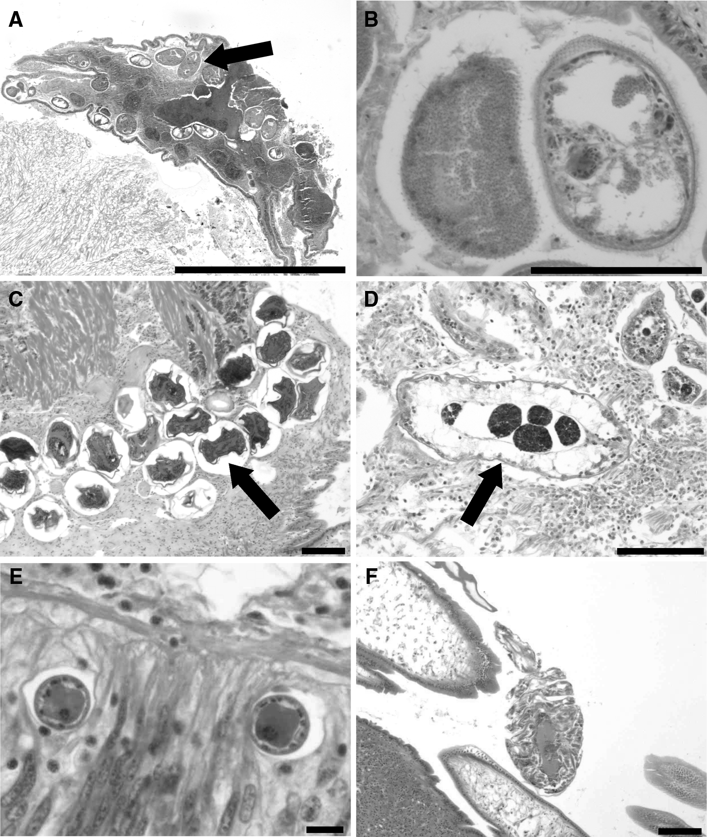
Gymnophallus (=Meiogymnophallus) minutus has been recorded on a number of occasions from cockle populations in Europe (Bowers & James, Reference Bowers and James1967). The parasite occurs enveloped by host tissue below the hinge area (Figure 5A) and stimulates the cockle to produce excessive ligament proteins, resulting in tissue folding around the parasite. Similar lesions have been reported in other bivalve species with a concomitant effect on survival (Campbell, Reference Campbell1985; Cremonte & Ituarte, Reference Cremonte and Ituarte2003; Mladineo & Peharda, Reference Mladineo and Peharda2009; Addinoa et al., Reference Addinoa, Lomovaskya, Cremonte and Iribarne2010). Bowers et al. (Reference Bowers, Bartoli, Russell-Pinto and James1996) suggested that this interfered with the close fitting of the tooth into the wedge shaped cavity by the hinge leading to an incomplete closure of the shell valves. Infected animals were more likely to be found close to or on the surface of the sediment with the open shell gaping upwards. James et al. (Reference James, Sannia, Bowers, Nelson-Smith and Bridges1977) and Goater (Reference Goater1993) reported mass mortalities of cockles infected with G. minutus in the Burry Inlet and Exe estuary respectively. However, Fermer et al. (Reference Fermer, Culloty, Kelly and O'Riordan2010, Reference Fermer, Culloty, Kelly and O'Riordan2011) suggest that there is no empirical evidence of an effect of the parasite on gaping, altered burrowing or direct mortality. The parasite is host to the microsporidian Unikaryon legeri (Figure 5B).
Histopathology of infections in Cerastoderma spp.: (A) low power view of cockle infected with Gymnophallus (=Meiogymnophallus) minutus (arrow). Note also the presence of numerous dead digeneans within the sporocyst as a result of hyperparasitism by Unikaryon legeri; (B) histological section through two G. minutus. Individual on left shows heavy infection with the hyperparasite U. legeri. In addition, note the presence of small spines along body wall on peripheral edges of both specimens, a typical feature of the parasite; (C) metacercaria of Himasthla sp. in the mantle of cockle (arrow). Note moderate host reaction surrounding the parasites that occur within a pearl; (D) daughter sporocyst of Monorchis parvus (arrow) containing developing germinal cells which eventually develop into metacercaria; (E) cross-section through two unidentified nematodes embedded within the epithelium of intestine. Note the lack of host response to the nematodes; (F) Herrmannella rostrata on the gills of cockle. Scale bar for A = 1 mm; B, C, D, E and F = 100 µm. Histological sections stained with haematoxylin and eosin (H&E).
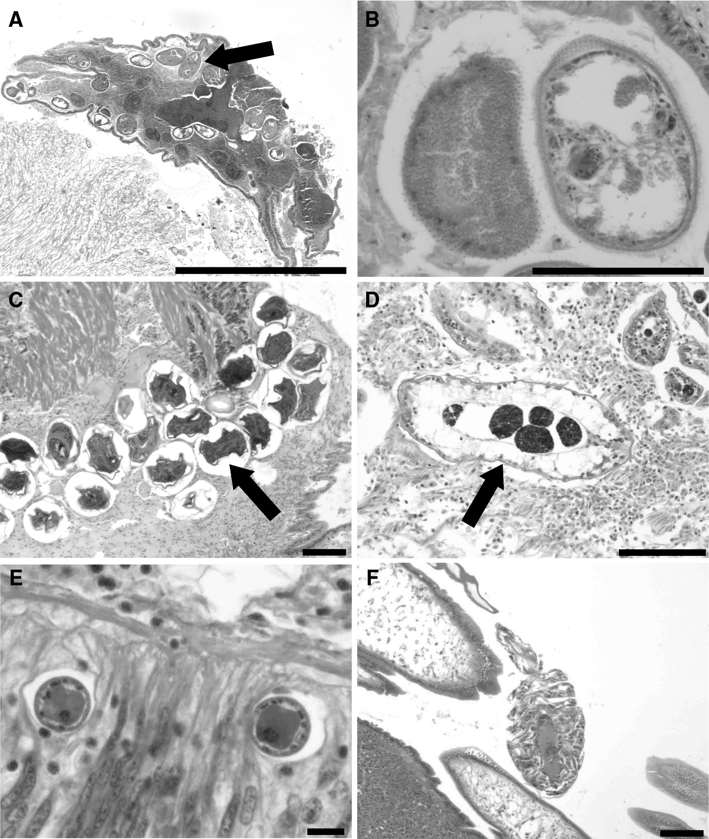
Four members of the genus Himasthla (family Echinostomatidae) occur as metacercaria in the foot and mantle of cockles (Figure 5C). Three of these, H. elongata (Mehlis, 1831), H. interrupta Loos-Frank, 1967 and H. continua Loos-Frank, 1967 are considered to be native to C. edule and C. glaucum and are identifiable by the presence of 29 collar spines. The life cycle of a fourth species, H. quissetensis (Miller & Northup, 1926) with 31 collar spines was described from Mya arenaria Linnaeus, 1758 in North America (Stunkard, Reference Stunkard1938) and has been reported in cockles in Europe (Bartoli & Gibson, Reference Bartoli and Gibson2007). de Montaudouin et al. (Reference de Montaudouin, Jensen, Desclaux, Wegeberg and Sajus2005) consider that the parasite is a recent introduction from the USA into Europe, although a ‘rare’ Himasthla sp. with 31 collar spines which may be conspecific with H. quissetensis has been noted previously in the North Sea (Lauckner, Reference Lauckner1971). The three native Himasthla spp. use either Hydrobia ulvae (Pennant, 1777) or Littorina littorea (Linnaeus, 1758) as first intermediate host whilst the non-native H. quissetensis has been recorded as cercaria in Nassarius reticulatus (Linnaeus, 1758) in Portugal (Russell-Pinto et al., Reference Russell-Pinto, Gonçalves and Bowers2006). They are able to infect spat as well as recently settled cockles around 4 mm in size and infect cockles in their first summer (Jensen et al., Reference Jensen, Castro and Bachelet1999; Wegeberg et al., Reference Wegeberg, de Montaudouin and Jensen1999; de Montaudouin et al., Reference de Montaudouin, Jensen, Desclaux, Wegeberg and Sajus2005). Transmission of the parasites occurs following predation on cockles by migratory birds (de Montaudouin et al., Reference de Montaudouin, Wegeberg, Jensen and Sauriau1998).
It has been difficult to unequivocally link the presence of Himasthla spp. directly to mortalities, despite both field and experimental studies. Wegeberg & Jensen (Reference Wegeberg and Jensen1999) held cockles under hypoxic conditions for 30 hours. Survival of cockles infected with H. elongata was significantly reduced compared with non-infected hosts. In contrast however, under normal conditions there was no effect of parasites on survival. This was supported by the studies of Desclaux et al. (Reference Desclaux, de Montaudouin and Bachelet2004) who showed that whilst the parasites had some direct impact on host survival, being responsible for up to 46% of the mortality, it was the synergistic effect of environmental conditions coupled with the presence of the parasite that caused the majority of deaths. Cockles infected with more than 10 H. quissetensis metacercaria are more likely to be found on the surface compared with uninfected or cockles with lower numbers of metacercaria (Desclaux et al., Reference Desclaux, de Montaudouin and Bachelet2002) which may be due to a mechanical interruption of the cockles ability to burrow and increasing time to burrow by up to 20 times (Lauckner, Reference Lauckner1984). As with other digeneans, H. interrupta can cause loss of flesh weight and body condition (Wegeberg & Jensen, Reference Wegeberg and Jensen2003); marginal reductions in growth rates occur in animals infected with Himasthla spp. (Javanshir et al., Reference Javanshir, Seyfabadi, Bachelet and Feghhi2007). Himasthla spp. infections in the foot of cockles are considered to destroy muscle fibres as they migrate through the muscle layers either through mechanical pressure or through the release of cercarial enzymes as well as causing pressure atrophy and haemocyte accumulations once embedded in the host (Desclaux et al., Reference Desclaux, de Montaudouin and Bachelet2004).
Cockles infected with the digenean Monorchis parvus Loos, 1902 (family Monorchiidae) (Figure 5D) are significantly smaller than healthy individuals, particularly in the early years (Sannia & James, Reference Sannia and James1978). However, it is considered to be relatively rare in the UK, being recorded sporadically in the Thames (at a prevalence of 1%) in 1975–1976, the north-east of England in three cockles (Lebour, Reference Lebour1912), Scotland (at a prevalence of 0.5%), and the south-east of England (at a prevalence of 0.15%). Boyden (Reference Boyden1970) reported the parasite in 0.07% of C. edule and 0.12% of C. glaucum. Infected cockles that were not eaten by the final host died within 12 months of being exposed to the parasite (Sannia & James, Reference Sannia and James1978). Jonsson & André (Reference Jonsson and André1992), following reports of a mass mortality of cockles in Sweden, examined animals from two localities. In addition to carrying out a histological study of these animals, the authors tested the burrowing capability of the cockles under experimental conditions. Significantly fewer infected individuals burrowed compared with their uninfected counterparts. Damage to the infected tissues was defined as ‘serious' and many individuals had no remaining muscular tissue in the foot and few gametes were detected. Jonsson & André (Reference Jonsson and André1992) report that heavily infected cockles ‘were slow to close the valves and gave off a putrid smell’ and that cockles gaping on the surface were often attacked by the gastropod N. reticulatus. They concluded that the parasite, which uses sparids (Diplodus spp.) as a final host (Bartoli et al., Reference Bartoli, Jousson and Russell-Pinto2000), was responsible for the reported mortalities in Swedish coastal areas.
Psilostomum brevicolle (Linton, 1928) (family Psilostomidae) occurs in the visceral mass of C. glaucum and C. edule along the Atlantic coast of Europe with a more northerly distribution and an apparent absence in the Mediterranean Sea (de Montaudouin et al., Reference de Montaudouin, Thieltges, Gam, Krakau, Pina, Bazairi, Dabouineau, Russell-Pinto and Jensen2009; Derbali et al., Reference Derbali, Jarboui and Ghorbel2009). Prevalence is variable between sites with no significant difference in prevalence or intensity between buried and unburied cockles suggesting that it is not a mortality driver for cockles (Desclaux et al., Reference Desclaux, de Montaudouin and Bachelet2002). Infection of cockles takes place in autumn in animals greater than 19 mm in length (de Montaudouin et al., Reference de Montaudouin, Kisielewski, Bachelet and Desclaux2000). No data exist on the pathology associated with this parasite or its impact on cockle hosts.
Renicola roscovita (Stunkard, 1932) (family Renicolidae) infect a number of bivalve species and has a preference for the palps of larger cockles (de Montaudouin et al., Reference de Montaudouin, Kisielewski, Bachelet and Desclaux2000; Thieltges & Reise, Reference Thieltges and Reise2006). Lauckner (Reference Lauckner1984) suggested that juvenile cockles were more likely to die as a result of infection due to the preference of the parasite to encyst in the gills and visceral mass of smaller cockles. Cockles are infected in their first summer when around 5 mm in size with accumulation of parasites continuing through the life of the host (de Montaudouin et al., Reference de Montaudouin, Kisielewski, Bachelet and Desclaux2000; Thieltges & Reise, Reference Thieltges and Reise2007; Thieltges, Reference Thieltges2008). Peak transmission occurs during the summer months and although maximum infectivity of cercariae occurs at 25°C, the optimum temperature for transmission is 20°C (Thieltges & Rick, Reference Thieltges and Rick2006). Cockles originating from low intertidal areas of the shore line have higher levels of R. roscovita compared with cockles from other parts of the shoreline, reflecting the higher densities of the first intermediate host H. ulvae in this area (Thieltges, Reference Thieltges2007).
The life cycle of Diphterostomum brusinae Stossich, 1888 (family Zoogonidae) was recently demonstrated by Pina et al. (Reference Pina, Tajdari, Russell-Pinto and Rodrigues2009b) using a molecular approach to link different life stages in each host. Sporocysts containing cercaria and encysted metacercaria occur within the gonads and digestive gland of N. reticulatus. Cercaria can encyst within the gastropod host for transmission direct to the fish host or can be released and infect C. edule where the metacercaria occur in the foot, mantle border and gills (Pina et al., Reference Pina, Tajdari, Russell-Pinto and Rodrigues2009b). Palombi (Reference Palombi1930) and Bartoli & Gibson (Reference Bartoli and Gibson2007) suggest that there is no second intermediate host and that transmission occurs directly from the gastropod to the fish hosts although Francisco et al. (Reference Francisco, Almeida, Castro, Pina, Russell-Pinto, Rodrigues and Santos2010a) have shown that Mytilus galloprovincialis Lamarck, 1819 can act as a second intermediate host for the parasite. Molecular primers for the ITS1 and for 18 rDNA are provided by Pina et al. (Reference Pina, Tajdari, Russell-Pinto and Rodrigues2009b) and Francisco et al. (Reference Francisco, Almeida, Castro, Pina, Russell-Pinto, Rodrigues and Santos2010a). No data exist on the impact of the parasite in cockles, although Francisco et al. (Reference Francisco, Hermida and Santos2010b) have shown that the parasite elicits a strong immune response consisting of haemocyte encapsulation in M. galloprovincialis.
Digenea of lesser concern that have not been shown to impact on the overall health of cockles include Lepocreadium pegorchis (Stossich, 1901) (family Lepocreadiidae), the freshwater digenean Asymphylodora demeli Markowski, 1935 (family Lissorchiidae) and Paratimonia gobii Prévôt & Bartoli, 1967 (family Monorchidae) (Reimer, Reference Reimer1973; Maillard, Reference Maillard1975a; Kesting et al., Reference Kesting, Gollasch and Zander1996; Zander, Reference Zander1998; Bartoli & Gibson, Reference Bartoli and Gibson2007; de Montaudouin et al., Reference de Montaudouin, Thieltges, Gam, Krakau, Pina, Bazairi, Dabouineau, Russell-Pinto and Jensen2009). Cercarial stages of the digenean Parorchis acanthus Nicoll, 1907 (family Philophthalmidae) encyst on a number of surfaces including the foot, mantle and shell of C. edule which erroneously led Lebour & Elmhirst (Reference Lebour and Elmhirst1922) to suggest that they may be intermediate hosts for this parasite.
PHYLUM PLATYHELMINTHES—CLASS CESTODA
Cestodes have a complex life cycle utilizing at least two hosts, usually an invertebrate and a vertebrate host (Gulyaev, Reference Gulyaev2008). In general, few bivalves have been reported as being hosts for cestodes (Maeno et al., Reference Maeno, Yurimoto, Nasu, Ito, Aishima, Matsuyama, Kamaishi, Oseko and Watanabe2006; Aguirre-Macedo et al., Reference Aguirre-Macedo, Sima-Alvarez, Román-Magaña and Guemez-Ricalde2007; Kim & Powell, Reference Kim and Powell2007; Park et al., Reference Park, Tsutsumi, Hong and Choi2008; Holland & Wilson, Reference Holland and Wilson2009). de Montaudouin et al. (Reference de Montaudouin, Kisielewski, Bachelet and Desclaux2000) reported the presence of an unidentified cestode (as a tentaculo-neoplerocercoid) in the foot of Cerastoderma edule from Arcachon Bay, France at a prevalence of 0.4% (two animals infected), whilst Vaullegeard (Reference Vaullegeard1900) listed ‘Cardium’ as a host for Echinobothrium typus van Beneden, 1849 (family Echinobothriidae). James et al. (Reference James, Sannia, Bowers, Nelson-Smith and Bridges1977) recorded Anatinella (=Monosaccanthus = Hymenolepis) brachycephala (Creplin, 1829) cysticercoid larvae in the haemolymph of the digestive gland and foot of C. edule from the Burry Inlet, South Wales. No prevalence data or pathology information were provided. Final host for this cestode was recorded as the oystercatcher.
PHYLUM NEMATODA
Life cycles of parasitic nematodes are normally complex, with at least three hosts, including bivalves in a small number of cases. However, reports of nematodes in both Cerastoderma edule and C. glaucum are rare. Unidentified ectocommensal nematodes occur in the mantle cavity and gills of C. edule and C. glaucum in the Wadden Sea and Mediterranean Sea respectively and do not elicit a host response (Culurgioni et al., Reference Culurgioni, D'Amico, De Murtas, Trotti and Figus2006; Figus et al., Reference Figus, Culurgioni, De Murtas and Trotti2006; Thieltges et al., Reference Thieltges, Krakau, Andresen, Fottner and Reise2006). Chao (Reference Chao2000), quoting a list of unpublished data from B. James, reported third stage larvae of Porrocaecum pectinis Cobb, 1930 in the digestive gland and gonads of cockles (Figure 5E). However, P. pectinis is a parasite of Argopecten gibbus (Linnaeus, 1758) from the south-eastern USA (Minchin, Reference Minchin2003) and thus its report in cockles must be considered dubious.
PHYLUM ARTHROPODA
The copepod Herrmannella rostrata Canu, 1891 occurs in the mantle cavity and gills (Figure 5F) of Cerastoderma edule from the British Isles, France, Spain and The Netherlands (Atkins, Reference Atkins1934; Stock, Reference Stock1993; Carballal et al., Reference Carballal, Iglesias, Santamarina, Ferro-Soto and Villalba2001; Faasse, Reference Faasse2003). Typically the parasite undergoes six naupliar stages, five copepodid stages and a single adult stage where the sexes are separate (Costanzo & Calafiore, Reference Costanzo and Calafiore1985); no pathology or detrimental impact has been noted for this parasite.
The intestine dwelling copepod Mytilicola intestinalis Steuer, 1902, occurs in a number of bivalve species, notably Mytilus spp. (Aguirre-Macedo & Kennedy, Reference Aguirre-Macedo and Kennedy1999; Buck et al., Reference Buck, Thieltges, Walter, Nehls and Rosenthal2005). Under low levels of infection the parasite is considered benign; numbers of more than 3 individuals per infected mussel appear to be detrimental to the host (Gee & Davey, Reference Gee and Davey1986; Watermann et al., Reference Watermann, Thomsen, Kolodzey, Daehne, Meemken, Pijanowska and Liebezeit2008). Local metaplasia of the intestinal lumen of a single cockle infected with a Mytilicola sp. in Galicia was noted by Carballal et al. (Reference Carballal, Iglesias, Santamarina, Ferro-Soto and Villalba2001). Overall prevalence of the parasite in the samples from 3 sites in Galicia, Spain was only 4%. Similarly low prevalence values were obtained by de Montaudouin et al. (Reference de Montaudouin, Kisielewski, Bachelet and Desclaux2000) in Arcachon Bay, France who showed that the parasite did not infect individuals smaller than 9 mm. Its presence in the Burry Inlet in South Wales was noted by James et al. (Reference James, Sannia, Bowers, Nelson-Smith and Bridges1977); no data on prevalence, intensity or impact were provided by these authors.
Pea crabs live commensally in a range of marine invertebrates, including bivalve molluscs, with the common Pinnotheres pisum (Linnaeus, 1767) infecting a number of bivalve species in Europe (Becker & Türkay, Reference Becker and Türkay2010). Pea crabs infecting other hosts can lead to reductions in growth and condition factor and impact gonadal development as well as damaging the gills (Kruczynski, Reference Kruczynski1972; Bierbaum & Ferson, Reference Bierbaum and Ferson1986; O'Beirn & Walker, Reference O'Beirn and Walker1999; Mercado-Silva, Reference Mercado-Silva2005; Sun et al., Reference Sun, Sun, Yuqi, Baowen and Weibo2006). However, no studies have been carried out on the impact of P. pisum on cockle growth or survival although it has been noted in the mantle cavity of up to 70% of C. edule from Morocco and at an unknown prevalence in the Burry Inlet (James et al., Reference James, Sannia, Bowers, Nelson-Smith and Bridges1977; Gam et al., Reference Gam, Bazaïri, Jensen and de Montaudouin2008). The pea crab, Afropinnotheres monodi Manning, 1993, originally described from Moroccan waters, has now been found in three native bivalve species, including C. glaucum, in the Gulf of Cadiz (Portugal) (Subida et al., Reference Subida, Arias, Drake, Garcia-Raso, Rodriguez and Cuesta2011).
PHYLUM NEMERTEA
The entocommensal nemertean Malacobdella grossa (Müller, 1776) is associated with a number of bivalves around the coastlines of Europe and the Pacific and Atlantic coasts of the Americas (Gibson, Reference Gibson1967). In the bivalve host, the nemertean either predates on small crustaceans entering the mantle cavity or those living commensally with its bivalve host (Gibson & Jennings, Reference Gibson and Jennings1969). Malacobdella grossa was noted in the mantle cavity of Cerastoderma edule from the Shetland Islands by Jones et al. (Reference Jones, Jones and James1979). Prevalence was close to 100% at some sites and cockles less than 15 mm were uninfected (Jones et al., Reference Jones, Jones and James1979). The relationship between the haplosporidian Haplosporidium malacobdellae Jennings & Gibson, 1968 in M. grossa and those occurring in cockles are unknown (McDermott, Reference McDermott2006). No further information on the impact of M. grossa on cockle health was noted and it has not been recorded since.
CONCLUSIONS AND FUTURE DIRECTIONS
This review has covered all the disease conditions, parasites and commensals of cockles, and considered the effect of these on both individual and population health. It has been clear that in a number of areas there are large gaps in our knowledge that require further studies. In particular, there has been a lack of taxonomic rigour applied to many studies on parasites and diseases and as a result, there has been a number of poorly described species and erroneous host records. The limited data on some parasite life cycles, in particular haplosporidians, Marteilia sp. and Digenea, should be addressed using a combination of molecular and experimental approaches and the pathological impact of infections should be considered through the use of histopathology and field and experimental studies. The role of plankton in the dissemination of selected cockle pathogens should be examined as this constitutes a major component of cockle diets. Use of both in vitro and in vivo models to assess impact of infections should be considered. These data along with life history information will be useful in determining impact of these parasites at an individual and population level and minimize risk of transfer of non-native parasites between sites. A clear understanding of the species and hosts involved along with the impact of these parasites will ensure that biologically relevant decisions are made in managing any cockle fishery. No data exist on infections of larval cockles or on the impact of these on survival although some studies have considered digenean infections of recently settled spat; this gap needs to be addressed.
ACKNOWLEDGEMENT
The authors thank the Welsh Government for providing financial support for the production of this review.