I. INTRODUCTION
Advanced oxidation by semiconductor photocatalysis is of increasing importance in the treatment of wastewater due to its ability to destroy a wide range of organic and inorganic pollutants without the generation of harmful byproducts.Reference Gogate and Pandit1 TiO2Reference Akpan and Hameed2 and ZnO are important semiconductor photocatalysts; however, the large-scale treatment of water with TiO2 is uneconomical. A suitable alternative is nanosized ZnO, which is available at low cost. ZnO has the same band gap energy (3.2 eV) and follows the same mechanism of photodegradation as TiO2.Reference Sakthivel, Neppolian, Shankar, Arabindoo, Palanichamy and Murugesan3 The advantage of semiconductor photocatalysis is that it can be carried out under ambient conditions and leads to complete mineralization of organic compounds. The use of ZnO has been demonstrated for the degradation of dyesReference Yu, Cai and Liu4, Reference Daneshvar, Salari and Khataee5 and other organic contaminants.Reference Hariharan6 Advanced oxidation processes (AOPs) generate powerful oxidizing OH radicals, which destroy pollutants in wastewater quickly and nonselectively.Reference Agustina, Ang and Vareek7 Among AOPs, semiconductor photocatalysis has emerged as an important destructive technology.
The performance of ZnO, catalysis,Reference Kantam, Kumar and Sridhar8 gas sensors,Reference Bie, Yan, Yin, Duan and Yuan9 piezoelectric devices,Reference Bdikin, Gracio, Ayouchi, Schwarz and Kholkin10 etc., depends on shape, size, and crystallinity, which in turn are dependent on the synthesis conditions. Many different synthetic routes have been used to control shape, size, and morphology by physicalReference Özgür, Alivov, Liu, Teke, Reshchikov, Doğan, Avrutin, Cho and Morkoç11 and chemical methods.Reference Wang, Shen, Wang, Gao, Kang, Tian, Lan and Zhang12–Reference Tseng, Lin, Chen and Chu14
In our recent work,Reference Atla, Chen, Yang, Chen, Sun, Lin, Maity, Pan and Cheng15 we reported a novel method to synthesize NiO nanomaterials by pneumatic foam fractionation. In which we showed a method to control and separate Ni-based crystal nanostructures with different physical properties by means of foam fractionation. As an extension of work, we now report the synthesis of ZnO by pneumatic foam fractionation method. The new route is simple, of low cost, and obtains a high-purity product. In our experiments, a well-controlled volume of gas was introduced into a definite volume of zinc nitrate and a surfactant, sodium dodecyl sulfate (SDS), and the foam produced was allowed to travel to a vertical column height and the foamate collected. The effect of flow rate on the synthesis of ZnO was studied. As a demonstration, cationic thiazine dye, methylene blue (MB), was chosen, and degradation studies were performed using ZnO obtained at different velocities. This method of using a SDS surfactant is also useful for obtaining hydrophobic ZnO at room temperature.
II. MATERIALS AND METHODS
A. Materials and reagents
Zn(NO3)2•6H2O was obtained from J.T. Baker (Philipsburg, NJ), SDS was purchased from Aldrich Chemicals, and ammonia 25% was obtained from Panreac Quimica S.A.U. (Barcelona, Spain). Milli-Q water (Billerica, MA) was used in all experiments.
B. Synthesis of the photocatalyst by foam fractionation
Zn(NO3)2•6H2O (5.49 g) and SDS (1.4 g) were dissolved in 200 mL of Milli-Q water so that the final concentration of Zn(NO3)2 and SDS in solution was 0.1 and 0.02 M, respectively. The concentration of SDS was higher than the critical micelle concentration (8.1 × 10⁻³ M).Reference Sastry, Gole, Banpurkar, Limaye and Ogale16 The feed solution containing the surfactant molecules and zinc nitrate entered the vertical column a significant distance above the base of the vessel, and gas was sparged through the base of the column to create a dispersion of rising bubbles. The gas was injected at rates of 0.2, 0.3, 0.5, and 1.0 (Nl/min) through a cylindrical ceramic sparger. The surfactant molecules along with zinc nitrate adsorbed to the surface of the rising bubbles and emerged from the liquid pool to form pneumatic foam. Foam was collected at a distance of 33 cm. A schematic of the experimental setup is shown in Fig. 1. Five milliliters of 25% ammonia was introduced into a small chamber (Fig. 1) in the jacketed vessel when the foam began to exit the column. This was considered as the start of the reaction.
Foam fractionation apparatus (a) column height (the height of the column was measured from above the feed solution) 33 cm, (b) the bulk solution containing Zn(NO3)2 and SDS (200 mL) (13 cm), (c) small container holding NH3•H2O, (d) manometer held at different pressures (Nl/min), (e) air pump; the total height of the vessel was 51 cm, with an internal diameter of 15.5 cm. The internal diameter of the bubble column was 2.7 cm.

The flow of air was stopped when the liquid level in the bulk solution reached the level of the sparger. The precipitate obtained was then separated by centrifugation at a speed of 3700 rpm for 30 min. The precipitate was washed thrice with Milli-Q water followed by ethanol for the complete removal of the surfactant. Finally, the precipitate was dried in an air oven for 6 h at 50 °C (as-synthesized). The precipitates were further calcined at a temperature of 420 °C for 4 h (calcined).
C. Characterization
X-ray diffraction (XRD) data were obtained on a Shimadzu x-ray diffractometer (model LabX XRD-6000; Kyoto, Japan) equipped with Ni-filtered CuKα (λ = 0.1541 nm, 40 kV A, 30 mA). Absorption spectra were recorded using a Shimadzu UV-3101PC scanning spectrophotometer. Scanning electron microscopy (SEM) was performed on a FE-SEM Hitachi S-4800 machine (Tokyo, Japan).
FTIR spectra of the samples were recorded on a Shimadzu 8300 FTIR spectrometer under ambient conditions using thin circular disks made by pressing a mixture of the sample and KBr.
The water contact angles of the samples were measured using a contact angle goniometer by the sessile drop method with a microsyringe at 25 °C.
D. Photocatalytic degradation of MB
Degradation of MB was conducted by a batch process using a photochemical reactor (PR2000; Panchum Scientific Corporation, Taiwan, ROC) mounted suitably with a magnetic stirrer and a fan to remove heat. Ultraviolet (UV) light (253 nm; Sankyo Denki, Kanagawa, Japan) was used as the energy source for photocatalysis and was provided by UV lamps fitted to the sides of the reactor. The photodegradation experiments were performed at ambient temperature (25–30 °C). In a typical experiment, 50 mL MB solution (0.5 mg/50 mL) was placed in a glass cylindrical reactor (of 100 mL capacity) and 0.05 g of ZnO was then added. The resulting reaction mixture was stirred magnetically to obtain a uniform suspension in the glass tube. All glass reactors containing the different photocatalysts were then placed in the photochemical reactor and exposed to UV light for 6.2 h.
E. Analysis of MB
In all experiments, samples were withdrawn at regular intervals and centrifuged. Quantitative measurement of the dye was carried out using a UV–visible spectrophotometer (Thermospectronic, Heλiosβ, 390 nm).
F. Photocatalyst recycling experiments
After photocatalytic degradation, the solution was centrifuged to remove the solid ZnO. The ZnO obtained was washed with ethanol and oven dried at 50 °C for 5 h. The ZnO material was then used again for the photocatalytic degradation of MB to ascertain its catalytic activity.
III. RESULTS AND DISCUSSION
Few preliminary experiments were carried out to synthesize ZnO by the conventional precipitation method and its performance was compared with that of foam fractionation method. The characterization and activity data are given in the supplementary content file. One of the advantages of synthesis of ZnO by foam fractionation at different velocity is in a single batch one can engineer the desired physicochemical properties for ZnO. Different flow rates provide different enrichment of surfactant with the column height, thus producing ZnO with different physicochemical properties. The different properties arise owing to the different physical and chemical properties under foam fractionation conditions. Whereas to achieve similar results in conventional method would require different batch reactions and taking different initial concentrations of the reagents. Foam fractionation is one of the ways to eliminate such multiple tasks. The characteristics of ZnO obtained by foam fractionation at different velocities are described in the following sections.
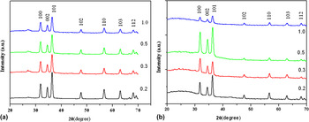
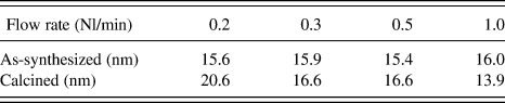
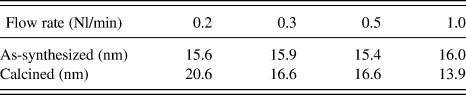
All particle samples were analyzed on a x-ray diffractometer to investigate the crystallinity and the phase of the samples (Fig. 2). The positions of the XRD peaks (as-synthesized) were in good agreement with those of the ZnO structure, but a small peak was also observed at 2θ = 27° [Fig. 2(a)]. This shows some deviation from the XRD pattern expected for pure crystalline ZnO. Choi et al.Reference Choi, Lichtenegger and Stucky17 also reported deviation in the XRD pattern of ZnO obtained by electrochemical synthesis from dilute SDS solution. On the other hand, the calcined ZnO showed good agreement with the hexagonal wurtzite structure of ZnO [Fig. 2(b)]. No peaks of Zn(OH)2 were observed in the XRD pattern under our experimental conditions. However, Samaele et al.Reference Samaele, Amornpitoksuk and Suwanboon18 reported the formation of Zn(OH)2, which was stabilized by SDS at different pH values. The average crystallite size of the particles determined by XRD line broadening measurement using the Debye–Scherrer equation is presented in Table I. The data show that the average crystallite size of the as-synthesized ZnO (∼15.5 nm) remained fairly stable with an increase in foam velocity from 0.2 to 1.0, while the particle size of calcined ZnO decreased (from 20.6 to 13.9 nm) with an increase in foam velocity from 0.2 to 1.0. The XRD of ZnO obtained by the conventional method (Fig. S1) showed that both the as-synthesized and calcined ZnO have several other peaks besides that of hexagonal wurtzite structure of ZnO. The peaks became sharper on calcination; this may be because of increased particle size and improved crystal quality.
XRD patterns of ZnO obtained at various flow rates (a) as-synthesized (b) calcined ZnO.

Particles size of ZnO calculated by the Debye–Scherrer equation.
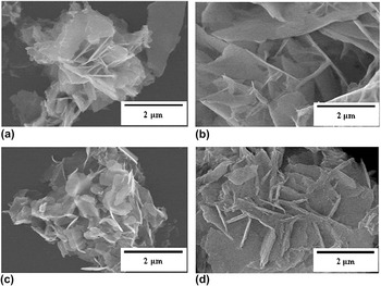
Morphological studies of the as-synthesized and calcined ZnO were carried out using scanning electron micrographs. The SEM micrographs (Fig. 3) showed the ZnO particles to be made up of aggregated plate-like structures. All ZnO particles obtained at different velocities had an aggregated plate-like structure; however, the thickness of the plates of the as-synthesized ZnO increased with increasing foam velocity. This increase in thickness might be strongly dependent on the solution surfactant concentration (SDS). Variation in the petal thickness of ZnO was also observed in our previous work with increasing biosurfactant used as a stabilizer for the synthesis of ZnO.Reference Reddy, Kuo, Atla, Chen, Chen, Shih, Chang, Maity and Chen19 During the travel of the foam at different velocities, the concentration of SDS changes with column height—this is because of the flow of the water between the bubbles that make up the foam, and the general driving forces are gravity and capillarity. Water flows through an interconnected network that consists of channels, also known as Plateau borders, where the flow is essentially unidirectional, and nodes, which are junctions of four channels. Concentration of surfactant plays an important role in the formation of different nanostructures. In our recent work, we showed the height of the foam column affected the degree of enrichment of the foamate and owing to the concentration gradient of surfactant across the column height different nanostructures were formed. The formation of various morphologies of Ni(OH)2, such as petal/coralloid and thin flake-like structures, took place at different concentration of surfactant under foam fractionation conditions.Reference Atla, Chen, Yang, Chen, Sun, Lin, Maity, Pan and Cheng15 The thickness of the ZnO plates was 42, 78, 51, and 109 nm, respectively, for foam velocities of 0.2, 0.3, 0.5, and 1.0 (Nl/min).
SEM images of as-synthesized ZnO obtained at different foam velocities. (a) 0.2, (b) 0.3, (c) 0.5, (d) 1.0.

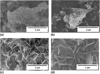
After calcination, the ZnO morphology was still one of aggregated platelets (Fig. 4). The ZnO obtained at a velocity of 0.2 consisted of aggregated irregular flakes with no sharp crystallographic edges; however, the ZnO obtained at a foam velocity of 1.0 was formed of flakes with crystallographic edges. The SEM images by conventional precipitation method (Fig. S2) showed that as-synthesized ZnO was composed of several small plates adhered together; however, on calcinations, the structure improved to form bigger particle plates.
SEM images of calcined ZnO obtained at different foam velocities. (a) 0.2, (b) 0.3, (c) 0.5, (d) 1.0.

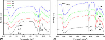
The IR spectra of the as-synthesized ZnO particles are shown in Fig. 5. The figure shows peaks arising from alkyl groups and sulfate head groups of SDS. The peaks at 2956, 2917, and 2850 cm−1 are due to –CH3 and –CH2 groups of SDS, while the peak at 1232 cm−1 is due to S = O stretching vibrations [Fig. 5(a)]. This shows that there exists an interaction between ZnO and the surfactant SDS. The alkyl sulfate ion is a potential ligand and has coordination properties and may play a role in the adsorption of these ions to the metal oxide surface. It was also seen from the IR intensity that ZnO particles obtained at high velocity (1.0) contained less surfactant, whereas with other velocities (0.2, 0.3, 0.5), the ZnO particles have a greater association with surfactant molecules. This is reasonable because as the foam travels with less velocity, it is less wet at the exit and will consequently be more enriched with surfactant. The spectra of as-synthesized ZnO were recorded after six repeated washings with water and ethanol. Michaelis et al.Reference Michaelis, Wöhrle, Rathousky and Wark20 reported SDS incorporated within ZnO films by the electrodeposition method. On calcination, the surfactant was completely removed from the ZnO surface [Fig. 5(b)]. The corresponding peaks of alkyl (2956, 2917, and 2850 cm−1) and sulfate (1232 cm−1) groups are absent in the calcined ZnO. Broad peaks at 3510 and 1672 cm−1 could be assigned to the O–H stretching vibration of the absorbed water on the ZnO surface.
FTIR spectra of ZnO obtained at different foam velocities (a) as-synthesized ZnO, (b) calcined ZnO.

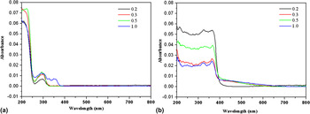
Figure 6 shows the absorbance spectra of as-synthesized ZnO nanoparticles obtained at different foam velocities. A strong absorbance of UV waves can be seen from 200 to 260 nm for as-synthesized ZnO [Fig. 6(a)]; in addition, there was another absorption from 260 to 320 nm. High-foam velocity (1.0) ZnO showed absorption of 320–400 nm UV irradiation, while other ZnO particles obtained did not show any absorption in this region. The absorption below 400 nm is assigned to the intrinsic band gap absorption of ZnO, due to electron transitions from the valence band to the conduction band. SDS forms a layer on the surface of ZnO thus reducing the UV absorption of modified ZnO at lower foam velocity. From IR results, we saw there is less SDS with high foam velocity as compared to lower foam velocity and hence (1.0) ZnO showed absorption of 320–400 nm UV irradiation. Because of the formation of barrier by the SDS around ZnO, it prevents direct contact of UV irradiation; this could be the reason of reduction of photocatalytic activity with 0.2, 0.3, and 0.5 ZnO in the degradation of MB, discussed in a later section.
UV absorbance of ZnO obtained at different foam velocities. (a) As-synthesized, (b) calcined.

The UV absorbance of the calcined ZnO is shown in Fig. 6(b). Figure 6(b) shows that the ZnO obtained at a velocity of 0.2 had the highest absorption, followed by the samples obtained at foam velocities of 0.5 and 0.3. Therefore, this sample would be more effective under UV light since it absorbs more energy as compared with the other samples.
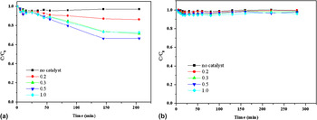
The performance of the as-synthesized ZnO/SDS hydrophobic material and the calcined ZnO for the photodegradation of MB was evaluated and compared. Figure 7 shows the concentration time profile obtained for MB in the dark for the as-synthesized and calcined ZnO particles. The as-synthesized ZnO [Fig. 7(a)] affected a decrease in the MB concentration for a period of time and then attained a steady state after 150 min, whereas the calcined ZnO [Fig. 7(b)] resulted in less than a 5% decrease in MB concentration. The major decrease with the as-synthesized ZnO can be attributed to the interaction of the organic moieties of ZnO/SDS with MB, which leads to more adsorption on the ZnO surface. The interfacial layer, the boundary between the solid ZnO and the aqueous solution containing MB, may be altered because of the presence of the SDS group, which may lead to more adsorption of MB. Because of the presence of SDS, ZnO was not uniformly dispersed in the MB solution—a phase separation was observed and particles were floating atop the MB solution.
C/C 0 versus time curves of MB in the dark with ZnO particles obtained at different foam velocities. (a) As-synthesized, (b) calcined.

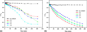
Figure 8 shows the photocatalytic degradation of MB via as-synthesized and calcined ZnO obtained at different flow rates. Both illumination and a catalyst were necessary for the complete destruction of MB. From the decomposition rate of MB with different ZnO samples, a difference in the photocatalytic activity was observed. The differing photocatalytic activity might be due to differences in the crystallinity, surface area, and particle morphology of the foam-fractionated ZnO. The results obtained are in accordance with the UV absorbance spectra obtained for the ZnO particles. In the case of as-synthesized ZnO [Fig. 8(a)], the ZnO obtained at a velocity of 0.1 exhibited a better catalytic activity than the other ZnO. It was observed that all catalysts resulted in an initial decrease in MB concentration, but after 250 min, the photodegradation of MB almost ceased. It is likely that the presence of SDS on the ZnO surface might hinder the progress of photocatalytic degradation. Pare et al.Reference Pare, Jonnalagadda, Tomar, Singh and Bhagwat21 also reported that in the photocatalytic degradation of acridine orange in the presence of anionic surfactants (SDS), the rate of degradation was retarded due to the preferential adsorption of the amphilic surfactant molecules on the photocatalyst surface. The calcined ZnO [Fig. 8(b)] showed good photocatalytic behavior as compared with the as-synthesized ZnO, and after 250 min, the calcined ZnO affected a linear increase in the degradation of MB. Prolonged irradiation caused the MB solution to turn to pale white, and this phenomenon implied that all parts of the auxochromic groups (methyl or methyl amine) degraded. ZnO in the presence of UV generates hydroxyl radicals, which degrade the pollutant. Among the calcined ZnO samples, the 0.2 ZnO, which exhibited good catalytic activity, was chosen for recyclability and stability experiments. The 0.2 ZnO calcined catalyst was tested in three consecutive recycles, and the catalyst retained its original activity. The XRD spectra of the spent catalyst showed a hexagonal wurtzite structure of ZnO, thus indicating a true heterogeneous catalyst (data not shown). The conventional method ZnO synthesis also exhibited similarly in the MB degradation (Fig. S3). The differences in the catalytic behavior of calcined ZnO can be related to the effect of SDS surfactant concentration at different foam flow rate. In our previous work,Reference Reddy, Kuo, Atla, Chen, Chen, Shih, Chang, Maity and Chen19 we showed that surfactin concentration affected the thickness and density of the petals in the rose-like ZnO structures, and thickness of the ZnO petals was found to influence the photocatalytic activity in the degradation of MB. The present study demonstrated that foam flow rate also resulted in the formation of nanostructues having different physicochemical properties. The advantage of using foam fractionation is that studies involving gas flow rate, reagents concentrations, and column height can yield different nanostructures having different physicochemical properties.
Photocatalytic degradation of MB with time. (a) As-synthesized ZnO, (b) calcined ZnO.

Several approaches have been reported in the literature to control the surface wettability of ZnO structures. Hydrophobic surfaces are needed for applications involving water repellency, prevention of the adhesion of snow, fog, and contamination, self-cleaning, and antifouling properties.Reference Blossey22 Wettability is controlled both by chemical composition and the geometric features of the surface.Reference Badre, Pauporté, Turmine and Lincot23 Alkyl-modified ZnO surfaces have been reported in the literature.Reference Hou, Zhou, Yu and Liu24, Reference Chen, Li, Zhu, Zhou, Zhao, Gao, Ma, Wang and Wang25 Our experiments with foam fractionation resulted in the formation of hydrophobic ZnO at room temperature. The contact angles of water on the ZnO/SDS surfaces are presented in Table II. The contact angle was above 90°, which demonstrated that the obtained ZnO was hydrophobic in nature. Pauporte and RathouskyReference Pauporte and Rathousky26 reported the wetting behavior of ZnO/SDS films and found that the water contact angle increased from 110 to 130° with an increase in SDS concentration. Our experiment with foam fractionation demonstrates that different foam rates produce ZnO material with differing hydrophobicity, as was evident from the water contact angle.
Contact angle of water on the ZnO/SDS surface.

IV. CONCLUSIONS
In this study, ZnO particles were successfully synthesized by pneumatic foam fractionation at different foam velocities. Using SDS as the surfactant, the as-synthesized ZnO was functionalized at room temperature. The water contact angle of the as-synthesized ZnO was above 90°, demonstrating the hydrophobic nature of the ZnO surface. The as-synthesized and calcined ZnO affected the photodecomposition of MB under UV irradiation. It was found that the calcined ZnO exhibited a better photocatalytic ability in the photodecomposition of MB than the as-synthesized ZnO. The calcined photocatalyst also showed good recyclability. It is likely that other hydrophobic oxide materials and metal oxides may be prepared by the foam fractionation method at different foam rates to produce different physicochemical properties from a single batch.
ACKNOWLEDGMENT
The authors are very grateful to the National Science Council of Taiwan for sponsoring this research.