Introduction
With the intensification of aquaculture, there has been a significant increase in the incidence of various kinds of diseases of fish. Antibiotics help greatly in controlling many bacterial diseases. However, there are many problems associated with the use of antibiotics. Vaccination is an effective method for disease prevention. However, effective vaccines are not available for most pathogens that affect aquaculture species. As an alternate measure to prevent fish diseases, immunostimulants have attracted more and more attention in the past decades.
Synthetic compounds, polysaccharides, vitamins, animal and plant extracts have been shown to enhance the non-specific immune response of fish (Sakai, Reference Sakai1999; Raa, Reference Raa, Cruz-Suárez, Ricque-Marie, Tapia-Salazar, Olvera-Novoa and Civera-Cerecedo2000). China has a long history of using herbs as immunostimulants in human beings and terrestrial animals. More than 200 kinds of Chinese herbs have been shown to have immunostimulanting effects; the major active components include polysaccharides, organic acids, alkaloids, glycosides and volatile oil (Liu, Reference Liu2002). Due to their natural origin, abundance and low toxicity, Chinese herbs meet the requirements for healthy aquaculture (Wang et al., Reference Wang, Wang, Li and Lin2006). Recently, there has been growing interest in using herbs to prevent and control diseases in fish culture.
Lentinan, isolated from the mushroom shiitake or Lentinus edodes, possesses a backbone of β-(1,3)-d-glucan and side chains of β-(1,6)-d-glucans. It has shown significant effects against tumours and viruses, possesses immune modulation activity and enhances interferon (IFN) secretion, etc. (T. Zhang et al., Reference Zhang, Liu, Wei and Zhang2003; Maruyama et al., Reference Maruyama, Sukekawa and Kaneko2006; F.M. Zhang et al., Reference Zhang, Zhang, Sun, Liu and Chen2006). Lentinan is a common immune booster used in humans and animals. However, few in vitro studies have been conducted on its immunomodulating effects. The objective of this research was to investigate the immunomodulating effect of lentinan on macrophages of the head kidney and peripheral blood leucocytes in carp, and to clarify the lentinan immunomodulating mechanism at the cellular and molecular level. The results might form the scientific basis for more intensive applications of lentinan in aquaculture.
Materials and methods
Materials
Common carp (Cyprinus carpio) was obtained from the Freshwater Fish Research Center (Chinese Academy of Fishery Sciences, China). Fish were reared at 25°C in a recirculation system and fed with commercial diets twice a day, using an automatic belt feeder. They were about 6 months old at the start of the experiment, with an average weight of 150 g. L-15 medium, lipopolysaccharide (LPS), Hanks balanced salt solution (HBSS), phytohaemagglutinin (Percoll), phobol myristate acetate (PMA), nitroblue tetrazolium (NBT), streptomycin/penicillin, heparin and 3-(4,5-dimethylthiazol-2-yl)-2,5-diphenyl tetrazolium bromide (MTT) were purchased from Sigma Company (St. Louis, Missouri, USA). Fetal calf serum (FCS) and cell culture plates were ordered from Gibco Company (Carlsbad, California, USA). RNAiso Reagent and ExScriptTM RT-PCR Kit (Perfect Real Time: SYBR Green I) were from TaKaRa Biotechnology (Dalian, China).
Immunostimulants
The crude flour of lentinan was purchased from Wuxi Zhengda Poultry Co. Ltd (Wuxi, China). The purification of lentinan was according to the modified method described by Ni et al. (Reference Ni, Su and Liu1998). An aliquot of 100 g of lentinan was soaked in 100 ml of distilled water, then boiled for 30 min. The mixture was centrifuged at 350 g for 10 min at 20°C. After removing the insoluble material the supernatant was further precipitated in 80% ethanol overnight. The mixture was then filtered through two layers of sterile gauze. The precipitate was dried at 40°C, ground and stored for use. The extract was then diluted to 1, 10, 100 and 1000 μg/ml in 5% FCS–L-15 medium containing 1% streptomycin (S)/penicillin (P), 0.2% heparin and 5% FCS.
The calibration curve was constructed following the method developed by Dubois et al. (Reference Dubois, Gilles and Hamilton1956) and the polysaccharide concentration was determined according to the linear equation. Ten milligrams of glucans were weighed into a 100 ml volumetric flask to which some deionized water was then added and the final volume was 100 ml. Volumes of the standard solution (0.2, 0.4, 0.6, 0.8 and 1.0 ml) were transferred to cuvettes and distilled water was added to each to a total volume of 2.0 ml. A volume of 1.0 ml of 5% phenol was added to each cuvette. After a gentle shaking, 5.0 ml H2SO4 was transferred to each test tube, followed by further shaking. The reaction was allowed to run for 10 min. The next process consisted of bathing the tubes in boiling water for 15 min, then cooling at room temperature. Samples and distilled water (negative control) were measured by spectrophotometry at 490 nm. All results were recorded and concentrations were considered as variables in the linear regression equation: y=1.0219+1.7225x (where x and y are concentration and absorbance, respectively). The correlation coefficient was r 2=0.999. Two milligrams of purified lentinan were weighed into a flask and dissolved as described above. Its absorbency was determined to be 2.638 according to the standard curve, and the glucose content was 93.82% determined by the linear regression equation.
Isolation of head kidney macrophages and peripheral blood leucocytes
Macrophages from the head kidney (HK) of carp were isolated according to the method described by Bayne (Reference Bayne1986). Carp were anaesthetized with 0.02% tricaine methane sulphonate (MS-222). Both head kidneys were removed using sterile tools and pushed through a 147 μm nylon mesh with L-15 medium containing 1% streptomycin/penicillin, 0.2% heparin and 0.1% FCS. The cell suspension was placed on 34/51% Percoll gradients and centrifuged at 350 g for 25 min at 4°C. The macrophage-enriched cells from the 34/51% Percoll interface were centrifuged as above and washed three times with the medium. Viable phagocytic cells, including neutrophils (about 10%) and macrophages (about 90%), were counted by trypan blue exclusion.
To prepare peripheral blood leucocytes, fish were anaesthetized with 0.02% MS-222 and the blood was removed from the caudal vein under sterile conditions. Cells were removed and loaded on the top of 60% Percoll and centrifuged at 350 g for 25 min at 4°C. The granulocyte-enriched cells from the 60% Percoll interface were centrifuged at 500 g for 5 min and washed three times with the medium. Viable granulocytes were counted by trypan blue exclusion.
Immune assays
Superoxide anion detection in the HK macrophages
The production of superoxide anions in macrophages was determined by the reduction of NBT. Under sterile conditions (such as a laminar hood), 100 μl aliquots of the collected macrophages were placed in eight replicate wells on a sterile 96-well microtitre plate and left for 2 h to allow for cell attachment. Plates were incubated at 25°C for 2 h. The cells were washed twice with HBSS, and 100 μl of NBT solution (1 mg/ml in HBSS), which included phorbol myristate acetate (1 μg/ml PMA), was added to each well and incubated for 60 min at 25°C. For the control, the macrophages were cultured in L-15 medium or LPS alone. After the removal of the medium from cells, NBT reduction was halted by the addition of methanol. The formazan in each well was dissolved in 120 μl of 2 mol/l KOH and 140 μl of dimethyl sulphoxide (DMSO), and the optical density (OD) was measured by a multiscan spectrophotometer at 620 nm. Eight replicate wells were used for each variable investigated.
Nitric oxide (NO) production by HK macrophages
Most of the steps of the procedure were the same as described above. The cells were cultured at 25°C for 96 h. The control group was incubated with medium or LPS alone. The culture supernatants (75 μl) were removed to another clean microtitre plate and Griss solution added. The OD was measured by a multiscan spectrophotometer at 540 nm. Eight replicate wells were used for each variable investigated.
Proliferation of peripheral blood leucocytes
For assay of the proliferation of peripheral blood leucocytes, the viable leucocytes were adjusted to 1×10⁷ cells/ml of 5% FCS–L-15 medium and 100 μl of the cells were added to wells of microtitre plates and incubated at 25°C for 24 h. Control leucocytes were cultured in L-15 medium or LPS alone. After the incubation period, 20 μl 5% MTT (5 mg/ml) was added to each well and incubated at 25°C for 4 h. The mazan crystals in each well were dissolved in 100 μl of DMSO. When the mazan crystals turned purple, indicating complete solubilization, the optical density (OD) was measured using a multiscan spectrophotometer at 492 nm. Eight replicate wells were used for each variable investigated.
Real-time polymerase chain reaction
To investigate the expression of the IL-1β gene in carp macrophages stimulated by lentinan, a real-time polymerase chain reaction (PCR) was carried out. The viable HK macrophages were adjusted to 1×10⁷ cells/ml and 600 μl of the cells were added to 24-well microtitre plates and incubated at 25°C for 24 h. Control cells were cultured in L-15 medium supplemented with 5% FCS. The mRNA was isolated from the HK macrophages stimulated by lentinan at different concentrations and cDNA synthesis was performed. Based on the carp IL-1β sequence in GenBank, special primers were designed and real-time quantitative PCR was carried out using β-actin as a reference gene. PCR primers were constructed from carp IL-1β gene (forward: CAA CAT TCG TGT CGA G; reverse: AAG TTT GGT TCG GG) and carp β-actin gene (forward: ACT ACC TCA TGA AGA TCC TG; reverse: TTG CTG ATC CAC ATC TGC TG). Fluorescence signal baseline and threshold were set manually for each run, generating a threshold cycle (CT) for each sample. Then, relative expression levels were calculated from the normalized CT value by the 2−ΔΔCT method (Livak and Schmittgen, Reference Livak and Schmittgen2001).
Statistics
The data were compared using Student's t-test and one-way analysis of variance (ANOVA). They were expressed as mean optical density (OD)±SD and differences were considered statistically significant at P<0.05 or 0.01.
Results
Superoxide anion detection in HK macrophages
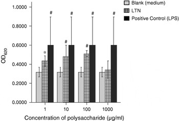
Results of detection of superoxide anions in C. carpio HK macrophages after incubation with different concentrations of lentinan for 24 h showed that the positive group (LPS group) could stimulate superoxide anions in macrophages (Fig. 1), suggesting that under in vitro conditions the cultured cells were normal. Compared with the blank group (incubated in L-15 medium), lentinan at 1 (P<0.05), 10 and 100 μg/ml (P<0.01) could significantly induce superoxide anions in macrophages (Fig. 1).
Detection of superoxide anions in Cyprinus carpio macrophages from the head of the kidney after 24 h incubation with lentinan (LTN) at different concentrations. Results expressed as means±SD in four fish. *P<0.05, #P<0.01. LPS, lipopolysaccharide.

Nitric oxide (NO) production in HK macrophages
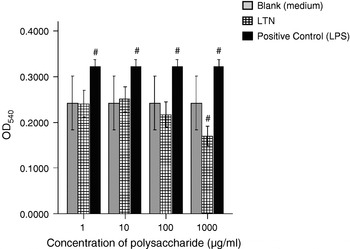
NO production in HK macrophages showed that lentinan at low doses had no evident effect on nitrogen burst activity of macrophages after 96 h incubation. However, this activity was inhibited by 1000 μg/ml (P<0.01) (Fig. 2). The positive control could stimulate NO production in macrophages (Fig. 2), which again indicated the normal conditions of cultured cellsin vitro.
Reduction of nitric oxide by Cyprinus carpio macrophages from the head of the kidney after 96 h incubation with lentinan (LTN) at different concentrations. Results expressed as means±SD in four fish. *P<0.05, #P<0.01. LPS, lipopolysaccharide.

Peripheral blood leucocyte proliferation
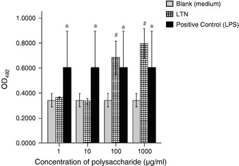
The proliferation of positive control groups after 24 h was markedly stimulated (Fig. 3). Compared to the blank group, proliferation of groups treated with lentinan at 100 and 1000 μg/ml (P<0.01) was highly improved (Fig. 3).
Proliferation of peripheral blood leucocytes after 24 h incubation with lentinan (LTN) at different concentrations. Results expressed as means±SD in four fish. *P<0.05, #P<0.01. LPS, lipopolysaccharide.

Real-time PCR for detecting IL-1β gene expression
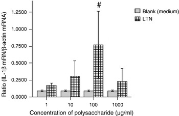
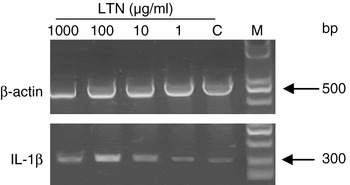
RNA extracted from HK cells which had been treated with various concentrations of polysaccharides was used for cDNA synthesis. A real-time quantitative PCR was carried out using β-actin as reference gene. The threshold cycle (CT) of all samples was collected and calculated by the 2–ΔΔCT method. Results showed that the expression of IL-1β from the groups treated with lentinan was significantly increased, compared to the blank group (Fig. 4). At the same time, the result of real-time PCR showed that lentinan at 100 μg/ml could significantly stimulate the expression of IL-1β mRNA (P<0.01) (Fig. 5).
Expression of IL-1β in macrophage cells of Cyprinus carpio after 24 h stimulation by lentinan (LTN) at different concentrations. Results expressed as means±SD in four fish. *P<0.05, #P<0.01.

The real-time PCR expression of IL-1β in macrophage cells following treatment with lentinan (LTN) at different concentrations for 24 h. M, 100 bp DNA marker; C, control cells (cultured in medium).

Discussion
Macrophages and peripheral blood leucocytes are the most important elements in the immune system. Not only can they phagocytose pathogens, but they also produce cytokines. Macrophages can also act as antigen-presenting cells. This study shows that lentinan could stimulate the proliferation of peripheral blood leucocytes and greatly induce superoxide anions in macrophages. Lentinan could modulate the non-specific immune responses of the immune cells. At suitable doses, it may increase resistance to infectious diseases by stimulating the macrophages and peripheral blood leucocytes.
Lentinan at low doses had no effect on nitric oxide production. However, at elevated doses, it inhibited the nitrogen burst activity of macrophages, suggesting that lentinan modulates the immune response in both directions. High doses of lentinan may cause a cytotoxic effect on the macrophages. Similar results were previously reported for a water extract of Astragalus, which could boost the proliferation of HK macrophages and enhance the nitric oxide production of microphage mixtures and granulocytes induced by LPS (Yin et al., Reference Yin, Geert and Li2004), but, alone, it had no effect on nitric oxide production. The mechanism of lentinan on the immune responses of macrophages may be related to the induction of inducible nitric oxide synthase (iNOS) and NO production (Ding et al., Reference Ding, Hou and Zhou2003).
Cytokines are simple polypeptides or glycoproteins that act as signalling molecules within the immune system (Zlotnik and Yoshie, Reference Zlotnik and Yoshie2000; Jin, Reference Jin2001). Interleukin-1β (IL-1β), produced by macrophages, activates T cells and macrophages and is considered to be one of the most important cytokines in the specific and non-specific immune responses. It has been shown that lentinan could enhance IL-2 and tumour necrosis factor-α (TNFα) mRNA expression in mouse spleen monocytes and human blood monocytes (Liu et al., Reference Liu, Li, Fanzuo, Lin and Gao1998). Ganoderma lucidum polysaccharide (GLP) injected intraperitoneally into male inbred BALB/c mice promoted a remarkable increase in the expression of IL-1α, IL-1β, TNFα and macrophage colony-stimulating factor (M-CSF) in spleenocytes and IL-1β, TNFα and M-CSF in macrophages (Ooi et al., Reference Ooi, Ooi and Fung2002). A water extract of Astragalus radix could stimulate IL-1α, IL-1β and IL-16 mRNA expression and Astragalus polysaccharides significantly induced NO production and iNOS transcription through the activation of nuclear factor-κB/Rel (NF-κB/Rel) in RAW 264.7 macrophage cells (Kun and Young, Reference Kun and Young2005; Young et al., Reference Young, Kyung and Chan2005). Lentinan could promote macrophage anti-microorganism activity and induce the expression of IL-12, TNF-α and interferon-γ (IFN-γ) (Cohen-Kupiec et al., Reference Cohen-Kupiec, Broglie, Friesem, Broglie and Chet1999). The present study showed that lentinan could enhance the expression of IL-1β, which further indicates that lentinan may increase disease resistance by enhancing the expression of cytokines.