Introduction
Coenurosis (gid or sturdy) is caused by Coenurus cerebralis, the bladderworm stage of Taenia multiceps, which develops predominantly in the brain or spinal cord of many mammalian species, including humans, and especially sheep (Ing et al., Reference Ing, Schantz and Turner1998; Sharma & Chauhan, Reference Sharma and Chauhan2006; Christodoulopoulos, Reference Christodoulopoulos2007; Christodoulopoulos et al., Reference Christodoulopoulos, Theodoropoulos and Petrakos2008). However, there have also been reports of Coenurus cysts in subcutaneous or intramuscular tissues and the abdominal cavity in ungulates but especially goats. In goats, these non-cerebral Coenurus cysts have been called Coenurus gaigeri, and in sheep they have been called Coenurus skrjabini (Verster, Reference Verster1969; Sharma & Chauhan, Reference Sharma and Chauhan2006; Schuster et al., Reference Schuster, Sivakumar and Wieckowsky2010). Meanwhile, the metacestodes of Taenia serialis cause cysts in subcutaneous and muscle tissues of hares and rabbits, known as Coenurus serialis (Verster, Reference Verster1969). The objective of the present study was to report the occurrence of non-cerebral coenurosis in sheep and to describe its macroscopic features.
Materials and methods
Collection and examination of sheep carcasses
An abattoir survey of non-cerebral coenurosis in sheep was performed in three abattoirs in the city of Al Ain of the United Arab Emirates (UAE) between October 2010 and May 2011. During this period, after the customary examination by the meat inspectors, any sheep carcass suspected of displaying non-cerebral coenurosis was brought to the attention of the authors for further investigation.
Any carcass identified for investigation was inspected by visual examination as well as by palpation. The suspected cyst was revealed by dissecting the surrounding tissue. If a Coenurus cyst was suspected, it was removed, placed in a plastic bag that was labelled and transferred in a portable fridge at 4–8°C to the laboratory within 1 h.
The following data were recorded: the age and the country of origin, the position of the Coenurus cysts in the body, the number of the cysts, as well as the presence of any other kind of cestode cysts found. The brain and the spinal cord were examined for the presence of any Coenurus cysts.
Examination of cysts
A cyst was initially identified as a Coenurus cyst based on the inclusion of a bladder that was filled with watery fluid and having a thin and transparent wall with numerous scolices attached to its inner surface. Upon laying the bladderworm on a flat surface, the number of scoleces and their arrangement in clusters were counted.
To confirm the identification, a piece of the larval membrane containing a cluster of scoleces was submerged in warm tap water (37–39°C) for 30–60 min to provoke evagination of the scolices. Subsequently, the cluster was placed on a slide, a cover slip was pressed tightly on it and finally the scoleces of the cluster were examined under a light microscope. The identification of the Coenurus larvae was based on the recognition of the rostellar hooks along with the four surrounding suckers in the evaginated scolices.
Results and discussion
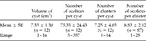
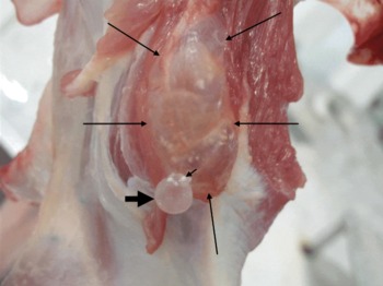
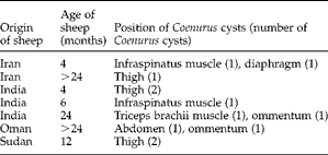
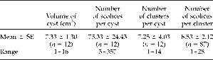
Only 7 sheep of 90,415 (0.008%) slaughtered were found to have non-cerebral Coenurus cysts. Three sheep originated in India, 2 from Iran, 1 from Oman and 1 from Sudan. All 12 cysts contained a single bladderworm with multiple scolices (table 1). The cysts were located in the triceps brachii muscle (fig. 1), the diaphragm, the infraspinatus muscle, the muscles of thigh and the abdomen, and in one case a Coenurus cyst was found attached to the omentum. One cyst of volume 1 cm3 was found in the subcutaneous tissue of the thigh. In the cases of infestation in the muscles of thigh and the abdomen, the cysts affected more than one muscle (table 2). No clinical evidence for non-cerebral coenurosis had been recorded during the antemortem veterinary inspection.
Macroscopic characteristics of Coenurus cysts found in sheep (N=7).
Coenurus cyst attached to the triceps brachii muscle. The outer membrane of the bladderworm was ruptured during skinning so the bladderworm protrudes and a cluster of scoleces is revealed. The margins of the Coenurus cyst (size: 7 × 3.86 cm) are indicated by the long, thin arrows, the bladderworm (diameter: 1.38 cm) is indicated by the thick, short arrow, and the cluster of scoleces is indicated by the thin, short arrow (a colour version of this figure can be found online at http://www.journals.cambridge.org/jhl).

The occurrence, the age of sheep and the location of Coenurus cysts.
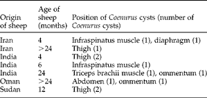
A prevalence of 2.6% has been reported for non-cerebral coenurosis in goats in Iran (Oryan et al., Reference Oryan, Nazifi, Sharifiyazdi and Ahmadnia2010). To the authors' knowledge, no epidemiological data are available for the situation in sheep. The most recent reports of non-cerebral coenurosis in sheep trace back to the 1930s (Schuster et al., Reference Schuster, Sivakumar and Wieckowsky2010). Verster (Reference Verster1969) reported that the disorder occurred only in an isolated area of Kazakhstan; however, the present data suggest that the disorder occurs in Asia, the Middle East and Africa. Interestingly, in the literature there are reports of the occurrence of non-cerebral coenurosis in goats in Asia, the Middle East and Africa (in India, Iran and Oman) (Sharma et al., Reference Sharma, Sanil, Agnihotri and Singh1995; Sharma & Chauhan, Reference Sharma and Chauhan2006; Oryan et al., Reference Oryan, Nazifi, Sharifiyazdi and Ahmadnia2010; Schuster et al., Reference Schuster, Sivakumar and Wieckowsky2010).
The Coenurus cysts found in the present study were of small size and contained few scoleces in comparison to the cyst size and number of scoleces reported for cerebral coenuri. It is accepted that cerebral coenuri in sheep can reach 95 cm3 in size and can contain up to 700 scolices (Boev et al., Reference Boev, Sokolova, Tazieva and Boev1964; Schuster et al., Reference Schuster, Sivakumar and Wieckowsky2010). The number of scolices and the size of cysts reported here for sheep non-cerebral coenurosis are similar to the findings for non-cerebral coenurosis of goats reported by Schuster et al. (Reference Schuster, Sivakumar and Wieckowsky2010), who found cyst sizes to vary between 1 and 40 cm3 and the number of scolices per cyst to be between 46 and 474.
In conclusion, non-cerebral coenurosis in sheep is a rare disorder. The taxonomic status of the non-cerebral Coenurus cysts remains to be determined by molecular techniques.
Acknowledgements
We acknowledge with many thanks the prompt and fruitful help of Mr M.S. Al-Shamsi, Head of the Public Slaughterhouse Section in Al Ain Municipality, Mr Z.M. Swareldahab, Director of the Meat Inspectors, and all the meat inspectors who participated in this research, especially Mr M.M.A. Eltahier.