Case
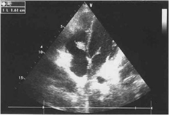
A 13-year-old girl with suspected systemic disease and an abnormality of the tricuspid valve, found on echocardiography, was admitted for further cardiac diagnostic workup. The patient’s 1-year history included high sedimentation rate (45 mm/hour), with low C-reactive protein and procalcitonin levels, high anti-nuclear antibodies titres (1:10,000), predominantly anti-Sjogren syndrome antigen A and B, progressive thrombocytopaenia, and recurrent facial rash. On echocardiography, a heterogeneous mass measuring 15×10 mm was found adjacent to the septal leaflet of the tricuspid valve, without significant blood flow compromise (Fig 1).
Abnormal mass adjacent to the tricuspid septal leaflet on echocardiography.

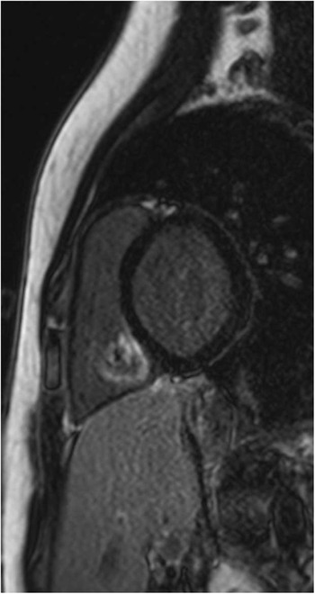
Owing to the uncertain nature of the echocardiography findings and in anticipation of detailed immunology results, cardiac magnetic resonance imaging was performed. Cardiac magnetic resonance imaging showed an abnormal, irregular, mobile mass below the septal leaflet of the tricuspid valve. In addition to an immobile oval part, an elongated part adjacent to the posterior section of the tricuspid annulus was also noted. The mass was iso-intense on T1-weighted images before contrast, showing no early contrast enhancement and unequivocal late enhancement with a hypo-intense centre at 20 to 30 minutes after contrast, whereas thrombus was excluded on inversion recovery images acquired with long inversion times (550 ms) (Fig 2). Subsequently, detailed immunology tests results became available, which included anti-nuclear antibodies of Sjogren syndrome antigen A (+++) and Sjogren syndrome antigen B (+) types, double-stranded deoxyribonucleic acid (+), IgG anti-cardiolipin antibodies (>120 U/ml), low C4 complement, reduced haemolytic complement activity, and hyper-gammaglobulinaemia, confirming the diagnosis of systemic lupus erythematosus, co-existing with anti-phospholipid syndrome. An infective process was excluded, after three negative microbiological blood tests, whereas the echo and cardiac magnetic resonance imaging findings supported the diagnosis of Libman–Sacks endocarditis as part of the disease.
Abnormal mass involving the tricuspid valve on magnetic resonance imaging. Phase-sensitive inversion recovery image of the mass is seen with late enhancement sparing the most central part.

Discussion
Libman–Sacks endocarditis was first described in 1924 as characterised by the presence of papillary, non-infective vegetations found predominantly on the inflow parts of the mitral and aortic valves. Much less frequently, vegetations involve other heart valves, chordae tendineae, endocardium, or papillary muscles. These changes usually do not cause any specific clinical symptoms, but long-term follow-up studies have shown the development and progression of valvular dysfunction.Reference Moyssakis, Tektonidou and Vasilliou 1 At present, these changes are found on echocardiography in ~10% of patients with systemic lupus.Reference Moyssakis, Tektonidou and Vasilliou 1 Unfortunately, due to the far lower incidence of systemic lupus erythematosus in children, no reliable statistics exist regarding echocardiographic changes in this population. The interrelation between the occurrence of valvular changes and duration of systemic disease, its severity, or treatment mode or duration has not been elucidated.Reference Mattos and Santiago 2 Nevertheless, there is evidence of the impact of anti-cardiolipin antibodies co-existing with systemic lupus.Reference Mattos and Santiago 2 , Reference Sharma, Lasic and Bornstein 3 It is believed that they promote thrombus formation on the damaged endocardium and increase the intensity of inflammatory reactions. Shapiro et alReference Shapiro, Gamble and Wiesner 4 used advanced microscopic examination to confirm the presence of complement and antibody deposits, including anti-cardiolipin, explaining the thickening of the valvar leaflets or cusps with a typical appearance of Libman–Sacks vegetations. In our case, we did not perform histological examinations. At present, given the increasingly recognised stronger relationship of Libman–Sacks inflammation with primary anti-phospholipid syndrome rather than systemic lupus, thromboembolic complications such as cerebral ischaemic events are likely to be diagnosed more frequently than symptomatic dysfunction of the heart valves or endocarditis.Reference Bulckaen, Puisieux and Bulckaen 5 In our case, the inflammatory process involved the tricuspid valve; therefore, potential complications related to pulmonary embolism could be expected. At present, the patient is undergoing treatment for the underlying disease, with steroids and anti-coagulants, and is showing very good clinical response. Follow-up echocardiography has shown significant regression of the cardiac mass.
Acknowledgement
None.
Financial Support
This research received no specific grant from any funding agency, commercial, or not-for-profit sectors.
Conflicts of Interest
None.