Endothelial cells lining the inner surface of microvessels form a semipermeable barrier that actively participates in blood–tissue exchange of plasma fluid, proteins and cells. The precise regulation of endothelial permeability is essential for maintaining circulatory homeostasis and the physiological function of different organs. As a result, microvascular barrier dysfunction and endothelial hyperpermeability represent crucial events in the development of a variety of disease processes, such as adult respiratory distress syndrome (ARDS), ischemia–reperfusion (I–R) injury, diabetic vascular complications, and tumour metastasis. Better insight into the molecular mechanisms underlying pathogenic conditions related to microvascular hyperpermeability is required for developing effective therapeutic strategies. Following intensive studies over the past few decades, it is now understood that endothelial permeability is mediated through a transcellular pathway (across cells) and a paracellular pathway (between cells), both of which are highly regulated by mechanical forces and biochemical signals.
Transcellular versus paracellular permeability
An important molecular mechanism underlying transcellular permeability is macromolecule transcytosis via caveoli – specialised plasmalemmal vesicles containing caveolin-1. The involvement of caveolin-1 in regulating cardiovascular functions associated with endothelial barrier properties has been demonstrated through studies using transgenic and knockout animals (Refs Reference Bauer1, Reference Gratton, Bernatchez and Sessa2, Reference Lin3, Reference Razani4). Upon binding to plasma proteins (of size >3 nm, e.g. albumin), the cell-surface docking protein gp60 interacts with caveolin-1 and signalling intermediaries, including a G protein and Src-family tyrosine kinase. This cascade results in the formation and release of albumin- and solute-containing caveolae from the apical membrane (Ref. Reference Mehta and Malik5). The vesicles are subsequently transported to the basal membrane and release their contents through exocytosis (Ref. Reference Mehta and Malik5). Recent ultrastructural evidence suggests that caveolae-like vesiculovacuolar organelles can interconnect to each other, forming secondary, grape-like structures that function as transmembrane channels for molecular trafficking across the cell (Ref. Reference Dvorak6). Receptors of endogenous permeability-enhancing agents have been identified on the surface of these channels, indicating the possibility of their participation in transcytosis-related endothelial permeability (Refs Reference Curry and Adamson7, Reference Dvorak8).
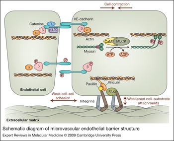
Despite the potential contribution of transcytosis to the basal permeability of the endothelium, paracellular flux of plasma fluid and proteins through endothelial cell–cell junctions has been emphasised for its pathophysiological importance in vascular inflammation during disease and injury. Among the different types of junction structures in the vascular endothelium, the tight junction and adherens junction are the best characterised with respect to their function in mediating cell–cell adhesion and thus barrier properties. Briefly, the tight junction is a zipper-like structure formed at the cell–cell contact area by a group of transmembrane proteins primarily expressed in the blood–brain barrier and retinal microvasculature, including claudins, occludins and zonular occludins (ZO-1 and ZO-2). The adherens junction has been identified in nearly all types of vascular beds, especially in the peripheral microvasculature (Fig. 1). Its molecular structure is based on VE-cadherin (vascular endothelial cadherin), a transmembrane receptor whose extracellular domain homophilically binds to the extracellular domain of another VE-cadherin molecule from an adjacent cell and whose intracellular domain is anchored to the cell cytoskeleton via a family of actin-binding proteins called catenins (α, β, γ and p120 catenins). The catenins not only serve as a structural linkage between VE-cadherin and the cytoskeleton, but also transduce biochemical signals for cell–cell communications (Refs Reference Iyer9, Reference Xiao10). Moreover, endothelial cells are tethered to the extracellular matrix through focal adhesions, which consist mainly of integrin transmembrane proteins and a family of actin-linking proteins including focal adhesion kinase (FAK), talin and paxillin (Ref. Reference Mehta and Malik5) (Fig. 1). The stability of this junction–cytoskeleton complex withstands fluid shear stress and is essential in maintaining the endothelial barrier function.
Schematic diagram of microvascular endothelial barrier structure. The barrier is formed by endothelial cells that connect to each other through the junctional adhesive molecule vascular endothelial (VE)-cadherin, which binds to another VE-cadherin molecule from an adjacent cell and connects to the actin cytoskeleton via a family of catenins (α, β, γ and p120). This endothelial lining is tethered to the extracellular matrix through focal adhesions mediated by transmembrane integrins composed of α and β subunits, focal adhesion kinase (FAK), and cytoskeleton-linking proteins including paxillin and vinculin. The integrity of this barrier is maintained by VE-cadherin-mediated cell–cell adhesions and focal-adhesion-supported cell–matrix attachment. A dynamic interaction among these structural elements controls the opening and closing of the paracellular pathways for fluid, proteins and cells to move across the endothelium. In particular, the Ca2+/calmodulin (CaM)-dependent myosin light chain kinase (MLCK) catalyses phosphorylation of myosin light chains (small red circles), triggering binding of the myosin heavy chain motor domains to actin and their cross-bridge movement. This reaction promotes cytoskeleton contraction and cell retraction. In parallel, phosphorylation of VE-cadherin and/or catenins may cause the junction complex to dissociate from its cytoskeletal anchor, leading to weakened cell–cell adhesion. The cytoskeletal and junctional responses act in concert causing paracellular hyperpermeability. These structural changes are caused by signalling reactions depicted in Figure 2.

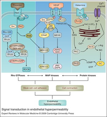
Many inflammatory mediators are capable of disrupting the interendothelial junction assembly, thereby causing endothelial hyperpermeability. More in-depth molecular analyses suggest that the mechanism underlying inflammation-induced endothelial paracellular hyperpermeability involves phosphorylation, internalisation or degradation of the junctional molecules (Ref. Reference Esser11). In addition, the junction–cytoskeleton complex participates in other cellular processes including molecular scaffolding, intracellular trafficking, transcription and apoptosis that may directly or indirectly alter vascular barrier function (Ref. Reference Dejana12). Regardless of the molecular details, however, essentially all permeability responses in the vascular endothelium are initiated with receptor occupancy followed by a series of intracellular signalling cascades, some of which are described below (Fig. 2).
Signal transduction in endothelial hyperpermeability. (Legend; see previous page for figure) Multiple cascades of intracellular signalling reactions are initiated when an inflammatory agonist binds to its respective receptor expressed on the endothelial surface [e.g. thrombin binds the protease-activated receptor 1 (PAR-1), histamine binds its receptor H1, and vascular endothelial growth factor (VEGF) binds its receptor VEGFR-2 (KDR)]. Occupancy of G-protein-coupled receptors activates RhoA and its effector kinase ROCK (left), or it triggers phospholipase (PLC)-catalysed protein kinase C (PKC) activation and elevated intracellular calcium, which stimulates nitric oxide production and cGMP-dependent protein kinase (PKG) activation (right). Agonist binding of receptor tyrosine kinase also activates the mitogen-activated protein (MAP) kinase cascades characterised by phosphorylation of extracellular-signal-regulated kinases (ERK1/2) (middle). The three pathways (Rho GTPases, MAP kinases and protein kinases) interact with each other, causing changes in the endothelial barrier structure. Abbreviations: cGMP, cyclic guanosine monophosphate; CRAF, Raf-1; DAG, diacylglycerol; ER, endoplasmic reticulum; GC, guanylate cyclase; GDP, guanosine diphosphate; GEF-H1, guanine-nucleotide-exchange factor H1; GRB2, growth factor receptor-bound protein 2; GTP, guanosine triphosphate; JNK, c-Jun N-terminal kinase; JNKK, c-Jun N-terminal kinase kinase; l-Arg, l-arginine; NO, nitric oxide; NOS, nitric oxide synthase; p115RhoGEF, 115 kDa guanine-nucleotide-exchange factor; p190RhoGAP, p190 Rho GTPase-activating protein; Ras, ras gene product; Rho-GDI, GDP dissociation inhibitor (GDI); Rho-GDl, Rho GDP-dissociation inhibitor 1; ROCK, Rho kinase; RTK, receptor tyrosine kinase; SAPK, stress-activated protein kinase; SEK, stress-activated protein kinase/ERK kinase; Sos, Son of sevenless; Src, src gene product.

Intracellular signal transduction
Cytosolic calcium
Many inflammatory agonists mediate endothelial hyperpermeability via a calcium (Ca2+)-dependent mechanism. The signalling cascade is triggered when an agonist binds to its respective receptor expressed on the endothelial surface, which activates phospholipase C (PLC) either directly (e.g. VEGF receptor) or via a G protein (e.g. histamine receptor) (Fig. 2). This culminates in inositol 1,4,5-trisphosphate [Ins(1,4,5)P 3] production and Ca2+ release from intracellular stores as well as influx via various channels. Elevated intracellular Ca2+ further targets cytoskeleton proteins or junction structures that determine the paracellular permeability property.
At the cytoskeleton, Ca2+ activates Ca2+/calmodulin-dependent myosin light chain kinase (MLCK), which phosphorylates myosin light chain (MLC) and promotes a cross-bridge movement between actin and myosin (Fig. 1). This interaction generates a contractile force that pulls neighbouring cells apart from each other (retraction), leading to intercellular gap formation (Ref. Reference Tiruppathi13). In parallel, Ca2+ stimulates nitric oxide synthase (NOS), which produces nitric oxide (NO) and subsequently cyclic GMP (cGMP); the latter activates cGMP-dependent protein kinase (PKG). The NO–PKG cascade is known to mediate endothelial barrier structural and functional responses to a wide spectrum of physiological and pathological mediators, including angiogenic growth factors and pro-inflammatory agents (Refs Reference Yuan14, Reference Yuan15). Additionally, intracellular Ca2+ has been shown to participate in the transcellular transport of macromolecules via caveoli, as demonstrated by Ca2+ waves in caveolin-1-rich cell edges under shear stress or ATP stimulation (Ref. Reference Isshiki16).
Protein kinase C
Protein kinase Cs (PKCs) are a family of serine/threonine kinases that differ in structure, expression, subcellular distribution, and function. Their activities are differentially regulated by Ca2+, diacylglycerol (DAG) and phospholipids. Some isoforms can be activated by synthetic compounds such as phorbol 12-myristate 13-acetate (PMA). The downstream signals of PKCs include the Rho family of GTPases, mitogen-activated protein kinases (MAPKs) and MLCK. More specifically, PKCs induce rapid phosphorylation of the Rho-GDP dissociation inhibitor (GDI), promoting Rho-GDP–GTP exchange and Rho-associated protein kinase (ROCK) activation coupled with increased actomyosin ATPase activity (Ref. Reference Maekawa17). With respect to the MAPK cascades, PKCs trigger Ras-dependent signalling through a sequential activation of a Ras protein, Raf protein, MAPK kinase (MEK1/2) and extracellular-signal-regulated kinase (ERK1/2) (Refs Reference Mehta, Rahman and Malik18, Reference Verin19). In particular, PKCs cause a time-dependent increase in the activity of Ras, which binds CRAF (Raf-1), a serine/threonine kinase capable of activating MEK1/2 and subsequently ERK1/2. Inhibition of Ras completely abolishes PMA-induced CRAF activity, and MEK1/2 inhibitors attenuate PMA-induced hyperpermeability, consistent with the role of PKC–MAPK signalling in endothelial barrier regulation (Ref. Reference Verin19).
Structurally, PKCs can also directly stimulate the cytoskeletal contractile machinery by phosphorylating MLCK, a key molecule triggering actin–myosin motor function. PKCs also interact with other cytoskeletal proteins known to participate in cell contraction, such as actinin, caldesmon, and the intermediate filament protein vimentin (Ref. Reference Stasek, Patterson and Garcia20). Furthermore, PKC activation correlates with disassembly of adherens junctions (Ref. Reference Sandoval21) and tight junctions (Ref. Reference Clarke, Soler and Mullin22).
Tyrosine kinases
Both receptor and nonreceptor tyrosine kinases participate in the signal transduction that mediates changes in endothelial barrier structure and function. As a representative of receptor tyrosine kinases, vascular endothelial growth factor (VEGF) receptor 2 (VEGFR-2; also named KDR) undergoes dimerisation and phosphorylation at multiple tyrosine residues upon ligand binding. This provides a molecular configuration for recruiting more intracellular signals and further stimulating the receptor-linked PLCγ. The downstream reactions include PKC activation, Ca2+ release, NO production, and MAPK signalling (Refs Reference Aramoto23, Reference Bates and Curry24, Reference Breslin25, Reference Lal26, Reference Varma27, Reference Wu28). In addition to these well-characterised pathways, VEGFR-2 has been shown to mediate endothelial hyperpermeability by activating p21-activated kinase (PAK) (Ref. Reference Stockton, Schaefer and Schwartz29) and by phosphorylating FAK, thereby triggering integrin signalling (Ref. Reference Wu30). In endothelial adherens junctions, both VE-cadherin and catenins (β-, γ- and p120-catenin) can be tyrosine phosphorylated by this receptor kinase in the presence of VEGF (Ref. Reference Esser11). Recently, it became evident that the internalisation of VE-cadherin is closely regulated by VEGF signalling in a β-arrestin-dependent manner (Ref. Reference Gavard and Gutkind31).
Among multiple nonreceptor tyrosine kinases, the Src family (c-Src, Fyn, Yes, Yrk, Lyn, Hck, Fgr, Blk, Lck) has been frequently implicated in the barrier-opening action of various inflammatory mediators and growth factors (Ref. Reference Eliceiri32). The mechanisms underlying Src-induced paracellular hyperpermeability may involve cytoskeleton contraction driven by MLCK phosphorylation (Ref. Reference Birukov33), and/or junction opening triggered by VE-cadherin and β-catenin phosphorylation (Ref. Reference Wu, Ustinova and Granger34). Furthermore, it has been demonstrated that Src-induced phosphorylation of caveolin-1 and dynamin is crucial for the signalling machinery in directing the polarised transportation of albumin and other solute through the transcellular pathway (Refs Reference Mehta and Malik5, Reference Shajahan35).
Rho GTPases
The activity of Rho GTPases is regulated through GDP–GTP cycling dependent on guanine-nucleotide-exchange factors. Three members of the Rho family (RhoA, Rac and Cdc42) have been characterised for their effects on endothelial permeability through distinct subcellular targeting and functions. In particular, RhoA promotes cell contractility and focal adhesion, whereas Cdc42 and Rac1 are major players in cell membrane protrusion or filopodia formation and thus cell migration (Ref. Reference Miao36). Under physiological conditions, changes in mechanical forces (e.g. shear stress) act to relay intracellular signals to regulate RhoA activity, thus maintaining endothelial integrity, especially at intercellular junctions (Ref. Reference Mammoto, Mammoto and Ingber37). The importance of increased RhoA activity in endothelial barrier dysfunction is supported by studies showing that RhoA and its effector kinase ROCK mediate endothelial hyperpermeability in response to histamine, thrombin, VEGF, neutrophils, and mechanical stimuli (Refs Reference Dvorak8, Reference Mammoto, Mammoto and Ingber37, Reference Breslin38, Reference Breslin and Yuan39, Reference Sun40, Reference van Nieuw Amerongen41). Upon activation, RhoA inhibits myosin-associated protein phosphatase by phosphorylating its inhibitory myosin phosphatase targeting (MYPT) subunit via ROCK, indirectly promoting MLC phosphorylation and actin–myosin contraction (Ref. Reference Patil and Bitar42). In addition, RhoA increases endothelial permeability by stimulating Ca2+ entry, which activates MLCK.
By contrast to the permeability-increasing effect of RhoA, Rac and Cdc42 have been generally accepted as barrier protectors capable of decreasing permeability. Although a recent study suggests that Rac contributes to VEGF-induced hyperpermeability (Ref. Reference Gavard and Gutkind31), activation of Rac is associated with a tightened barrier after treatment with sphingosine 1-phosphate (Ref. Reference Garcia43) and hepatocyte growth factor (Ref. Reference Liu44), Likewise, Cdc42 has been shown to play a critical role in re-annealing adherens junctions and restoring barrier function in endothelial cells during the recovery phase of inflammatory injury (Ref. Reference Kouklis45). While the detailed mechanisms by which these Rho members protect endothelial barrier properties remain elusive, their effects may involve PAK-dependent cytoskeleton redistribution or junction reorganisation (Ref. Reference Garcia43).
cAMP
The adenine nucleotide is a conserved, ubiquitous intracellular second messenger that has been well recognised for its barrier protection function (Refs Reference Yuan14, Reference Moore46). Several mechanisms have been proposed to explain the action of cyclic adenosine monophosphate (cAMP) on endothelial barrier structure. First, increased intracellular cAMP activates its cognate protein kinase, PKA, which by stabilising the actin cytoskeleton counteracts the cell retractile force, thus preventing intercellular gap formation (Ref. Reference Moore46). Second, activated PKA can inhibit MLCK activity, which in turn leads to decreased MLC phosphorylation and minimises actin–myosin contraction. Finally, cAMP inhibits membrane phospholipid hydrolysis, thereby reducing DAG production and PKC activity. In accordance with these protective effects of cAMP at cell–cell junctions, previous studies also show that cAMP analogues induce a diffuse distribution of focal adhesions of endothelium and improved barrier function (Ref. Reference Lampugnani47).
Hyperpermeability mediators
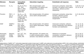
Increased circulating levels of immune cells (e.g. neutrophils) and pro-inflammatory cytokines [e.g. interleukins (ILs) and tumour necrosis factors (TNFs)] or other soluble mediators (e.g. histamine, thrombin and VEGF) are a hallmark of host defence against injury. Most of these mediators produce vasoactive and cytotoxic effects by directly targeting the vascular endothelium and activating the aforementioned signalling pathways. With recent intensive studies, the list of mediators inducing endothelial hyperpermeability continues to grow. In this review, we focus on some typical acute inflammatory mediators and discuss their effects and mechanisms in endothelial hyperpermeability (Table 1).
Acute inflammatory stimuli capable of inducing endothelial hyperpermeability

Abbreviations: CaMKII, Ca2+/calmodulin-dependent protein kinase type II; cAMP, cyclic adenosine monophosphate; eNOS, nitric oxide synthase, endothelial; EPCR, endothelial protein C receptor; ERK1/2, extracellular-signal-regulated kinases; FAK, focal adhesion kinase; Gα12/13, guanine-nucleotide-binding protein Gα12/13; Gi, guanine-nucleotide-binding protein G(i) subunit alpha-I; Gq, guanine-nucleotide-binding protein G(q) subuint alpha-q; Gs, guanine-nucleotide-binding protein G(s) subunit alpha short; ICAM, intracellular adhesion molecule; IL-1, interleukin 1; MAPK, mitogen-activated protein kinase; MEK1/2, MAP kinase kinase or ERK kinase; MLC, myosin light chain; MMP, matrix metalloproteinase; p115RhoGEF, 115 kDa guanine-nucleotide-exchange factor; PAR-1, -3, -4, proteinase-activated receptor 1, 3, 4; PI3K, phosphoinositide 3-kinase; PKA, protein kinase A; PKC, protein kinase C; PKG, cGMP-dependent protein kinase; PLC, phospholipase C, ROCK, Rho kinase; ROS, reactive oxygen species; TNF, tumour necrosis factor; VCAM, vascular cell adhesion molecule; VE-cadherin, vascular endothelial cadherin; VEGF, vascular endothelial growth factor; VEGFR-2, vascular endothelial growth factor receptor 2 (also known as KDR).
Histamine
Histamine has long been known as an oedematogenic factor contributing to microvascular leakage in the acute inflammatory response associated with trauma, burns, allergy and certain types of infectious disease. Endothelial paracellular hyperpermeability resulting from intercellular gaps has been accepted as the major cellular mechanism underlying histamine-induced barrier dysfunction. Four subtypes of G-protein-coupled receptors (H1, H2, H3 and H4) have been attributed to histamine action (Refs Reference Hill48, Reference Parsons and Ganellin49), among which H1 is considered the most important with respect to vascular permeability (Ref. Reference Clough, Bennett and Church50). It is well established that histamine binding to Gq-coupled H1 receptor activates PLCβ and elevates intracellular Ca2+, which increases MLCK activity and triggers actin–myosin contraction (Refs Reference Yuan15, Reference Yuan51). Other downstream reactions include PKG-dependent MAPK activation (Ref. Reference Wu, Yuan and Granger52). At the junctional level, histamine signalling causes phosphorylation and disruption of components of the adherens junction and tight junction (Refs Reference Mehta and Malik5, Reference Guo53, Reference Shasby54).
Thrombin
This procoagulant serine protease is well known for its endothelial hyperpermeability effect, especially in vitro. Its receptors belong to the family of protease-activated receptors (PARs). Of four known isoforms, PAR-1, -3 and -4 are activated by thrombin and PAR-2 is activated by trypsin, whereas PAR-1 is best characterised with respect to endothelial permeability. Similar to the histamine H1 receptor, PAR-1 signals through Gq-coupled Ca2+ mobilisation, PKC activation, and MAPK signalling (Refs Reference Guo53, Reference Bogatcheva, Garcia and Verin55, Reference Borbiev56). In addition to Gq, other G proteins may participate in PAR-1 signalling as well. For example, PAR-1 coupling of Gα12/13 has been shown to activate p115RhoGEF (Ref. Reference Birukova57), a RhoA activator that also functions downstream of PKC (Refs Reference Mehta, Rahman and Malik18, Reference Holinstat58). Furthermore, through the PAR-1 cascades, thrombin is able to activate multiple protein kinases including Src, FAK, PKC and MAPK, as well as to stimulate their crosstalk (Ref. Reference Grand, Turnell and Grabham59).
Vascular endothelial growth factor
VEGF is a glycoprotein originally identified as a vascular permeability factor based on the fact that it caused interstitial accumulation of intravenously injected dyes and ascites in vivo (Ref. Reference Senger60). Subsequently, its role in pathological angiogenesis has been extensively studied with respect to diabetic retinopathy, I–R injury, and tumour development and metastasis, all of which involve altered endothelial permeability (Ref. Reference Weis and Cheresh61). Three types of VEGF receptors have been characterised. Although a recent study using venom VEGF (TfsvVEGF) suggests that VEGFR-1 is involved in endothelial permeability responses (Ref. Reference Takahashi62), VEGFR-2 is still considered as the primary receptor mediating the hyperpermeability action of VEGF (Refs Reference Sun40, Reference Zachary63). As indicated above, the intracellular signal transduction triggered by this receptor tyrosine kinase involves complex interactions among multiple signalling molecules and structural proteins (Refs Reference Aramoto23, Reference Bates and Curry24, Reference Breslin25, Reference Lal26, Reference Varma27, Reference Wu28, Reference Stockton, Schaefer and Schwartz29, Reference Wu30). While there has been ultrastructural evidence supporting VEGF-induced transcellular permeability, recent studies suggest that VEGF also affects paracellular permeability by promoting cytoskeleton contraction, focal adhesion dynamics, and cell–cell junction opening (Refs Reference Wu30, Reference Sun40, Reference Behzadian64).
Neutrophils and neutrophil-released cytokines
In response to injurious or inflammatory stimulation, neutrophils undergo a series of kinetic changes characterised by adherence to the venular endothelium, followed by transendothelial migration and chemotaxis to the site of stimulation (Refs Reference Kelly, Hwang and Kubes65, Reference Kubes66, Reference Liu67, Reference Rao68, Reference Rao69, Reference Rosenkranz and Mayadas70, Reference Zarbock and Ley71), a process often accompanied by plasma fluid and protein leakage (Ref. Reference Sarelius72). While the leak response is conventionally attributed to mechanical disruption of the endothelial barrier as a result of neutrophil transmigration, an emerging paradigm emphasises a dynamic and reversible interaction between the endothelium and vasoactive mediators released from neutrophils during adhesion and migration (Ref. Reference Yuan73). In this regard, activated neutrophils produce a variety of hyperpermeability factors, including oxidants, cytokines, lipid metabolites, leukotrienes and proteases. These factors directly or indirectly target the endothelium, inducing a series of biochemical and conformational changes in the barrier. It has been shown that microvascular endothelial cells play an active role in response to neutrophil activation through a Src- and RhoA-dependent endothelial cell–cell interaction, characterised by VE-cadherin and β-catenin phosphorylation and adherens junction disorganisation; the response is coupled with MLC phosphorylation-dependent cytoskeleton contraction (Refs Reference Breslin38, Reference Breslin and Yuan39, Reference Yuan73, Reference Tinsley74). Consistently, many studies have demonstrated that neutrophil-released factors, such as oxidants and cytokines, cause endothelial hyperpermeability by activating the contractile machinery and opening cell–cell junctions through signalling pathways that involve Src, RhoA, PLC, Ca2+, MAPK and MLCK (Refs Reference Chiba75, Reference Sumagin, Lomakina and Sarelius76, Reference Usatyuk77, Reference Usatyuk78).
Two of the best-studied cytokines released by activated leukocytes and endothelial cells, with respect to their impact on the endothelial barrier, are TNF and IL-1 (Ref. Reference Mehta and Malik5). TNF and IL-1 augment endothelial permeability and facilitate leukocyte infiltration in tissues when injected locally. This is mainly mediated through upregulation of endothelial adhesion molecules, such as intercellular adhesion molecule 1 (ICAM-1), vascular cell adhesion molecule 1 (VCAM-1) and E-selectin that afterwards interact with their ligands on the activated leukocytes to mediate cell arrest and subsequent transendothelial migration (Refs Reference Mehta and Malik5, Reference Blackwell and Christman79, Reference Reutershan and Ley80). Recent studies have also implicated PKC, RhoA/ROCK, transient receptor potential canonical 1 (TRPC1), and matrix metalloproteinases (MMPs) in TNF-induced endothelial hyperpermeability (Ref. Reference Mehta and Malik5). These subcellular mediators can influence paracellular permeability at the level of adherens junction disorganisation as well as actin cytoskeleton reorganisation.
It has recently been shown that ICAM-1-mediated Src-dependent caveolin-1 phosphorylation is crucial for neutrophil-induced pulmonary vascular hypermeability. This finding suggests a potential role of transcellular permeability in neutrophil-mediated inflammatory responses (Ref. Reference Hu81).
Clinical relevance
Vascular inflammation contributes to the development of a variety of diseases or pathological processes, including trauma, burns, I–R injury, sepsis, diabetes, atherosclerosis, thrombosis, and tumour development and metastasis. Below is a brief discussion of some clinical situations directly relevant to endothelial barrier injury.
Adult respiratory distress syndrome
ARDS is a severe pulmonary condition resulting from a major burn, trauma, sepsis or acute pancreatitis. It is characterised by diffuse pulmonary inflammation, leukocyte infiltration, alveolar-capillary barrier dysfunction, and plasma extravasation. The pathogenesis of ARDS involves multiple pro-inflammatory factors, such as histamine, TNF-α, IL-1 and IL-6 (Refs Reference Bhatia and Moochhala82, Reference Groeneveld83). The plasma level of histamine is significantly increased in patients and experimental animals with septic shock (Refs Reference Hinshaw, Jordan and Vick84, Reference Nagy85). Besides a strong bronchoconstrictive effect, histamine increases pulmonary microvascular permeability, leading to lung oedema (Ref. Reference Breil86). This injury can be augmented by TNF-α and IL-1β produced from activated leukocytes during the development of ARDS (Ref. Reference Bhatia and Moochhala82). The levels of TNF-α in serum and bronchoaveolar lavage fluid are substantially increased in patients (Refs Reference Bauer87, Reference Hamacher88), whereas blocking TNF-α ameliorates pulmonary oedema in animal models of ARDS (Ref. Reference Windsor89). The mechanisms by which TNF-α causes barrier injury include actomyosin-contraction-driven intercellular gap formation, as well as overexpression of other pro-inflammatory cytokines (e.g. IL-1β) and adhesion molecules (e.g. ICAM-1) mediated by the transcription factor NF-κB (Refs Reference Blackwell and Christman79, Reference Reutershan and Ley80). IL-1β is also found in bronchoaveolar lavage fluid from ARDS patients, although its detrimental role in the disease is challenged by an in vitro study showing that IL-1β contributes to the repairing process of injured alveolar epithelium (Ref. Reference Geiser90).
Ischaemia–reperfusion injury
I–R injury refers to tissue damage after blood supply returns to previously ischaemic areas, typically observed under the clinical conditions of myocardial infarction and organ transplantation. The injury is often accompanied by intracellular Ca2+ stress and accumulation of reactive oxygen species (ROS), which can disrupt endothelial cell–cell junctions and cause microvascular hyperpermeability (Refs Reference Gilmont91, Reference Wang92). Furthermore, ROS has been implicated in the initiation and progress of the inflammatory response to I–R injury by upregulating leukotriene B4, thromboxane A2, and endothelial adhesion molecules that induce leukocyte activation and chemotaxis (Ref. Reference Aktan and Yalcin93). Also, oxidative stress increases VEGF production and VEGF receptor expression in endothelial cells (Ref. Reference Chua, Hamdy and Chua94). Although it is commonly accepted that the upregulated VEGF signalling benefits collateral vessel formation after myocardial infarction, recent studies using animal models of I–R injury suggest that VEGF may aggravate pulmonary oedema and cerebral haemorrhagic transformation (Refs Reference Abumiya95, Reference Godzich96). In the gastrointestinal system following ischaemia, mucosal mast cells are activated by free radicals and subsequently release histamine, thus inducing microvascular permeability and leukocyte infiltration (Ref. Reference Nosal'ova97). Another pathway leading to I–R injury is complement activation, which not only upregulates pro-inflammatory cytokines but also activates leukocytes and endothelial cells (Ref. Reference Collard98). As a consequence, these cells release more oxidants, vasoactive factors and proteases (elastases and cathepsin G) that directly target the microvascular barrier resulting in plasma leakage and tissue oedema (Refs Reference Carden and Granger99, Reference Hirahashi100). Accumulated evidence further suggests that MMPs are also significantly upregulated in response to oxidative stress and contribute to I–R injury in multiple organs through increasing endothelial permeability (Ref. Reference Alexander and Elrod101). Since MMPs have long been associated with degradation of extracellular matrix components, the reduced cell–matrix interaction (i.e. cell detachment) and subsequent apoptosis may partially confer microvascular barrier dysfunction.
Diabetes
Diabetes mellitus represents a group of metabolic disorders associated with defects in either insulin production or utilisation. Without proper treatments, diabetes results in a series of complications that affect multiple end organs, manifested as retinopathy, cardiomyopathy, nephropathy, and cerebral and peripheral vascular disease. Most of these problems are initiated with abnormal microcirculatory function and endothelial barrier injury; as an example, both adherens junctions and tight junctions are diminished in the retinal and cerebral microvasculature of diabetic patients (Refs Reference Chehade, Haas and Mooradian102, Reference Davidson103). Although the precise mechanism remains elusive, it is commonly believed that the injury is caused by polyol flux, oxidative stress, advanced glycation endproducts (AGEs) and upregulated DAG–PKC signalling, all stemming from hyperglycaemia. In diabetes, the adapted metabolism of excessive intracellular glucose results in overconsumption of nicotinamide adenine dinucleotide phosphate oxidase (NADPH) and glutathione, leading to oxidative stress that compromises vascular barrier function (Ref. Reference Lee and Chung104). In parallel, AGEs at high concentration induce microvascular hyperpermeability by binding to their receptors on the endothelium as well as leukocytes (Refs Reference Bonnardel-Phu105, Reference Otero106), where they upregulate pro-inflammatory cytokines such as TNF-α and IL-1, thereby initiating multiple inflammatory cascades (Refs Reference Yuan15, Reference Schmidt107, Reference Yamagishi108). AGEs may alternatively induce endothelial barrier damage by modifying extracellular matrix proteins (Ref. Reference Brownlee109). Also, in hyperglycaemia increased de novo synthesis of DAG can activate the PKC pathway. Many PKC isoforms, including PKCα and β, promote endothelial permeability (Refs Reference Harrington110, Reference Tinsley, Teasdale and Yuan111, Reference Yuan112). Accordingly, selective inhibition of PKCβ attenuates microvascular leakage in the retina, kidney and heart of diabetic patients and animals (Refs Reference Yuan15, Reference Yuan112, Reference Guo113). Of particular interest, VEGF and its receptor VEGFR-2 are also upregulated during diabetes, which may play a critical role in the development of microvascular complications, especially diabetic retinopathy (Ref. Reference Caldwell114).
Cancer
Tumour growth requires excessive supply of oxygen and nutrients and active removal of metabolic wastes. This extremely high level of blood–tissue exchange is supported by a dense microvasculature developed through pathological angiogenesis. Unlike other microvessels, tumour microvessels have thin walls, with defective and leaky endothelium, partially due to under developed endothelial cell–cell junctions and discontinuous smooth muscles and pericytes (Refs Reference Hashizume115, Reference Morikawa116). The hyperpermeability property of the endothelium plays a crucial role in the initial development and continued growth of tumours as well as in tumour metastasis, as it facilitates cell transmigration and plasma accumulation in the matrix to support new vessel formation. This process is characterised by a highly orchestrated cellular response requiring interactions of multiple growth factors, adhesion molecules, and matrix proteins (Ref. Reference Piulats and Mitjans117). Typically, VEGF is considered a key signal for endothelial barrier breakdown that allows endothelial cell migration and matrix-supported capillary growth. In cancer tissues, especially breast cancer and other types of cancer, an excessive amount of VEGF is detected (Ref. Reference Anan118) and its expression level correlates with the degree of tumour malignancy (Ref. Reference Dvorak119), whereas inhibition of VEGF signalling suppresses tumour angiogenesis and malignant progression (Refs Reference Lee120, Reference Yuan121). Previously, it has been demonstrated that VEGF increases endothelial permeability and promotes angiogenesis via complex endothelial cell–cell and cell–matrix interactions (Ref. Reference Wu122). Recent experiments with breast cancer cell and endothelial monolayer cocultures further indicate that matrix proteins, such as MMP-2, can be activated upon tumour cell attachment to the endothelium or after VEGF stimulation. This effect is accompanied by increased endothelial permeability and tumour cell transendothelial migration (Ref. Reference Kargozaran123).
Therapeutic implications
As described above, under the pathophysiological conditions of inflammation, microvascular hyperpermeability is elicited by circulating mediators and growth factors that can bind to their endothelial receptors and trigger further signalling reactions in the barrier structure. Selective blockades of pro-inflammatory agents have been used in patients with trauma and inflammatory injury. Further inhibition of their downstream signalling or terminal effectors represents a promising area of drug development for prevention and treatment of vascular disease. Below are examples of potential therapies relevant to vascular endothelial hyperpermeability.
Antihistamine therapy
Histamine receptor antagonists have been widely used in the treatment of allergic reactions. Most of them competitively bind H1 receptors, thereby blocking histamine-induced hypersensitive responses and endothelial hyperpermeability. In animal models, the H1 receptor antagonist diphenhydramine reduces lung microvascular permeability and ameliorates pulmonary oedema (Ref. Reference Brigham, Bowers and Owen124). Another histamine antagonist, loratadine, exerts protective effects on microvascular barrier function during I–R injury (Ref. Reference Nosal'ova97). When administered simultaneously, the H1 receptor antagonist diphenhydramine and the H2 receptor antagonist cimetidine prevent I–R-induced leukocyte infiltration (Ref. Reference Cordeiro125). Clinically, however, single blockades of histamine receptors often demonstrate a limited utility for they do not block the complex interactions and redundant effects of multiple inflammatory agents and pathways elaborated under injurious conditions. In the cases of severe anaphylaxis and angio-oedema, glucocorticoids are usually used in combination with antihistamine drugs. Glucocorticoids not only suppress T helper 1 (Th1)-mediated cellular immunity and systemic inflammatory response but also inhibit histamine release from mast cells (Refs Reference Elenkov126, Reference Svedmyr127).
Anti-VEGF therapy
Given the importance of VEGF in angiogenesis-associated diseases, such as tumourigenesis and diabetic retinopathy, a considerable research effort has been devoted to developing novel strategies that inhibit the VEGF pathway. Current products mainly include anti-VEGF antibodies, VEGF traps (genetically engineered soluble VEGF receptors), and VEGF receptor blockades. Animal experiments demonstrate that VEGF neutralising antibodies can suppress microvascular permeability and angiogenesis in tumours, thereby reducing tumour size (Ref. Reference Yuan121). Bevacizumab, the first anti-VEGF agent for cancer therapy approved by the US Food and Drug Administration, not only inhibits tumour growth in a dose-dependent fashion (Ref. Reference Gerber128) but also produces an antimetastatic effect (Refs Reference Lee120, Reference Zhang129). This drug has been used along with chemotherapy in patients with lung, renal or colorectal cancer. In addition to cancer therapy, anti-VEGF strategies have proven beneficial in other diseases or disorders associated with microvascular hyperpermeability. For example, in experimental diabetic retinopathy, administration of anti-VEGF antibodies or VEGF traps significantly reduces the abnormal growth of microvessels and hyperpermeability in the retina (Refs Reference Aiello130, Reference Robinson131). In patients with diabetic macular oedema, the therapeutic effect of anti-VEGF agents has been tested using fluorescein angiograph and optical coherence tomography (Ref. Reference Haritoglou132). Subsequently, several VEGF receptor antagonists have been developed and are currently under preclinical or clinical trials. They act either as pan-inhibitors of VEGF receptors (e.g. pazopanib), or as specific inhibitors of VEGFR-2 (e.g. ZM323881). In animal studies, ZM323881 effectively blocks the acute vascular leak response caused by VEGF (Refs Reference Haritoglou132, Reference Whittles133). In a Phase I trial, pazopanib demonstrates an inhibitory effect on tumour growth in patients with hypernephroma.
In addition to the strategies specifically targeting VEGF/VEGFR-2, a barrier-protective effect against VEGF-induced damage can derive from other approaches, including administration of angiopoietin-1 and inhibition of PLC, PKC, PKG, intracellular Ca2+, MEK and Src (Refs Reference Yuan14, Reference Wu28, Reference Wu, Yuan and Granger52, Reference van Nieuw Amerongen and van Hinsbergh134). Although both angiopoietin-1 and VEGF are proangiogenic factors, angiopoietin-1 promotes vessel maturation and inhibits VEGF-induced endothelial hyperpermeability. A recent study suggests that the protective effect mediated by angiopoietin-1 is through inhibition of Src activation (Ref. Reference Gavard, Patel and Gutkind135). In support of this, it has been demonstrated that VEGF-induced vascular barrier dysfunction is specifically blocked in Src-deficient mice, with sparing of normal angiogenesis (Ref. Reference Eliceiri32). Further evidence shows that topical application of a novel VEGFR-2/Src-kinase inhibitor suppresses VEGF-mediated retinal vascular hyperpermeability in animal models (Ref. Reference Scheppke136).
Activated protein C
Activated protein C (APC) is an endogenous anticoagulation factor that has recently been shown to possess anti-inflammatory and anti-apoptotic functions (Refs Reference Dhainaut, Yan and Claessens137, Reference Riewald138). Recombinant APC [DrotAA; drotrecogin-alpha (activated)] has been used in critical care for patients with severe sepsis and disseminated intravascular coagulation. The mechanism underlying the dual functions of APC is a subject of current investigation. It is generally accepted that the beneficial effect of APC is mediated by endothelial protein C receptor (EPCR)-dependent cleavage of PAR-1 on the vascular endothelial surface (Refs Reference Riewald138, Reference Bae139, Reference Feistritzer and Riewald140). Binding of APC to EPCR switches the PAR-1 signalling downstream of thrombin to a protective pathway through coupling of PAR-1 to a Gi protein (Refs Reference Bae139, Reference Feistritzer and Riewald140). It has been shown that soluble APC–EPCR complexes with proteinase-3 and integrins expressed on leukocytes (Ref. Reference Kurosawa, Esmon and Stearns-Kurosawa141), and that APC downregulates adhesion molecules such as ICAM-1, VCAM-1 and E-selectin expressed on endothelial cells, thereby inhibiting vascular inflammation (Refs Reference Franscini142, Reference Joyce143). In animals, DrotAA significantly attenuates smoke-induced lung microvascular hyperpermeability (Ref. Reference Wong144). It also ameliorates pulmonary oedema caused by I–R injury (Ref. Reference Teke145).
PKC inhibitors
In light of the fact that PKC plays a central role in the endothelial response to various pro-inflammatory factors, targeting this common signalling molecule with site specificity may become an effective adjunct therapy for treating vascular inflammatory injury. In fact, it has been well documented that PKC inhibitors significantly reduce microvascular hyperpermeability caused by cytokines, vasoactive agents and growth factors (Ref. Reference Wu28). In diabetic patients and animals, oral administration of PKCβ inhibitors (e.g. LY333531 or Ruboxistaurin) prevents microvascular dysfunction and delays the progress of retinopathy and nephropathy (Refs Reference Aiello146, Reference Ishii147). In a series of studies with porcine models of diabetes, it was demonstrated that PKC expression and enzymatic activity (especially that of the βII isoform) was upregulated in the heart and coronary system during early stages of diabetes. The abnormalities correlate with increased coronary microvascular permeability (Refs Reference Yuan112, Reference Guo113), which is significantly attenuated by PKCβ inhibitors (Ref. Reference Yuan112). These results support the therapeutic potential of targeting PKCβ for treating diabetic microvascular complications.
Rho inhibitors
Similar to PKCs, the RhoA–ROCK pathway has been frequently implicated in the pathological progression of endothelial barrier dysfunction associated with ARDS, I–R injury and diabetes mellitus. In experimental models of ARDS, systemic administration of a ROCK inhibitor, Y-27632, substantially reduces pulmonary microvascular permeability and lung injury (Refs Reference Chiba75, Reference Koksel148). Fasudil, a ROCK inhibitor, has been evaluated in clinical trials for treating pulmonary hypertension, cerebral vasospasm, and angina (Ref. Reference Olson149). A recent study demonstrates that fasudil inhibits VEGF-induced angiogenesis (Ref. Reference Yin150). In diabetic animals, fasudil ameliorates microvascular injury associated with retinopathy and nephropathy (Refs Reference Arita151, Reference Peng152). In addition, some drugs that are clinically used for treating hypertension and hypercholesterolaemia have been shown to protect vascular endothelial barrier function through RhoA inhibition. For example, simvastatin can decrease thrombin-induced endothelial dysfunction by attenuating RhoA activation and stress-fibre formation (Ref. Reference van Nieuw Amerongen153). Moreover, simvastatin reduces VEGF-induced glomerular endothelial hyperpermeability (Ref. Reference Zeng154). However, compared with other selective inhibitors of RhoA signalling such as Y-27632, statins do not seem to deliver any immediate or rapid barrier protection and their downstream effectors are relatively nonspecific as a result of a broad targeting of multiple Rho GTPases.
Future directions
Microvascular barrier dysfunction is a significant problem confronted in the clinical treatments for various diseases. In vitro and animal studies in this field have led to a better understanding of the molecular mechanisms underlying endothelial hyperpermeability, and corresponding strategies have been developed for clinical applications as described in this review. However, many puzzles still remain to be elucidated. Of particular significance is the fact that although several mediators (such as VEGF, histamine and thrombin) share similar signalling cascades in triggering endothelial hyperpermeability, their temporal effects and the permeability-recovery rates are rather different. This heterogeneity may be attributed to differential regulation of binding, trafficking, internalisation, and/or desensitisation of their receptors on the plasma membrane. It is also possible that different levels of counter-regulatory signalling (e.g. cAMP) are activated concurrently with the hyperpermeability signalling. Furthermore, despite the efficacy of antagonising individual mediator-induced hyperpermeability in certain pathological states, only moderate effectiveness is demonstrated under more complicated inflammatory conditions where multiple mediators are involved and interact with each other. Therefore, targeting their common terminal effectors (e.g. RhoA/ROCK) may represent a promising strategy against vascular injury. Within this context, site-specific drug delivery needs to be developed. A good strategy should improve the barrier function in the affected vascular bed while sparing other tissues from unwanted effects. A similar issue applies to treating cancers, where microvascular hyperpermeability may favour the delivery of chemotherapeutic drugs. Research efforts to further understand endothelial barrier structure and function should ultimately lead to improved treatment of vascular inflammation.
Acknowledgements and funding
This work is supported by the NIH grants HL61507, HL70752, HL73324 and HL84542. We thank Ms Olesya Litovka for excellent artwork and assistance in the manuscript preparation. We also acknowledge the peer reviewers for their valuable comments.