INTRODUCTION
The genus Sotalia is currently represented by two species: Sotalia guianensis (van Bénéden, 1864), which comprises marine populations of the gray dolphin (Guiana dolphin or estuarine dolphin), and Sotalia fluviatilis (Gervais, 1853) also known as tucuxi, for riverine populations in the Amazon basin. Until recently, this genus was known as monospecific, supported by a study on the morphometry of sincranium, where consistent differences could not be found between marine and fluvial samples (Borobia, Reference Borobia1989). However, this interpretation was later revised based on studies on geometric morphometry and classical morphology involving the sincranium (Monteiro-Filho et al., Reference Monteiro-Filho, Monteiro and Reis2002; Fettuccia et al., Reference Fettuccia, da Silva and Simões-Lopes2009), as well as molecular biology (Cunha et al., Reference Cunha, da Silva, Lailson-Brito, Santos, Flores, Martin, Azevedo, Fragoso, Zanelatto and Solé-Cava2005; Caballero et al., Reference Caballero, Trujillo, Vianna, Barrios-Garrido, Montiel, Beltrán-Pedreros, Marmontel, Santos, Rossi-Santos, Santos and Baker2007), supporting the validation of the two species. The apparent contradiction between the studies in morphometry of the sincranium could be explained simply by the source of variation that was analysed in the different studies. The most significant variations between the marine and fluvial species are mainly in the basicranium region. This was not evaluated in the Borobia (Reference Borobia1989) study, given that the standard measures (sensu Perrin, Reference Perrin1975) for Delphinidae do not take into consideration this anatomical region.
On the other hand, variations in the postcranium are currently largely ignored in Delphinidae. With the exception of variations in the vertebral formula (Buchholtz, Reference Buchholtz2001; Buchholtz & Schur, Reference Buchholtz and Schur2004), a few comparisons of the postcranium were conducted between the species (Arvy & Pilleri, Reference Arvy and Pilleri1977; Pretto et al., Reference Pretto, Oliveira, Graipel and Simões-Lopes2009), although none of them compared the two species in question directly.
The osteological variations in the postcranium skeleton of S. guianensis are thanks to Williams (Reference William1928), Carvalho (Reference Carvalho1963), Menezes & Simões-Lopes (Reference Menezes and Simões-Lopes1996), Ávila et al. (Reference Ávila, Alves-Júnior, Parente, Vaz and Monteiro-Neto2002), Fettuccia & Simões-Lopes (Reference Fettuccia and Simões-Lopes2004), Fettuccia (Reference Fettuccia2006), Simões-Lopes & Menezes (Reference Simões-Lopes, Menezes, Monteiro-Filho and Monteiro2008) and Pretto et al. (Reference Pretto, Oliveira, Graipel and Simões-Lopes2009), although there are no comparisons between the populations distributed along the coast. For S. fluviatilis there are no detailed studies in the postcranial region, but only citations of the vertebral formula (da Silva & Best, Reference da Silva, Best, Ridgway and Harrison1994, Reference da Silva and Best1996). Comparisons between the two species are limited to the cranial region (Borobia, Reference Borobia1989; Monteiro-Filho et al., Reference Monteiro-Filho, Monteiro and Reis2002, Fettuccia et al., Reference Fettuccia, da Silva and Simões-Lopes2009).
In this study, an osteological comparison of sternum and appendicular skeleton of two Sotalia species were presented by means of traditional morphology.
MATERIALS AND METHODS
Sixty-five specimens of Sotalia from northern, north-eastern and southern regions of Brazil were analysed, and were held at the following collections in Brazil: Mammal Collection of Instituto Nacional de Pesquisas da Amazônia (INPA), Manaus, Amazonas (Sotalia fluviatilis, N = 23); Osteological Archive of the Association of Research and Preservation of Aquatic Ecosystems (AQUASIS), Caucaia, Ceará (Sotalia guianensis, N = 20); Laboratory of Aquatic Mammals of the Federal University of Santa Catarina (LAMAQ, UFSC), Florianópolis, Santa Catarina (S. guianensis, N = 22).
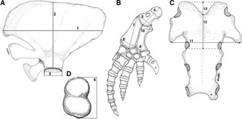
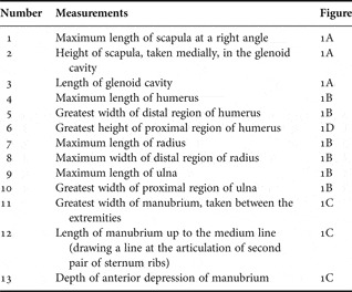
The sternum, scapula and the left pectoral flipper (humerus, radius and ulna) (Figure 1) were measured. The conventional linear measurements were taken according to Perrin (Reference Perrin1975), Menezes (Reference Menezes1998) and Simões-Lopes & Menezes (Reference Simões-Lopes, Menezes, Monteiro-Filho and Monteiro2008) (Table 1; Figure 1) with a 200 mm calliper (0.1 mm precision). In some incomplete samples, measurements were taken from the right hand side, since the appendicular skeleton is symmetrical.
Bone structures, showing the measurements used: (A) left scapula of Sotalia fluviatilis; (B) left flipper of Sotalia guianensis; (C) sternum of S. fluviatilis; (D) right humerus of S. guianensis in proximal view. Illustration of the flipper adapted from Simões-Lopes & Menezes (Reference Simões-Lopes, Menezes, Monteiro-Filho and Monteiro2008).

List of measurements of sternum and appendicular skeleton according to Perrin (Reference Perrin1975) and Menezes (Reference Menezes1998).

In order to eliminate ontogenetic variation only adult and subadult individuals previously classified according to the fusion stages of the cranial sutures (Dawbin et al., Reference Dawbin, Noble and Fraser1970; Ito & Miyazaki, Reference Ito and Miyazaki1990) and the vertebrae were considered (Perrin, Reference Perrin1975; Fettuccia & Simões-Lopes, Reference Fettuccia and Simões-Lopes2004). Individuals were considered sub-adults when they presented: (1) parts of the sutures mostly fused (occipital, basioccipital and pterygoid bones); (2) dental alveoli and intra-alveolar septa fully developed; and (3) most of the cervical and caudal vertebrae with fused epiphysis (showing the epiphyseal line slightly or completely invisible). The latter feature was included only if the vertebrae in the caudal and cervical regions fused before the thoracic and lumbar region (Lockyer & Goodall, Reference Lockyer and Goodall1988). In animals where most of the cervical and caudal vertebrae were already fused, the former features previously mentioned were observed. Considering the absence of osteological sexual dimorphism in Sotalia (Borobia, Reference Borobia1989, Monteiro-Filho et al., Reference Monteiro-Filho, Monteiro and Reis2002) all individuals were pooled for analysis (Table 2).
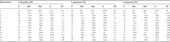
Descriptive statistics of postcranium of adults and sub-adults of Sotalia fluviatilis and Sotalia guianensis from northern (AM—INPA), north-eastern (CE—AQUASIS) and southern regions of Brazil (SC—UFSC). N, total number; Min, minimum; Max, maximum; X, average; SD, standard deviation; AM, Amazonas; CE, Ceará; SC, Santa Catarina. Measurements are in centimetres.

The morphometric comparisons were evaluated by a canonical variate analysis (CVA) (Past, available in: http://folk.uio.no/ohammer/past/) and Kruskal–Wallis analysis. The CVA has been used in studies of geographical variation and interspecific differentiation in order to eliminate the size variation of individuals within samples (Reis et al. Reference Reis, Pessôa and Strauss1990; Garavello et al., Reference Garavello, Reis and Strauss1991, Reference Garavello, Reis and Strauss1992). This is important considering the size difference between species, where S. guianensis is larger than S. fluviatilis.
The Kruskal–Wallis analysis was evaluated on BioEstat Program (5.0), free software. The Dunn test was performed a posteriori for multiple comparisons between groups (2–2).
RESULTS
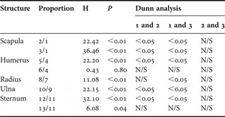
It was observed that the distal portion of the radius is delimited by three faces in the two species. Considering adults and subadults, the bones of forelimb are similar between the two species in the overall shape of humerus, radius and ulna. However, the width/length relationship of these three bones was larger in Sotalia fluviatilis (Table 3). The Kruskal–Wallis test did not show any significant difference between the species for the relationship between the height of proximal region and the length of humerus (Table 4).
Proportion of postcranium measurements for the samples analysed in adults and sub-adults of Sotalia fluviatilis and Sotalia guianensis from north (AM—INPA), north-east (CE—AQUASIS) and south of Brazil (SC—UFSC). Averages of the values proportionally greater in S. fluviatilis are highlighted in bold. N, total number, Min, minimum; Max, maximum; X, average SD, standard deviation; AM, Amazonas; CE, Ceará; SC, Santa Catarina. Measurements are in centimetres.

Results of the Kruskal–Wallis test for proportions of postcranium measurements of Sotalia fluviatilis and Sotalia guianensis. (1) S. fluviatilis (AM—INPA); (2 and 3) S. guianensis (CE—AQUASIS) and (SC—UFSC). N/S, no significant difference; AM, Amazonas; CE, Ceará; SC, Santa Catarina. Measurements as in Table 1.

The glenoid fossa of scapula was proportionally greater in S. fluviatilis in relation to its length and the maximum height. The measurements of scapula showed a significant difference between Sotalia guianensis and S. fluviatilis regarding the maximum length/height of scapula and the length of glenoid fossa (Tables 3 & 4). In Table 3 it is possible to note that the height of scapula is smaller in the north-eastern specimens (S. guianensis, Ceará), revealing a more triangular shaped scapula.
The general shape of sternum varied only slightly between the two species. Comparisons between the length and width of the manubrium indicate that this bone is proportionally larger in S. guianensis (Kruskal–Wallis: P < 0.01). The depth of the anterior depression of manubrium did not present any difference between the species. In both species a variable occurrence of foramina in different sites of sternum, and the development of lateral processes was observed. The length of the manubrium was considered subjective in the specimens where the manubrium was still not fused to the first sternebra, in which case the variation must be considered with caution.
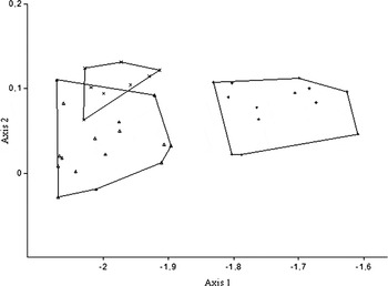
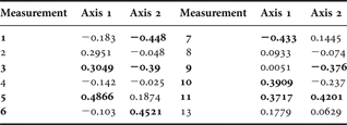
The CVA showed an evident separation between the marine and riverine species and an overlap of the marine populations (Figure 2). Axis 1 showed 87.2% of the variations and axis 2 showed 12.7% of the variations. The eigenvalues are presented in Table 5. In this analysis, 13 riverine specimens from Amazonas and 24 marine specimens (16 samples from Ceará and eight from Santa Catarina) were considered.
Projection of axis 1 and axis 2 of the canonical variate analysis based on the postcranium measurements in the three samples analysed (Amazonas (AM); Ceará (CE); Santa Catarina (SC)). Sotalia fluviatilis: AM (+); Sotalia guianensis: CE (Δ); SC (x).

Values of the canonical variate analysis of postcranium of Sotalia fluviatilis and Sotalia guianensis. Values in bold indicate the variates that best demonstrate the differences between the two species (measurements: (1) maximum length of scapula; (3) length of glenoid cavity; (5) greatest width of distal region of humerus; (6) greatest height of proximal region of humerus; (7) maximum length of radius; (9) maximum length of ulna; (10) maximum width of proximal region of ulna; (11) maximum width of manubrium). Measurement 12 was removed from the analysis.
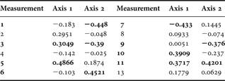
According to the CVA, the differences between species are mainly related to the maximum length of scapula (measurement 1: Table 4), length of glenoid fossa of scapula (measurement 3), greater width of distal region of humerus (measurement 5), greater height of proximal region of humerus (measurement 6), maximum length of radius (measurement 7), maximum width of proximal region of ulna (measurement10) and the maximum width of manubrium (measurement 11). The CVA confirms the differences previously observed in the proportions between the bones of the forelimb.
DISCUSSION
Morphological differences observed in the sternum and appendicular skeleton between the two species corroborate data from the literature for studies of the syncranium. Previous studies on cranium morphology (Monteiro-Filho et al., Reference Monteiro-Filho, Monteiro and Reis2002; Fettuccia, Reference Fettuccia2006; Fettuccia et al., Reference Fettuccia, da Silva and Simões-Lopes2009) and genetics (Cunha et al., Reference Cunha, da Silva, Lailson-Brito, Santos, Flores, Martin, Azevedo, Fragoso, Zanelatto and Solé-Cava2005; Caballero et al., Reference Caballero, Trujillo, Vianna, Barrios-Garrido, Montiel, Beltrán-Pedreros, Marmontel, Santos, Rossi-Santos, Santos and Baker2007) showed differences between the two species. Cunha et al. (Reference Cunha, da Silva, Lailson-Brito, Santos, Flores, Martin, Azevedo, Fragoso, Zanelatto and Solé-Cava2005) also presented molecular differences between marine populations along the coast of Brazil, therefore demonstrating that there are two distinct groups: one in the north-east and another in the south-east/south. Contrary to the expected, and with the exception of the scapula, the postcranium structures (humerus, radius, ulna and sternum) clearly showed an overlap between the populations in Ceará and Santa Catarina, thus showing that these structures do not differ between populations in the north-east and south of Brazil. This result confirms the findings by Monteiro-Filho et al. (Reference Monteiro-Filho, Monteiro and Reis2002) on the skulls of specimens of different areas from the Brazilian coast.
Morphological features of the humerus, radius and ulna of S. fluviatilis are comparable to the descriptions of S. guianensis made by Simões-Lopes & Menezes (Reference Simões-Lopes, Menezes, Monteiro-Filho and Monteiro2008). The distal portion of the radius delimited by three surfaces, a typical characteristic of S. guianensis in Delphinidae described by Menezes & Simões-Lopes (Reference Menezes and Simões-Lopes1996), was also observed in S. fluviatilis. Despite the morphological similarity, some measures (or proportions) showed significant difference between the two species.
Regarding the scapula, it is known that the proportions between the glenoid fossa and the maximum length vary throughout ontogenetic development (Menezes, Reference Menezes1998). Immature individuals of S. guianensis possess a glenoid fossa proportionally larger than mature individuals (Menezes, Reference Menezes1998). When comparing the two species, S. fluviatilis presents a glenoid fossa greater than S. guianensis, inferring that this is a juvenile character kept in the adult stages of the riverine species. According to Menezes (Reference Menezes1998), the glenoid fossa is wide in immature individuals and tends to get proportionally smaller throughout development. Menezes (Reference Menezes1998) noted that in S. guianensis specimens from the south of Brazil, the glenoid fossa in foetus is equivalent to 24% to 28% of the total length of the scapula. During the first year, this proportion decreases to 18% and down to 14% in mature animals. In this study, the percentage observed was 19% in adult specimens of S. fluviatilis and even lower percentages (16.6% to 15.7%) in S. guianensis for specimens from Ceará State and Santa Catarina State, respectively. Simões-Lopes & Menezes (Reference Simões-Lopes, Menezes, Monteiro-Filho and Monteiro2008) described the glenoid fossa in S. guianensis as oval and shallow. It is known that S. fluviatilis is smaller than S. guianensis with adult length of up to 1.52 m (da Silva & Best, Reference da Silva, Best, Ridgway and Harrison1994, 1996) and 2.20 m (Flores, Reference Flores, Perrin, Würsig and Thewissen2002) respectively. Thus, the fact that S. fluviatilis has a proportionally larger glenoid fossa suggests that this species requires a greater amplitude of movements to the humerus and consequently a wider range of movements to the flipper as a whole. In the sympatric species Inia geoffrensis (de Blainville, 1817) commonly known as pink river dolphin, the humerus is in contact with the glenoid cavity and with the sternum, so that almost the entire head of the humerus is fitted between these two bones. This position favours a wide movement of the flipper, therefore facilitating significantly during swimming (Klima et al., Reference Klima, Oelschläger and Wünsch1980). The fact that S. fluviatilis presents a larger glenoid fossa could be advantageous in terms of the ability to manoeuvre in the Amazonian environment that, as a submerged forest, is full of obstacles.
The sternum in both species showed to be extremely variable in terms of asymmetries, fenestrae and projections. The variation in the shape of the sternum between individuals, and even between genders, is known for many cetacean species (Arvy & Pilleri, Reference Arvy and Pilleri1977). However it is possible to note sufficient morphological differences for the distinction of the species (Arvy & Pilleri, Reference Arvy and Pilleri1977). According to Simões-Lopes & Menezes (Reference Simões-Lopes, Menezes, Monteiro-Filho and Monteiro2008) during ontogenetic development, the sternebra fuse together as a single piece, showing good variability and still maintaining features of diagnosable value. The sternum could be either perforated or not, present one or two lateral processes, be asymmetric or show great variation on its outline (Perrin, Reference Perrin1975; Arvy & Pilleri, Reference Arvy and Pilleri1977; Simões-Lopes & Menezes, Reference Simões-Lopes, Menezes, Monteiro-Filho and Monteiro2008).
In this study, the morphological analysis of the sternum and appendicular skeleton showed significant differences between the two species of Sotalia, including adaptive differences in relation to scapula of S. fluviatilis. These results provide basic information about these strutures for the first time for S. fluviatilis.
ACKNOWLEDGEMENTS
The authors thank Carlos Edward (UFAM), Fernando Rosas (INPA), Deise Nishimura and Daniel Pimpão for their helpful suggestions on improving the manuscript. Special thanks to Associação de Pesquisa e Preservação de Ecossistemas Aquáticos (AQUASIS). This work was supported by grants from CNPq and IEB (Beca Program).