INTRODUCTION
Invasions in marine habitats have been rapidly increasing and contributing to the alteration of ecosystem structure and functions, affecting baseline abiotic conditions and the resident biota (Grosholz, Reference Grosholz2002). In marine environments, ships and other structures have been recognized worldwide as the main source of fouling invaders being responsible for increasing of rates of species' introductions (Carlton, Reference Carlton1989; Carlton & Geller, Reference Carlton and Geller1993). The azooxanthellate sun coral Tubastraea Lesson, 1829 is a scleractinian of the family Dendrophylliidae. Tubastraea coccinea Lesson, 1829 is a more common coral with a circumtropical distribution (Cairns, Reference Cairns2000) and has been recognized as an invasive fouling species (Cairns, Reference Cairns2000; Castro & Pires, Reference Castro and Pires2001; Fenner, Reference Fenner2001; De Paula & Creed, Reference De Paula and Creed2004, Reference De Paula and Creed2005; Fenner & Banks, Reference Fenner and Banks2004; Sammarco et al., Reference Sammarco, Atchison and Boland2004).
Tubastraea coccinea and T. tagusensis Wells, 1982 were introduced into the south-western Atlantic on oil platforms (De Paula & Creed, Reference De Paula and Creed2004, Reference De Paula and Creed2005) where, since the late 1980s, they have been colonizing rocky shores on the south-east coast of Brazil (Castro & Pires, Reference Castro and Pires2001). These invasive corals have been observed overgrowing and competing with native organisms, such as sponges, ascidians, calcareous algae and native corals, including the endemic Mussismilia hispida (see Creed, Reference Creed2006). Highest densities have been observed in shallower waters, between 1.0 and 2.0 m depth (De Paula & Creed, Reference De Paula and Creed2005). Both introduced Tubastraea spp. have demonstrated the ability to rapidly expand their ranges along the south-western Brazilian coast. On these shores mean densities of 52.9 colonies m−2 for T. tagusensis and 25.5 colonies m−2 for T. coccinea have been reported, and a strong association between adult and recruitment densities has been found at small spatial scales (<1 m) (Creed & De Paula, Reference Creed and De Paula2007).
Studies of reproduction and larval biology are fundamental to a better understanding of the life histories and ecology of populations of scleractinians (Fadlallah, Reference Fadlallah1983; Harrison & Wallace, Reference Harrison, Wallace and Dubinsky1990). The successful establishment and spread of invaders increases with the number of release events and individuals released (Kolar & Lodge, Reference Kolar and Lodge2001). An extraordinary diversity of reproductive characteristics of scleractinians has been reported, including their mode and pattern of reproduction, gametogenesis, timing and early development (Szmant-Froelich et al., Reference Szmant-Froelich, Reutter and Riggs1985; Babcock et al., Reference Babcock, Bull, Harrison, Heyward, Oliver, Wallace and Willis1986; Szmant, Reference Szmant1986; Richmond & Hunter, Reference Richmond and Hunter1990; Harrison, Reference Harrison, Dubinsky and Stambler2011). The timing, frequency, duration, intensity and spatial distribution of these events determine the success of reproduction (Szmant, Reference Szmant1986).
According to Ayre & Resing (Reference Ayre and Resing1986) T. coccinea is a hermaphroditic coral and has been recognized as a brooding species in Hawaii (Edmondson, Reference Edmondson1929, Reference Edmondson1946; Richmond, Reference Richmond and Birkeland1997), in the Great Barrier Reef, Australia (Harrison, Reference Harrison1985; Ayre & Resing, Reference Ayre and Resing1986) and Costa Rica, Panamá and the Galápagos Islands (Glynn et al., Reference Glynn, Colley, Maté, Cortés, Guzman, Bailey, Feingold and Enochs2008). Most brooding species have multiple gametogenic cycles each year (Harrison & Wallace Reference Harrison, Wallace and Dubinsky1990).
These invasions gave us an interesting opportunity to study how two related species which have undergone differentiated selective pressures but today are in an identical environment react. Studies of these species in novel environments may reveal different reproductive strategies to the donor regions due to selection through the vector bottleneck, or we may find similar patterns. Because T. coccinea and T. tagusensis may be transported by vectors such as oil platforms, ship bottoms, dock pilings, buoys and other man-made objects (Cairns, Reference Cairns2000; Fenner, Reference Fenner2001; De Paula & Creed, Reference De Paula and Creed2004, Reference De Paula and Creed2005; Fenner & Banks, Reference Fenner and Banks2004; Sammarco et al., Reference Sammarco, Atchison and Boland2004) the expansion and maintenance of these alien populations could be increased by a successful reproductive strategy. Furthermore, the reproductive characteristics of these two invader species must be directly associated with their potential range expansion in newly colonized areas. Therefore, information about the reproductive biology of T. coccinea and T. tagusensis is fundamental to modelling these population expansions in the south-western Atlantic and to identifying their ability to successfully invade new areas.
The present study investigates the sexual reproduction of the invasive corals T. coccinea and Tubastraea tagusensis, where they occur sympatrically in the south-western Atlantic (due to a two-species invasion). We provide new information on gonad arrangement, sexual pattern, mode and cycle of reproduction, fecundity, gametogenesis, embryogenesis, planulation and settlement, as well as synchrony and timing of reproduction. We aim to enhance our current knowledge of scleractinian reproduction, in order to provide another piece in the jigsaw puzzle of information necessary to model colonization and range expansion of these deleterious invasive species, besides supporting control and management programmes that have been implemented.
MATERIALS AND METHODS
Sampling
Colonies of Tubastraea coccinea and T. tagusensis were collected by SCUBA diving, on rocky shores of Ilha dos Macacos (23°04′S 44°13′W) and Ponta Leste (23°03′S 44°14′W), Ilha Grande Bay (south-western coast of Brazil, 22°50′–23°20′S 44°00′–44°45′W). Four to six colonies of T. coccinea and T. tagusensis were sampled monthly from May 2000 to May 2001. The specimens were taken from the shallow subtidal to 4–7 m depth. Sea surface temperature varied from 21 to 32°C during the sampling period, and the tidal range in the region is 1.4 m (for more detailed description of the studied area, see De Paula and Creed, Reference De Paula and Creed2005). Colonies of 4–10 cm in diameter (10–40 polyps) were collected and were estimated to be >2 y (Authors unpublished data).
Histological procedures
Colonies were fixed in 10% formalin and three central polyps of three randomly chosen colonies of each species from each month were prepared for histological examination. The fragments were decalcified in a solution of 10% formic acid and 5% formaline solution. Tissues were washed in running tap water for 24 h, then dehydrated in an alcohol series and cleaned in xylene for routine paraffin embedding. Serial longitudinal or transversal 4.0–6.0 µm sections were obtained. Four to twelve slides were made from each block. Sections were stained using Mallory's triple stain (Pantin, 1848).
A gamete and embryo/planulae development classification for Tubastraea was adapted from Szmant et al. (1985) and Pires et al. (Reference Pires, Castro and Ratto1999). Measurements of different developmental stages of gametes, embryos and planulae were taken using an eyepiece micrometer on an Olympus BH2 microscope and a stereomicroscope Zeiss SV-6. The largest axes of oocytes were measured, when a nucleolus was present in the histological section. Photomicrographs were obtained using a Canon EOS Digital Rebel camera. The studied material was deposited in the Cnidaria collection of the Museu Nacional/Universidade Federal do Rio de Janeiro (MNRJ).
Gonadal arrangement and fecundity (number of oocytes per mesentery)
Colonies were collected monthly from August to December 2005, at depths of 1–3 m from Ponta Leste. Three central polyps of ten colonies from each month were chosen for histological preparation and 6 µm of two transverse serial sections were cut from different levels of the polyps, from the aboral portion of the polyps. In order to verify the number of oocytes per mesentery, new colonies were collected in 2005 (September, October and November 2005). These months were previously identified as reproductive peak periods, based in the examination of specimens collected from May 2000 to May 2001. Colonies were decalcified as above and one central polyp of ten colonies of each species, from each month, was selected and examined under the dissection stereomicroscope (Zeiss SV-6). Maximum oral disc diameter and distance between the oral and aboral ends were measured in all polyps, in order to correlate the number of oocytes per polyp with polyp volume and internal polyp area.
Planulation and larval development
Based on previously obtained data of the planulation periods of T. coccinea and T. tagusensis at Ilha Grande Bay in specimens collected in 2001–2002, four colonies (5–15 cm) of each species were collected in March 2006 from Ponta Leste and four additional colonies (5–7.5 cm) of T. coccinea were sampled from Arraial do Cabo (Ilha dos Porcos, 22°57′S 42°00′W). The latter locality is a second point of introduction with only T. coccinea and is located approximately 300 km to the north of the principal study site. Two colonies of each species were fixed in formalin and decalcified, as described above. Two colonies of each species were maintained alive in aquaria, and glass slides were disposed on the bottom for larval settlement. Planulae and embryos were observed under stereomicroscope and subsequently processed for histological analyses. Larval development was observed and photographed over two weeks. About 20–30 planulae were observed until all had either metamorphosed or died. Measurements and photomicrographs of different developmental stages of the planulae were obtained using a stereomicroscope Zeiss SV-6 and a Canon EOS Digital Rebel camera.
RESULTS
Reprodutive mode and pattern of sexual reproduction
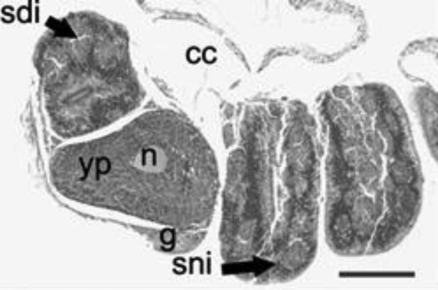
In the south-western Atlantic both Tubastraea coccinea and T. tagusensis presented the same gametogenic and development characteristics. Both species were considered simultaneous hermaphrodites—the female and male gametes occurred simultaneously in the same polyp (Figure 1). However, although most colonies showed only oocytes, none had only testes. Spermary was observed in 8.3% of T. coccinea colonies and in 5.5% colonies of T. tagusensis. Both species are brooders and the embryogenesis and the planulae development occur in the gastrovascular cavity. Tubastraea spp. oocytes, embryos and planulae observed in histological sections and by stereomicroscope (living and fixed in formalin) were yellow to red, and spermatic cysts were observed only in histological material. All examined colonies had sexual cells, suggesting a continuous gonad development.
Tubastraea coccinea (September 2000). Longitudinal histological section showing an hermaphroditic mesentery with spermaries and one oocyte. cc, coelenteric cavity; g, gastrodermis; n, nucleus; yp, yolk plates; sni, spermatogonia; sdi, spermatids. Scale bar: 100 µm.

Gametogenesis
The earliest stages of gamete development of both species were observed in the mesenterial gastrodermis. Oogenesis and spermatogenesis were classified into three approximate developmental stages, according to histological characteristics and relative sizes of oocytes and spermaries. Stage I represented the beginning of development, Stage II an intermediate step and Stage III represented mature cells. Gametogenesis was similar in both species. Overlapping cohorts of oocytes and spermatic cysts were observed in a single polyp. Gametogenesis and embryogenesis also overlapped.
OOGENESIS
Stage I
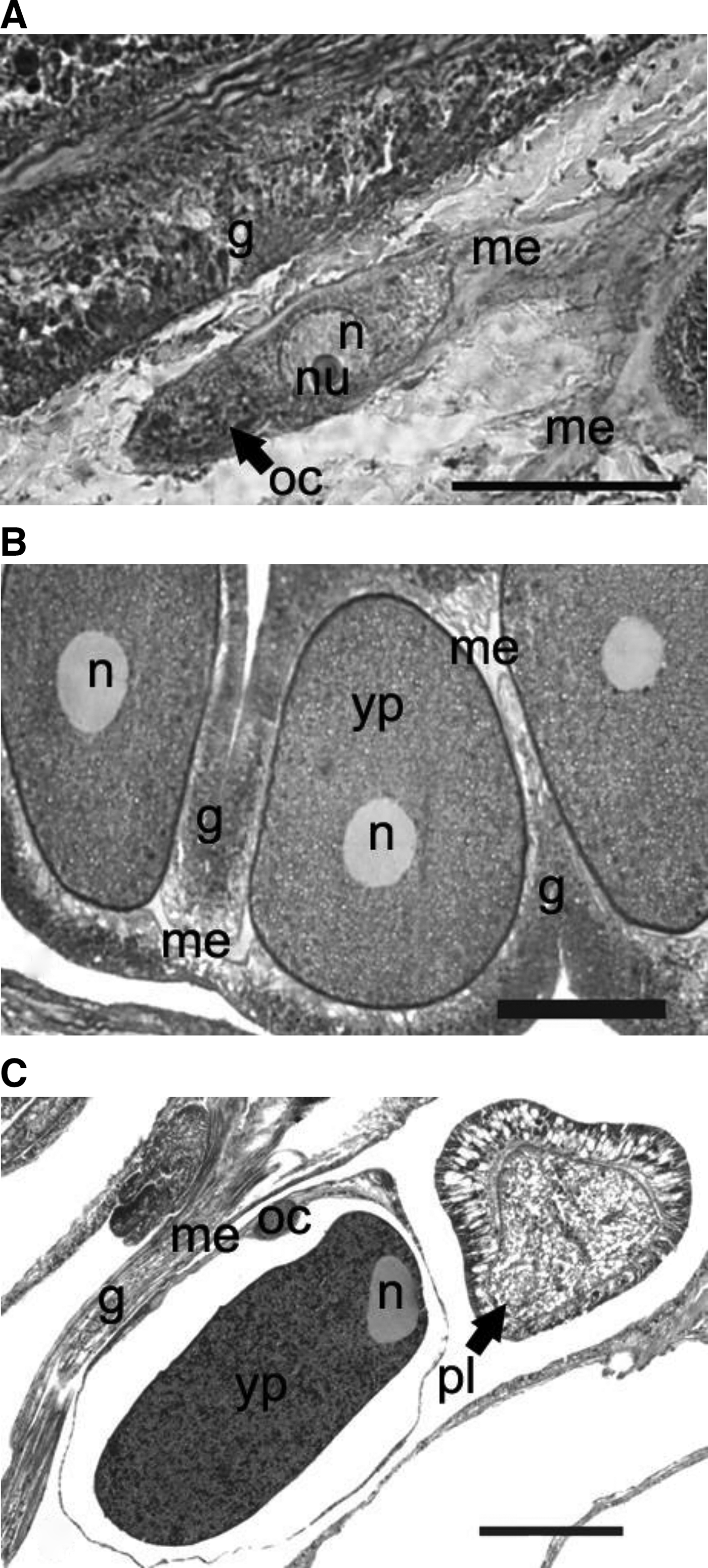
The early oogonia were characterized as differentiated elongated interstitial cells, which ranged from 10 to 150 µm in diameter and had an enlarged round or ellipsoidal nucleus (high ratio of cytoplasm to nucleus). The interstitial cells gradually migrated out from the gastrodermis to the blue mesogleal layer, where these cells were then recognized as ovoid or spherical oocytes. The oocyte's cytoplasm stained purple and usually presented a pinkish spherical nucleus with a bright-red nucleolus (Figure 2A).
Oogenesis of Tubastraea coccinea and Tubastraea tagusensis from Ilha Grande Bay, Rio de Janeiro, Brazil. Longitudinal histological sections. (A) Tubastraea coccinea (November 2003) oocyte Stage I. Small oocyte with a large nucleus: cytoplasm ratio, inside the mesoglea; (B) Tubastraea coccinea (May 2000) oocyte Stage II. Vitellogenic oocyte with a spherical nucleus located centrally, homogeneous cytoplasm; (C) Tubastraea tagusensis (January 1998) oocyte Stage III. Mature female gamete with numerous lipid vesicles and a peripheral nucleus. A transverse section of a late gastrula is also seen. oc, oocyte; g, gastrodermis; me, mesoglea; n, nucleus; nu, nucleolus; yp, yolk plates; pl, planulae (late gastrula). Scale bars: A, 8 µm; B, 100 µm; C, 130 µm.
Stage II
Oocytes in Stage II were surrounded by mesogleal layer, and had a denser cytoplasm, which stained reddish, with an increased amount of yolk vesicles. A purple stained central nucleus contained an orange stained nucleolus. The measurements of oocytes ranged from 130 to 500 µm (Figure 2B).
Stage III
Oocytes in Stage III enlarged in volume due to the yolk deposition. As oocytes grew, the nucleus gradually migrated towards the peripheral portion of the cell. Mature oocytes reached their full size with nucleus and nucleolus located at the outer portion of the cell, close to the plasma membrane. The cytoplasm stained orange–red, due the presence of numerous lipid and yellow yolk vesicles. The prominent nucleus stained a light purple (Figure 2C). Some oocytes equivalent in size to Stage III had a degenerated appearance (without a defined outline and few cytoplasm), seemed to be reabsorbed among other mature oocytes and the phagocytes stained purplish. This phenomenon is quite common and was previous described in other papers (Harrison & Wallace, Reference Harrison, Wallace and Dubinsky1990). Successful oocytes may obtain nutrients from these ‘ghost cells’ through phagocytosis (Rinckevich & Loya, 1979). Stage III oocytes had diameters that ranged from 400 to 1.252 µm. The maximum diameter of oocyte in Stage III of T. tagusensis (1252.5 µm) was greater than T. coccinea (901.8 µm).
SPERMATOGENESIS
Stage I
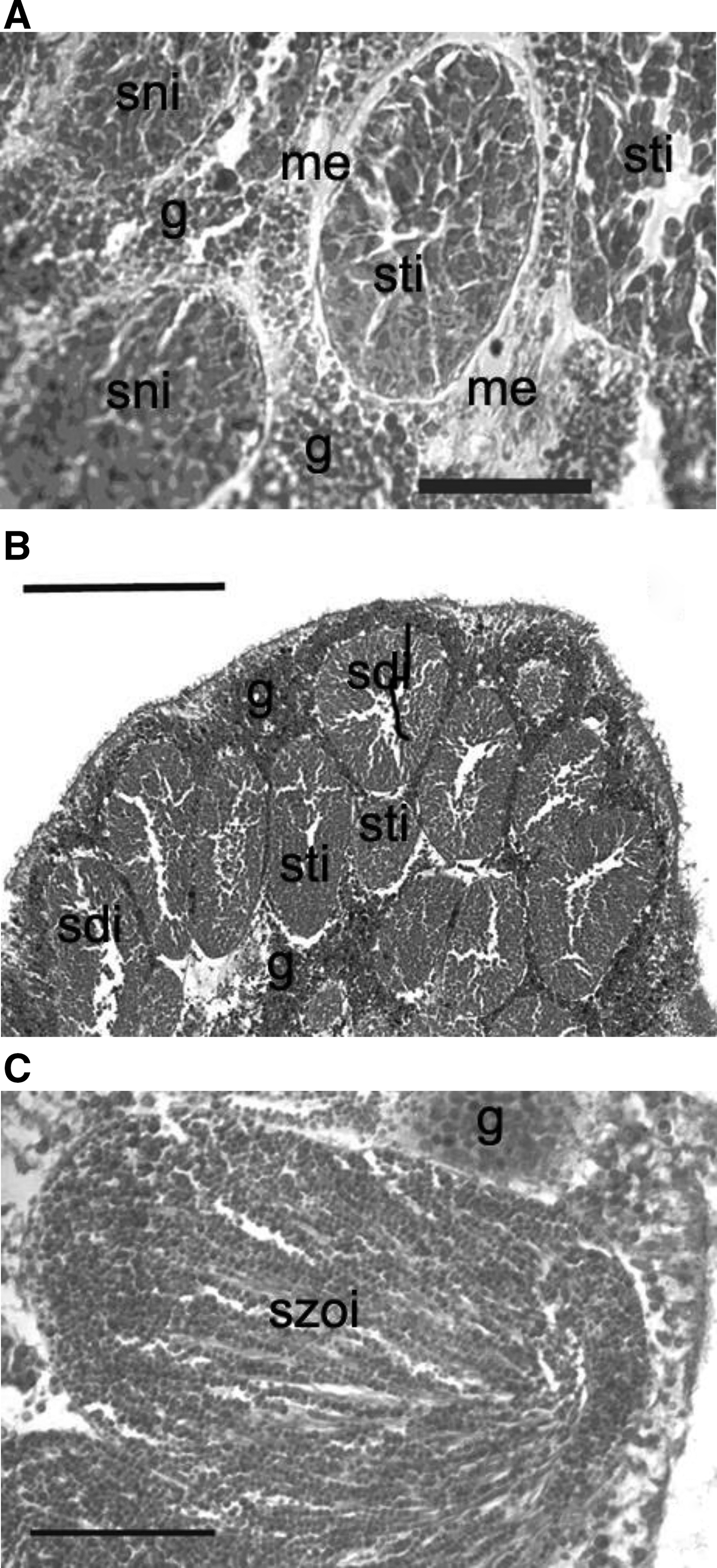
Primary spermatic cysts were formed as small clusters of interstitial cells in the gastrodermis. They stained dark-purple, ranging from 32 to 65 µm in the largest diameter (Figure 3A).
Spermatogenesis of Tubastraea coccinea from Ilha Grande Bay, Rio de Janeiro, Brazil. Longitudinal histological sections. (A) Spermatic cyst I (November 2003). Immature cells densely packed located in the gastrodermis; (B) spermatic cyst II (September 2000). Spermatids can be observed centrally where a cavity was formed; (C) spermatic cyst III (November 2003). Cluster of spermatozoa showing a bouquet-like arrangement of sperm. g, gastrodermis; me, mesoglea; sni, spermatogonia; sdi, spermatids; sti, spermatocytes; szoi, spermatozoa. Scale bars: A, 22 µm; B, 130 µm; C, 160 µm.
Stage II
Spermatocytes increased in number and size, and spermatic cysts ranging from 60 to 300 µm in diameter. They started to concentrate and presented a granular texture. Cysts in Stage II usually presented a central space, without cells and stained dark-pink to magenta (Figure 3B). During further development these spaces joined together to form eventually one lumen in the central region of the cyst. At this stage some tails were visible in the interior of the lumen.
Stage III
Mature cysts presented aggregates of spermatozoa with round nuclei and visible flagella. The spermaries ranged from 270 to 700 µm in diameter and stained pink to red. As the number of visible tails increased, they concentrated in small clusters that were arranged radially in the cysts. The typical arrangement of mature cysts had the appearance of ‘bouquets’ (Figure 3C). Nuclei were smaller than those observed previously.
Gonad arrangement and fecundity
Both species had the same arrangement of gonads in the mesenteries. Gonads were observed in complete and incomplete mesenteries, between the mesenterial filaments and the retractor muscle bands. Gonads were concentrated at the aboral portion of the polyp. Female gonads developed as single strings of cells in the mesoglea of each two adjacent mesenteries (one pair) and could be easily distinguished in dissections. Male gonads were only observed through histology and the spermaries were always adjacent to the female cells (Figure 1). Tubastraea coccinea polyps had 36–48 mesenteries and T. tagusensis had 36 mesenteries. Both species presented 12 complete pairs of mesenteries.
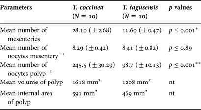
Although no significant differences in average values of number of oocytes per mesenteries was observed in the two studied species (T. coccinea, 8.29 ±0.42 SE and T. tagusensis, 8.41 ±0.82 SE, Student's t-test P = 0,89), the mean values of mesenteries (complete and incomplete) per polyp of T. coccinea was greater (28.1 ±2.68 SE) than T. tagusensis (11.6 ±0.47 SE). Therefore the total number of oocytes per polyp of T. coccinea was higher (245.5 ±30.29 SE) than T. tagusensis (98.7 ±10.13 SE) (Table 1). Only T. tagusensis had a positive correlation between oocyte number and polyp volume (Spearman's r = 0.73, N = 10, P = 0.016) or polyp area (Spearman's r = 0.73, N = 10, P = 0.016) (Table 1).
Fecundity parameters of Tubastraea coccinea and Tubastraea tagusensis examined in colonies under dissection (September–November 2005) in Ilha Grande Bay, Brazil.
*, Student's t-test; **, Mann–Whitney Wilcoxon; nt, not tested
Embryogenesis and planulae
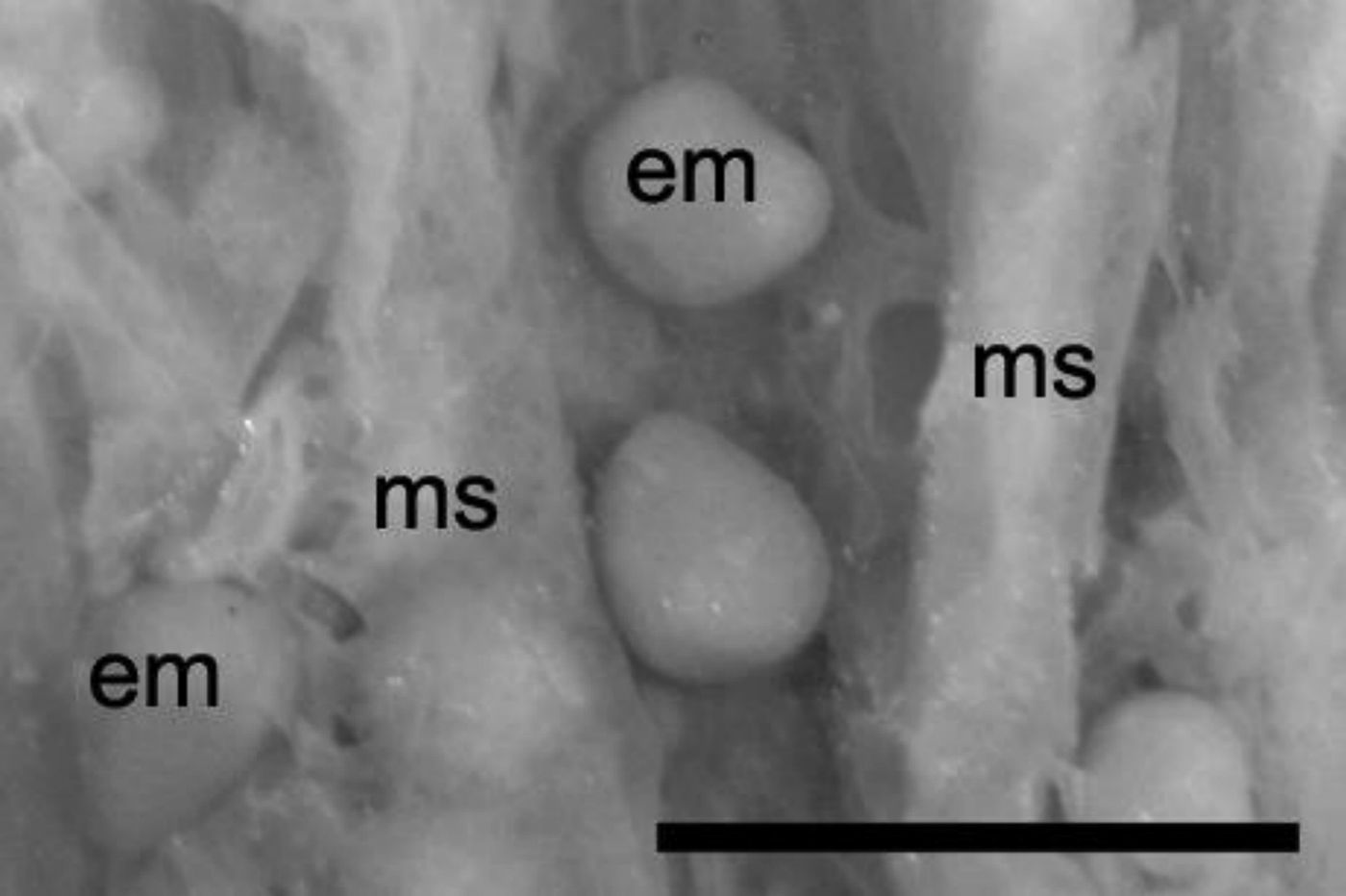
Embryos and planulae were found in the gastrovascular cavity, observed using a stereomicroscope without histological stain (Figure 4) at the base of the polyps. Embryos presented the same staining properties as mature oocytes seen in the histological preparations (Figure 5). Planulae were also observed close to the mouth aperture.
Tubastraea tagusensis (April 2001). Dissected material showing embryos inside the coelenteric cavity. em, embryos; ms, mesentery. Scale bar = 2 mm.
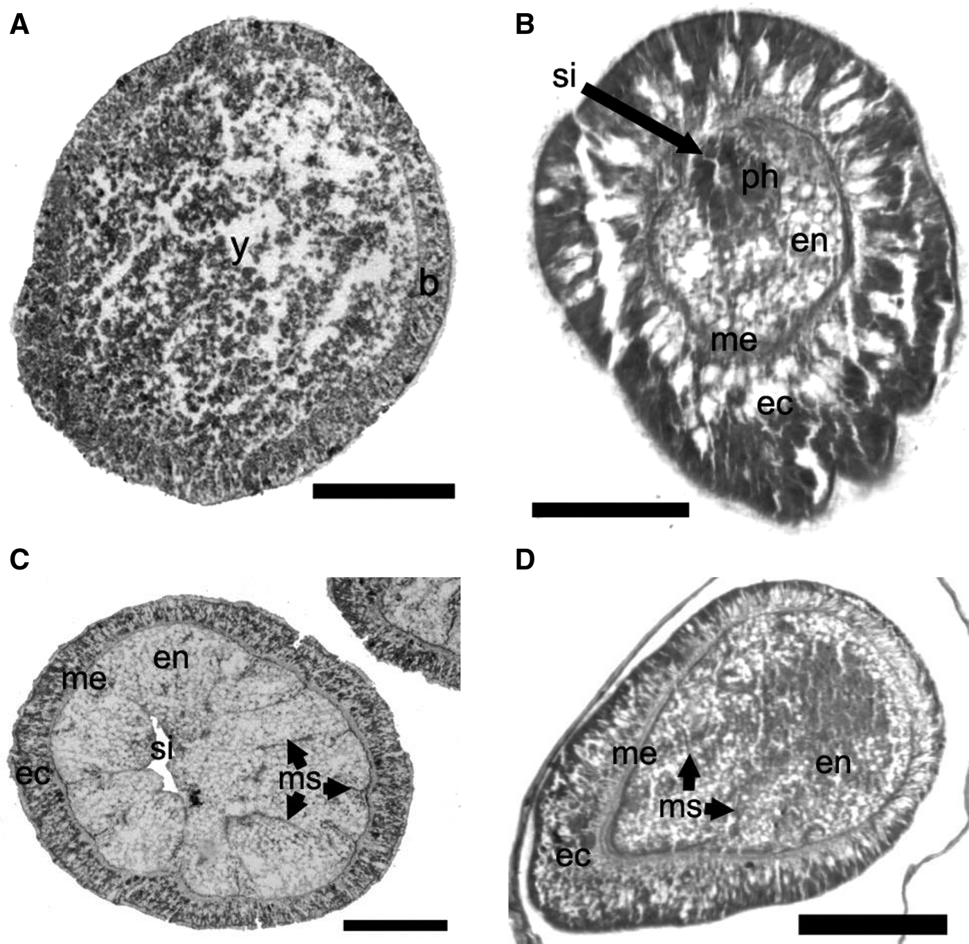
Embryonic and planula development of Tubastraea spp. (A) Stereoblastula (early stage). Cleavaged superficial layer surrounds the central yolk mass; (B) stereogastrula. The ectoderm was distinct from the endoderm separated by the mesoglea. The stomodeal invagination is also shown; (C) early planulae transverse section. Mesenterial filament has begun to differentiate; ectodermal cells have begun to multiply; (D) early planulae longitudinal section. Ectoderm and endoderm layers were similar to those of planulae at more advanced stages. b, blastoderm; y, yolk; me, mesoglea; ec, ectoderm; en, endoderm; ph, pharynx; si, stomodeal invagination; ms, mesentery. Scale bars: A–C, 62 µm; D, 250 µm.
Embryonic development was also subdivided into three stages. Stage I. Early-stage embryos were stereoblastula (solid and lacked blastocels) or blastula-like. They presented a poorly defined cellular layer covering an indistinct yolky mass dispersed in the cytoplasm and were smaller than mature oocytes (180–400 µm). Three cell layers became visible surrounding a central yolk mass (Figure 5A). Stage II. Embryos began to grow and the red-staining yolk material was more concentrated in the cytoplasm. The ectoderm layer was clearly differentiated (cells distinctly columnar in shape) and a defined mesogleal layer was separate from the endodermal mass. Diameters of embryos in Stage II ranged from 180 to 600 µm, and some cnidocysts were seen at the ectoderm. The stereogastrula were oval in shape and had developed an oral pore and an initial gastrovascular cavity. Differentiation of the stomodeum had begun, with the proliferation of ectoderm cells and their invagination towards the center of the embryo (Figure 5B). Stage III. The embryos began to elongate and resembled early stage planulae. The early planulae had a mesentery differentiation, which started via the apposition of endoderm cells and afterwards with the formation of the mesenterial filaments (Figure 5C, D).
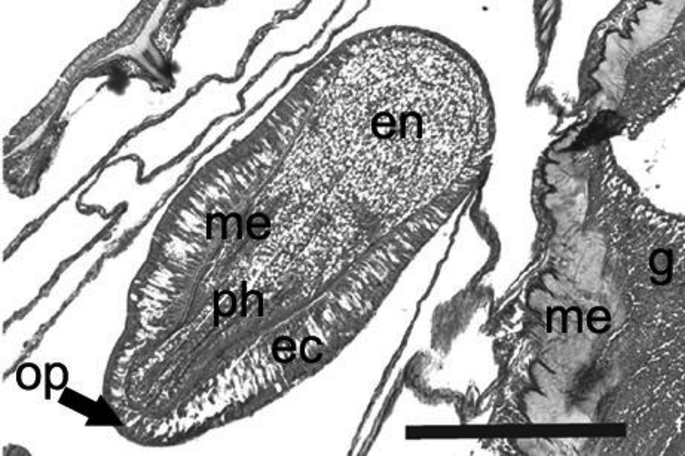
Early larvae had a well-developed gastrovascular cavity and an ectoderm and endoderm layers similar to those of planulae at more advanced stages. The developing planulae found in the gastrovascular cavity had completed ontogenesis, with a differentiated stomodeum and pharynx. Planulae from the inside the polyps showed a gastrovascular cavity divided by mesenteric septa with filaments and their largest axes ranged from 1 to 2.5 mm (Figure 6).
Tubastraea tagusensis (January 1998). Planulae close to the mouth showing oral pole and pharynx. The coelenteric cavity divided into compartments by the mesenteric septa. g, gastrodermis; me, mesoglea; ec, ectoderm; en, endoderm; ph, pharynx; op, oral pole. Scale bar: 500 µm.
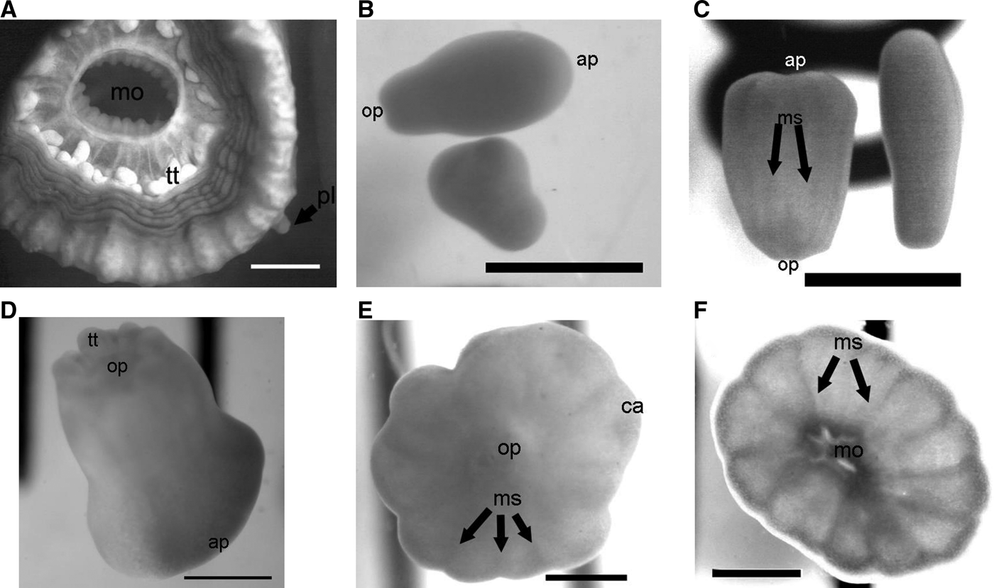
The number of mesenteries continued to increase and usually six complete pairs were present by the time planulation began (Figure 7).
Tubastraea tagusensis (March 2006). Larval release, settlement and recruit metamorphosis. Aspects of morphology and metamorphosis in larval development were observed in aquaria by stereomicroscope. (A) Planula fixed on oral disc of parental polyps; (B) semi-contracted, drop-shaped swimming planula; (C) cylindrically-shaped swimming planula and a contracted-shape with visible mesenteries; (D) a pear-shaped planula, with visible tentacles; (E) 2-d-old recruit with oral pole and calcareous base; (F) 5-d-old polyp, with visible mouth and mesenteries; pl, planulae; mo, mouth; tt, tentacles; op, oral pole; ap, aboral pole; ms, mesentery; ca, calcareous base. Scale bars: A, 3 mm; B,C, 1 mm; D–F, 0.5 mm.
Planulation, settlement and metamorphosis
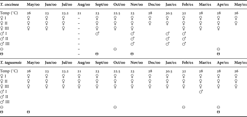
Field and laboratorial observations of planulae release indicated that it occurred at various times, with apparent preference for night periods. Colonies of T. tagusensis were observed releasing planulae in the field in April 2001, in the laboratory in March, April and August 2006, and histological sections indicated the presence of planulae in samples from January 1998 and August 2005. Colonies of T. coccinea were observed releasing planulae in the field in July 2003 and November 2000, in the laboratory in March, April, July and August 2006, and planulae were observed in histological sections of samples collected in May 2000 and September 2000 (Table 2).
Summary of gamete stages, planulae and embryo presence in histological preparations of Tubastraea coccinea and T. tagusensis (May 2000 to May 2001) from Ilha Grande Bay, Brazil. ♀, oocytes; ♂, cysts; ⊖, embryos; Θ, planulae; (–), not collected.
In field and in aquaria, planulae were released through the mouth. They were yellow to red, measuring between 1 and 3 mm in length and were seen moving over the colony and swimming close to the water surface. Some planulae fixed to parental polyps close to their oral disc areas after release. They stayed in these areas for a few hours and returned to the water column (Figure 7A). After release, larvae were buoyant and were observed mostly on the water surface. The planulae were initially drop shaped (Figure 7B), but sometimes became temporarily more cylindrical (sausage shaped) (Figure 7C). These phases presented active swimming, through ciliary movement. They also showed rotation around the oral–aboral axis. A bifurcation at the planulae aboral end was also observed (Figure 7B). After swimming throughout the water column, planulae explored the bottom and settled. Planulae development and metamorphosis were completed on average in three to seven days. When they started swimming near the bottom the planulae were pear-shaped and were larger at the base. A pore could be observed on the more flattened side (Figure 7D). Some planulae did not settle after the planulation cycle was finished, but remained on the bottom and seemed to metamorphose but did not fix and died.
The planulation in aquaria lasted about one week, with a peak liberation in the third and fourth days and planulae continued swimming for about 18 d. Settlement was observed on the horizontal glass bottom and vertical sides of the aquaria. The calcareous basal plates appeared 2 d after settlement (Figure 7E). A complete new polyp was observed 3–5 d after settlement (Figure 7F). Some settled metamorphosed recruits, if disturbed, were capable of detaching, swam again and reattached on the substrate.
Temporal patterns of reproduction
GAMETOGENIC CYCLES
Gametogenesis was synchronous among colonies of the same species, along the continuous reproductive cycles. A subtle asynchrony was observed between T. coccinea and T. tagusensis in relation to the presence of cysts and the distribution of different stages of oocytes (Table 2). However, embryos were seen in both species in October 2000 and April 2001 and planulae were present in colonies of T. coccinea and T. tagusensis collected in April 2001.
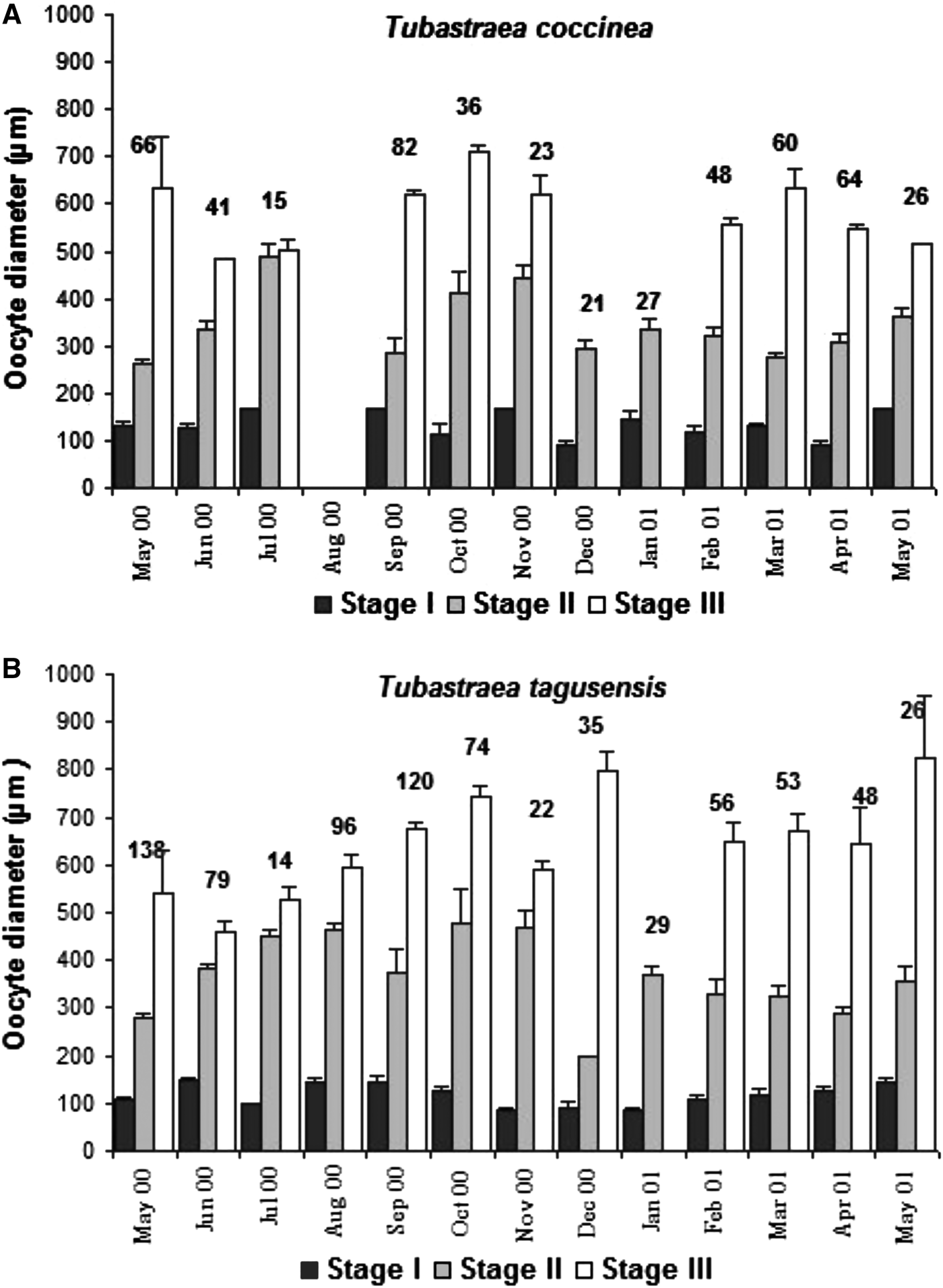
Tubastraea coccinea presented two peaks in oocyte diameters: September–November and February–April (Figure 8). Embryos were seen in colonies in October 2000 and April 2001. Planulae were present in colonies collected in August 2006, September 2000, November 2000 and April 2001 (Table 2).
Oocyte size (diameter) distribution in different stages of development (I, II and III) sampled monthly throughout the year (2000–2001). Data are mean ±SE. Values above bars indicate the number of oocytes on three stages observed in each month.
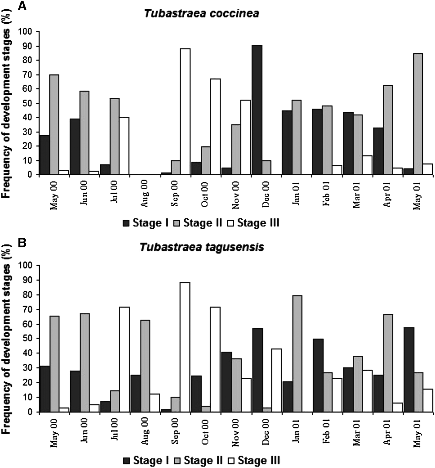
Spermaries were observed in September 2000, November 2000 and January and February 2001 (Table 2). In the same period that oocyte diameters increased in size (September–November) the abundance of mature oocytes (Stage III) was higher (Figure 9) and colonies collected in December 2000 and January 2001 lacked mature oocytes (Table 2).
Frequency of Tubastraea coccinea and T. tagusensis oocytes in different stages (I, II and III) May 2000–May 2001.
Tubastraea tagusensis also presented an increase in size of female gonad cells in September which extended until December (Figure 8), when oocytes were largest and an increase in abundance of mature oocytes was found (Figure 9). Another period of larger oocytes occurred from February to May (Figure 8). Planulae were observed in April 2001, May 2001 and August 2005 and 2006, and the spermaries occurred in March 2001 (Table 2). Although planulae and spermaries had not been observed throughout September–December, embryos were found in October 2000 and February and April 2001 and only immature oocytes Stages I and II) were found in January 2000 (Table 2).
The two observed peaks of growth of Tubastraea oocytes suggest the occurrence of at least two reproductive periods per year, and the occurrence of several planulation events in periods of three to four months (September–December and February–May). Moreover, Tubastraea tagusensis had precocious reproduction, since oocytes II were observed in 3-month-old individuals with only three polyps.
OOCYTE STAGE FREQUENCY
The percentage of each oocyte stage differed along the year in T. coccinea and T. tagusensis (Figure 9). Tubastraea coccinea had more mature oocytes (Stage III) from September to November 2000 (Figure 9) and in December 2000 no stage III oocytes were observed (Figures 8, 9). Tubastraea tagusensis presented higher percentages of mature oocytes in July, September and October 2000 (Figure 9). In January of 2001 no Stage III oocytes were observed (Figures 8, 9).
DISCUSSION
The quick expansion and establishment of Tubastraea through the Caribbean and in Brazil seems be due to the same characteristics as those suggested by Fine et al. (Reference Fine, Zibrowius and Loya2001) for Oculina patagonica, another scleractinian coral, which has successfully invaded the Mediterranean (Zibrowius, Reference Zibrowius1992): (1) ability to reproduce using different strategies; (2) high production of offspring; (3) early reproductive age; and (4) high growth rate. De Paula & Creed (unpublished data) observed that Tubastraea grew faster and had a higher rate of increment in number of polyps by budding. Additionally, invasive Tubastraea species studied here seem to be able to exploit seasonal opportunities in local environmental conditions. They can reach very high densities, of more than 50 colonies 0.25 colonies.m−2 (De Paula & Creed, Reference De Paula and Creed2005), probably because their reproductive features allow the production of a great number of offspring.
Despite the circumtropical distribution of Tubastraea coccinea in tropical shallow waters (Cairns, Reference Cairns2000) and its recognized ability to invade and quickly spread in new habitats (Cairns, Reference Cairns2000; De Paula & Creed, Reference De Paula and Creed2004, Reference De Paula and Creed2005; Fenner & Banks, Reference Fenner and Banks2004; Sammarco et al., Reference Sammarco, Atchison and Boland2004), studies or reports of the reproductive biology of T. coccinea are limited (Glynn et al., Reference Glynn, Colley, Maté, Cortés, Guzman, Bailey, Feingold and Enochs2008), and this study represents the first investigation of the reproductive biology of T. tagusensis. Some information about mode, patterns and seasonality of reproduction of T. coccinea are available (Edmondson, Reference Edmondson1929, Reference Edmondson1946; Harrison, Reference Harrison1985; Ayre & Resing, Reference Ayre and Resing1986; Richmond, Reference Richmond and Birkeland1997; Glynn et al., Reference Glynn, Colley, Maté, Cortés, Guzman, Bailey, Feingold and Enochs2008).
Tubastraea coccinea and T. tagusensis are simultaneous hermaphrodites and brooders. Balanophyllia europea, which also belongs to Dendrophylliidae, has a similar strategy (Goffredo & Teló, Reference Goffredo and Teló1998). According to Goffredo et al. (Reference Goffredo, Radetić, Airi and Zaccanti2005) the reproductive mode and trends in sexuality of dendrophylliids should be gonochorism and brooding, since broadcasting was found in 39% and hermaphroditism in 27% of the species studied in this family.
Among a total of 444 scleractinian species cited in Harrison (Reference Harrison, Dubinsky and Stambler2011), 61 brood embryos and planulae, including T. coccinea (a synonym for T. aurea), corresponding to Dendrophylliidae corals are cited 23 species, four are hermaphroditic and ten brood planulae. Ayre & Resing (Reference Ayre and Resing1986) also considered T. coccinea as a hermaphroditic species. Harrison (Reference Harrison1985) reported that both modes of reproduction were observed within a population of T. aurea in the Great Barrier Reef. In Brazil, T. coccinea and T. tagusensis are hermaphrodites, however, male and female gametes occurred together only in 8.3% of T. coccinea colonies and in 5.5% colonies of T. tagusensis examined. A similar proportion of hermaphroditic colonies were recently found in T. coccinea (12.9%) from the equatorial eastern Pacific (Glynn et al., Reference Glynn, Colley, Maté, Cortés, Guzman, Bailey, Feingold and Enochs2008). Due to the reduced presence of spermaries in both species, the observation of embryos and planula at different times and the evidence of asexual or parthenogenetic production of planulae, as registered by Ayre & Resing (Reference Ayre and Resing1986) for T. coccinea, asexual reproduction may be important for the quick colonization and range expansion of Tubastraea along the Brazilian coast.
Sperm of hermaphroditic coral species may be endowed with greater swimming potential to assist sperm movement away from eggs of the same coral to avoid self-fertilization and promote outcrossing (Harrison & Wallace, Reference Harrison, Wallace and Dubinsky1990). This does not seem important in T. coccinea which can brood asexually-produced planulae (Ayre & Resing, Reference Ayre and Resing1986) and, like a simultaneous hermaphrodite, can self-fertilize, an advantageous strategy for quickly colonizing new areas when few colonies (potential partnerships) are available (Tomalison, Reference Tomalison1966).
The two observed peaks of growth of oocytes and the presence of embryos/planulae suggest the occurrence of two reproductive periods during the studied period (September–December and February–May). Gametogenic processes in corals are generally cyclic and species that brood planulae usually have multiple gametogenic cycles (Harrison & Wallace, Reference Harrison, Wallace and Dubinsky1990). Gametogenesis is usually synchronized within each colony or solitary coral, and at least partially among members of breeding populations (Harrison & Wallace, Reference Harrison, Wallace and Dubinsky1990). The seasonal production of planulae may be the result of selection under conditions that maximize the number of juveniles that grow to sexual maturity (Fadlallah & Pearse, Reference Fadlallah and Pearse1982). Some studies suggest that the reproductive cycle is mainly regulated by seasonal variations in water temperature and photoperiod (Babcock et al., Reference Babcock, Bull, Harrison, Heyward, Oliver, Wallace and Willis1986; Oliver et al., Reference Oliver, Babcock, Harrison and Willis1988; Harrison & Wallace, Reference Harrison, Wallace and Dubinsky1990). The period during which germ cells are released varies geographically (Harrison & Wallace, Reference Harrison, Wallace and Dubinsky1990; Richmond & Hunter, Reference Richmond and Hunter1990). Comparison of the timing of gamete/planulae release among localities within coral species may reveal population responses to different environmental conditions (Oliver et al., Reference Oliver, Babcock, Harrison and Willis1988; Harrison & Wallace, Reference Harrison, Wallace and Dubinsky1990; Babcock et al., Reference Babcock, Wills and Simpson1994). Glynn et al. (Reference Glynn, Colley, Maté, Cortés, Guzman, Bailey, Feingold and Enochs2008) observed great variation in the release of planulae of T. coccinea between Panama and the Galapagos Archipelago: higher fecundity was estimated during the wet season in Panama and a significant decline in planulae was observed during the cool season in the Galapagos Archipelago. Here too the release of planulae may be influenced by temperature, as an inflow of cold and nutrient-rich South Atlantic Central Waters (SACW) occurs during the summer in Ilha Grande Bay.
The high abundance of mature oocytes (Stage III) in T. coccinea and the presence of planulae during the period from September to November 2000 and from February to April 2001 suggested a concentration of planulation events at these times. This evidence is also supported by the absence of mature oocytes in colonies collected on December 2000 and on January 2001. The presence of planulae in samples of T. tagusensis collected in April 2001, May 2001 and the occurrence of immature spermary on March 2001 indicated planulation events in this period. Planulae and spermaries were not observed throughout September–December. Embryos were found in October 2000 and in February and April 2001, and only immature oocytes (Stages I and II) were found in January 2000, suggesting that planulae release had already occurred by January 2000. All colonies of both species presented ripe gonads, and the number of oocytes examined per month, per polyp was also high in both: T. tagusensis, with a maximum number of 138 in May 2000 and T. coccinea with 82 oocytes in samples collected in September 2000.
Tubastraea produced large eggs, with T. coccinea close to values observed in the eastern Pacific (Glynn et al., Reference Glynn, Colley, Maté, Cortés, Guzman, Bailey, Feingold and Enochs2008). The mean number of mesenteries polyp−1 of T. coccinea was greater than in T. tagusensis and similar to that found in T. coccinea by Glynn et al. (Reference Glynn, Colley, Maté, Cortés, Guzman, Bailey, Feingold and Enochs2008). The total number of oocytes polyp−1 of T. coccinea was greater than the values found by Glynn et al. (Reference Glynn, Colley, Maté, Cortés, Guzman, Bailey, Feingold and Enochs2008), however, these authors only counted mature oocytes. The number of oocytes mesenteries−1 observed in T. coccinea and T. tagusensis were similar to those observed in T. coccinea from the eastern Pacific (Glynn et al., Reference Glynn, Colley, Maté, Cortés, Guzman, Bailey, Feingold and Enochs2008). The same high fecundity has been reported in other scleractinian coral species that occur in Brazil, but Siderastrea stellata produces lower numbers of small oocytes (Lins de Barros et al., 2003), and Mussismilia braziliensis, despite producing an equivalent number of oocytes per mesentery, presented a lower number of oocytes per polyp and, consequently, per colony (Pires et al., Reference Pires, Castro and Ratto1999, Reference Pires, Segal and Caparelli2010) than other aquatic invasive species such as O. patagonica and Dikerogammarus haemobaphes (see Fine et al., Reference Fine, Zibrowius and Loya2001; Bacela et al., Reference Bacela, Konopacka and Grabowski2009).
Both species in the south-western Atlantic developed large planulae, competent for 18 d. Paz-García et al. (Reference Paz-García, Reyes-Bonilla, Gonzaléz-Peralta and Sánchez-Alcántara2007) observed brilliant yellow T. coccinea planulae, between 3 and 5 mm long, moving around the parental polyp. Harrison & Wallace (Reference Harrison, Wallace and Dubinsky1990) reported that T. coccinea produces large planulae (approximately 1 mm diameter or long) through internal fertilization, that can remain competent to settle for 3–14 d in the water column. Edmondson (Reference Edmondson1929, Reference Edmondson1946) described Dendrophyllia manni planulae (synonym of T. coccinea) as elongate–oval in shape and about 2 mm in length. Like an adult polyp, the planula is brightly coloured, ranging from orange to brick-red. The planulae of T. coccinea and T. tagusensis here examined were larger than Balanophyllia europaea (2.15 mm) and smaller than B. elegans (3–5 mm), which are other brooder corals that belong to the same family (Fadlallah & Pearse, Reference Fadlallah and Pearse1982; Goffredo & Zaccanti, Reference Goffredo and Zaccanti2004). Another Dendrophylliidae, Leptopsammia pruvoti had elongated and cylindrical orange–yellow planulae, with a maximum diameter ranging from 695 to 1595 µm (Goffredo et al., Reference Goffredo, Radetić, Airi and Zaccanti2005). Field and laboratory observations indicate planulae release at various times, with an apparent preference for the night, as observed in T. coccinea by Glynn et al. (Reference Glynn, Colley, Maté, Cortés, Guzman, Bailey, Feingold and Enochs2008) in the eastern Pacific.
In aquaria many planulae of both species settled a few hours after release, but mean settlement time was 3–5 d. Glynn et al. (Reference Glynn, Colley, Maté, Cortés, Guzman, Bailey, Feingold and Enochs2008) observed that T. coccinea planulae settlement occurred from 1 to 3 d. In the laboratory planulae of Balanophyllia elegans attached to settling plates and began metamorphosis in 3 d (Gerrodette, Reference Gerrodette1981). Settlement-competency periods of 1–7 d are typical of tropical corals (Edmondson, Reference Edmondson1946). Tubastraea coccinea and T. tagusensis larvae had a competency period in aquaria of 18 d and settled close to the parental colony. The same occurs in the brooded larvae of Favia fragum, which also settle near parents (Carlon, Reference Carlon2002). A strong correlation between adult field density and recruitment density of Tubastraea coccinea and T. tagusensis at scales of less than 1 m has been demonstrated by Creed & De Paula (Reference Creed and De Paula2007). Glynn et al. (Reference Glynn, Colley, Maté, Cortés, Guzman, Bailey, Feingold and Enochs2008) also registered that T. coccinea typically aggregated spatially, with numerous small individuals around a few large adult colonies. We observed newly settled individuals very close to parental colonies. This observation and the pattern of aggregation around parental colonies in the field suggest that, despite the competency period of 18 d, the planktonic phase may be extremely short, with minimal settlement distances. This information is important as we can cross adult and recruitment densities in models of temporal and spatial patterns of larval recruitment and thus predict the potential size of future populations. Dispersal affects the amount and frequency of genetic exchange, and it may also have important consequences for an invasive species in establishment and spread. Larvae of B. elegans dispersed a mean distance of <0.5 m before settlement, indicating that its larval dispersal in the family may be very limited. However, since limited dispersal is neither a necessary nor a sufficient condition for aggregation, inferences about patterns of settlement cannot be made based on spatial distribution of adult colonies alone (Gerrodette, Reference Gerrodette1981).
Brooding species tend to become reproductive much earlier (1–2 y) when compared with the broadcast spawners (Harrison & Wallace, Reference Harrison, Wallace and Dubinsky1990). Fenner & Banks (Reference Fenner and Banks2004) reported that T. coccinea begins larval release at about 18 months. We found very early onset of reproduction, with Stage II oocyte in colonies of T. tagusensis established on previously deployed artificial substrata and with a known age of about 3 months (A.F. De Paula, persional observation). Glynn et al. (Reference Glynn, Colley, Maté, Cortés, Guzman, Bailey, Feingold and Enochs2008) found a similar precocious maturity in T. coccinea colonies of two polyps with mature ova and planulae.
Szmant (Reference Szmant1986) studied the reproductive strategies of corals from Porto Rico and suggested that brooder corals have a similar strategy: they have smaller colonies, multiple reproductive cycles per year and can occupy less favourable habitats because of their higher recruitment rates. Both species of Tubastraea established in Brazil seem to follow this model. As Tubastraea reaches high densities in Ilha Grande Bay, Brazil (up to 1132 colonies m−2, De Paula & Creed, Reference De Paula and Creed2005), such patterns may be enhanced by short dispersal distances of the planulae that promote an aggregated pattern of settlement near parental colonies (De Paula & Creed, Reference De Paula and Creed2005). These Tubastraea populations have great potential for range expansion in Brazil and in other regions where they have established, such as the Caribbean and Gulf of Mexico (Cairns, Reference Cairns2000). The strategies related to early sexual maturity, high fecundity and almost continuous larval production throughout the year, as here demonstrated, as well as the ability of both species to recruit onto different substrata (Creed & De Paula, Reference Creed and De Paula2007), contribute to successful expansion. According to Bacela et al. (Reference Bacela, Konopacka and Grabowski2009) these features suggest a high plasticity of species reproductive strategies. In addition, it is not known if Tubastraea spp. can self-fertilize; if they can, hybrids may increase their invasion abilities (Sloop et al., Reference Sloop, Ayres and Strong2009).
The potential for larval dispersal is increased even further because these organisms are fouling fauna on mobile oil and gas platforms in Brazil (Castro & Pires, Reference Castro and Pires2001; De Paula & Creed, Reference De Paula and Creed2004, Reference De Paula and Creed2005), the Gulf of Mexico and Caribbean (Cairns, Reference Cairns2000; Fenner, Reference Fenner2001; Fenner & Banks, Reference Fenner and Banks2004; Sammarco et al., Reference Sammarco, Atchison and Boland2004). These can be considered as numerous point sources of larvae capable of transport by local currents, or as stepping stones in the model suggests by Kimura & Weiss (Reference Kimura and Weiss1964). This study is the first investigation of gametogenesis and temporal patterns of reproduction of the invasive species T. coccinea and T. tagusensis, and has demonstrated that the reproductive biology of the species contributes to their successful colonization, dispersion, expansion to new habitats and population consolidation. These data will be useful for the improvement of management policies currently being implemented to control and eradicate these alien species.
ACKNOWLEDGEMENTS
We thank those who helped during the fieldwork. The ‘Centro de Estudos Ambientais e Desenvolvimento Sustentável’ of the Universidade do Estado do Rio de Janeiro, which provided field work support and laboratory facilities.
FINANCIAL SUPPORT
A.F.P. was the recipient of a grant from the The Nature Conservation, D.O.P. thanks the ‘Conselho Nacional de Desenvolvimento Científico e Tecnológico (CNPq)’ for a research fellowship and funding. J.C.C. thanks the Programa de Incentivo à Produção Científica, Técnica e Artística, CNPq and the Fundação Carlos Chagas Filho de Amparo à Pesquisa do Estado do Rio de Janeiro (Grant number E-25/170669/2004) for financial support.