Introduction
In most eukaryotic cells, oxidative phosphorylation is the main source of energy, wherein a complex of membrane proteins located in the inner mitochondrial membrane is able to generate large amounts of energy stored in the form of adenosine triphosphate (ATP).Reference Skulachev 1 , Reference Leverve 2 Mitochondria are dynamic organelles whose roles in cell function originated in part from their prokaryotic ancestor (likely an α-proteobacterium) eons ago. The ability of those prokaryotes to provide energy to eukaryotes in an aerobic environment helped drive the evolution of those early one-celled eukaryotes into the multiplicity of multicellular forms that dominate life on earth today.Reference Hagberg, Mallard, Rousset and Thornton 3 – Reference Ernster and Schatz 5
In addition to their important function as the ‘powerhouses’ of eukaryotic organisms,Reference Shaughnessy, McAllister and Worth 6 – Reference Sztark, Payen and Piriou 8 mitochondria play a role in the pathogenesis of certain chronic non-communicable diseases (NCD). In addition, many clinical and experimental studies have demonstrated a close relationship between nutritional status during embryonic development and the occurrence of metabolic impairment in the adult brain. The close correlation between poor early nutrition and subsequent metabolic dysfunction has led investigators to speculate that early insult to the mitochondrion is a key causative factor in the eventual occurrence of disease. This review focuses in particular on evidence of neural dysfunction associated with developmental undernutrition that results in damage to mitochondrial function. To place these subjects in proper context, we will first describe to the role of the mitochondrion in reactive oxygen species (ROS) production and oxidative stress, and then explore how ROS production (mitochondrial and non-mitochondrial) and nutrition-dependent mitochondrial damage specifically contribute to the development of neurogenic disease.
Oxidative phosphorylation products: ATP and ROS
Energy production in mitochondria depends mainly upon a proton motive force generated by the electron transport chain (ETC), which transfers electrons through reduced cofactors, NADH and FADH2, derived from either the oxidation of acetyl-CoA derived from the tricarboxylic acid (TCA) cycle or β-oxidation of fatty acids to molecular oxygen (O2) as a final electron acceptor. The energy generated by the flow of electrons through the ETC is used to transport protons outward across the inner mitochondrial membrane, and the influx of those protons into the matrix through the ATP synthase complex is used to generate ATP from ADP+Pi. When the ETC becomes highly saturated with electrons, excess electrons can be directly transferred to O2 to generate the superoxide anion (∙O2 −), which can be further reduced to a hydroxyl radical (OH∙), an oxidizing agent even more damaging to cells than ∙O2 −.Reference Melov 9 In animals living in an aerobic environment, mitochondria are the major source of ROS, whose production depends essentially on O2 concentration and the electron flow velocity.Reference Turrens, Freeman, Levitt and Crapo 10 , Reference Boveris, Oshino and Chance 11 Due to fluctuations in cellular respiration, the amounts of O2 available to the ETC also fluctuate and consequently the generation of ROS can vary considerably among different tissues.Reference Turrens 12 – Reference Halliwell 16
Mitochondria, on the other hand, also have a high antioxidant capacity residing in both enzymatic and non-enzymatic systems. The role of these antioxidant systems is to convert the ROS into harmless molecules, or at least into less reactive species.Reference Halliwell 17 The enzymatic antioxidant system employs an enzymatic cascade in which each enzyme uses the product from the prior reaction as a substrate for use by the next enzyme [i.e. ∙O2 − conversion to hydrogen peroxide (H2O2) by superoxide dismutase (SOD); then H2O2 to H2O by either catalase (CAT) or glutathione peroxidase (GPx)], while the non-enzymatic antioxidant system relies on molecules such as reduced glutathione (GSH) that are capable of donating H+, to stabilize the reactive species.Reference Halliwell 18 – Reference Jackson, Papa and Bolanos 20 Furthermore, H2O2 and organic peroxides can be reduced by thioredoxins and peroxiredoxins that use thiol compounds (SH) as their source of electrons.Reference Conrad, Schick and Angeli 21 , Reference Perkins, Nelson, Parsonage, Poole and Karplus 22
An imbalance between ROS production and removal in favor of retention of the oxidant compounds results in a condition of oxidative stress, characterized by oxidative damage to lipids, proteins, DNA that are causal to several clinical abnormalities.Reference Hirooka 23 – Reference Fisher-Wellman, Bell and Bloomer 26 In this review, we focus on altered mitochondrial ROS production and oxidative imbalance triggered by protein restriction during fetal development and its relation to the occurrence of specific neural disorders later in life.
Superoxide production sources
Superoxide, in most cases, is the first ROS produced, and it can be formed by auto-oxidizable reactions of non-radical molecules, both in mitochondrial enzymatic sites and non-mitochondrial enzymatic reactions.Reference Powers and Jackson 27
Two non-mitochondrial enzymatic reactions involve NADPH oxidase and xanthine oxidases. The first of these is a protein complex composed of membrane-associated cytochrome (b558) containing the subunits (gp91phox and p22phox), plus regulatory subunits localized in cytosol (p47phox, p40phox e p67phox) and a small G protein (Rac1 or Rac2). Although this enzyme complex is best recognized in phagocytic cells such as neutrophils, other cell types also produce ∙O2 − through NADPH oxidase activity.Reference Li and Shah 28 Xanthine oxidase also results in the non-mitochondrial production of superoxide, and is often activated following ischemia reperfusion, wherein hypoxanthine and xanthine components are oxidized to urate with concomitant ∙O2 − production.Reference Pritsos 29 For more information about those sources of ROS, see the reviews by Cantu-Medellin and Kelly and Bedard and Krause.Reference Cantu-Medellin and Kelley 30 , Reference Bedard and Krause 31
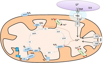
The mitochondrial monovalent reduction of O2 to ∙O2 − is thermodynamically favored and is regulated by two factors. The first is the concentration of electron carrier in proteins in a redox form and the second is the proportion of these proteins that are able to react with O2.Reference Murphy 32 Although complexes I and III are the major sources of mitochondrial ROS,Reference Turrens 13 there are additional mitochondrial sites that are able to produce ROSReference Murphy 32 – Reference Quinlan, Orr and Perevoshchikova 34 (see Fig. 1). Some of these are described below:
-
(1) Pyruvate dehydrogenase (PDH) is a mitochondrial enzymatic complex with three main catalytic componentsReference Fisher-Wellman, Gilliam and Lin 35 responsible for catalyzing the conversion of pyruvate to acetyl-CoA. It is proposed that the rate of ROS production from the PDH complex increased as the NAD(P)H/NAD(P)+ pool reduce.Reference Brautigam, Wynn, Chuang and Chuang 36 , Reference Ambrus, Nemeria and Torocsik 37
-
(2) 2-oxoglutarate dehydrogenase is another important mitochondrial enzymatic complex present in the Krebs (TCA) cycle that is able to produce ROS through NADH oxidation.Reference Tretter and Adam-Vizi 38 The mechanism relies on a third enzymatic element, in which the flavin from dihydrolipoamide dehydrogenase can generate large amounts of ROS in the mitochondrial matrix as consequence of NADPH/NAD+ ratio.Reference Starkov, Fiskum and Chinopoulos 39
-
(3) Mitochondrial glycerol 3-phosphate dehydrogenase (mGPDH) is a coenzyme located in the outer surface of the inner mitochondrial membrane that is able to transfer reduced cytosolic factors to the mitochondrial ETC.Reference Orr, Quinlan, Perevoshchikova and Brand 40 In addition to the ROS generated from mGPDH, oxidation of glycerol 3-phosphate can drive electrons both to complex IV and to complex I, leading to additional ROS production from these mitochondrial sources/sites.Reference Tretter, Takacs, Hegedus and Adam-Vizi 41
-
(4) Electron transferring flavoprotein Q oxidoreductase (ETF-QOR). During fatty acid oxidation mitochondrial acyl-CoA dehydrogenase transfers electrons to ETF, which is then oxidized by ETF-QOR by donating electrons to the ubiquinone (UQ) pool.Reference St-Pierre, Buckingham, Roebuck and Brand 42 Once the ratio of reduced ubiquinone (UQH2) and UQ becomes elevated, the electron leak increases ROS generation.Reference Perevoshchikova, Quinlan, Orr, Gerencser and Brand 43
-
(5) Monoamine oxidase (MAO) is a flavoenzyme located in the outer mitochondrial membrane that deaminates biogenic amines in the central and peripheral nervous systems and bloodReference Di Lisa, Kaludercic, Carpi, Menabo and Giorgio 44 , Reference Youdim, Edmondson and Tipton 45 in two-step reactions. In the first reaction, the flavin prosthetic group is reduced and produces aldehyde and ammonium. In the second, the reduced flavin is oxidized to form H2O2.Reference Tipton, Boyce, O’Sullivan, Davey and Healy 46 , Reference Toninello, Salvi, Pietrangeli and Mondovi 47
-
(6) Flavin site in complex II. Although the estimated ∙O2 − generation by this complex is ordinarily low, in a condition of low levels of succinate and diminished activities of complexes I and III, the flavin site complex can produce both superoxide and H2O2 at high rates.Reference Turrens 13 The mechanism proposed for this is based on the electron leak achieved by flavin in the semi- or fully reduced state.Reference Quinlan, Orr and Perevoshchikova 34
-
(7) Flavin prosthetic group in complex I. This process relies on the flavin mononucleotide (FMN) binding site, whose full reduction during forward electron flow from NADH induces electron leak to O2, producing ∙O2 −.Reference Herrero and Barja 48
-
(8) Ubiquinone site in complex I. This source of ∙O2 − is associated with the reduction of UQ to UQH2 by a substrate such as succinate, glycerol 3-phosphate or acyl-CoA. However, the electrons can also be driven reversely from UQH2 to NAD+, thereby generating ∙O2 − at high rates.Reference Brand 33 , Reference Lambert and Brand 49
-
(9) Outer ubiquinone site in complex III. The ∙O2 − production in this complex is based upon the electron transfer mechanism called Q cycle. Electron carriers into this complex gather the electrons from UQH2 to water-soluble cytochrome c in a sequential process that results in the formation of an unstable semiquinone UQ− that can reduce O2 to superoxide.Reference Figueira, Barros and Camargo 14
Schematic representation of the mitochondrial sites of reactive oxygen species (ROS) production and Ca2+-related ROS increase. In light blue, sites of ROS production: MAO, monoamine oxidase; PDH, pyruvate dehydrogenase; OGDH, oxoglutarate dehydrogenase; SDH, succinate dehydrogenase; ETF-QOR, electron transferring flavoprotein Q oxidoreductase; mGPDH, mitochondrial glycerol 3-phosphate dehydrogenase; electron transport chain complexes I and III. In gray, the proteins responsible for Ca2+ influx: RyR, ryanodine receptor; MCU, mitochondrial Ca2+ uniporter and RaM, rapid mode of calcium uptake. Dashed red lines indicate what enzymes have their ROS production stimulated by Ca2+ overload: OGDH and PDH. In light green, the proteins responsible for Ca2+ efflux: NCLX, Ca2+/Na+ exchanger and LETM1, Ca2+/H+ antiporter. Dark blue represents the other mitochondrial complexes; white ellipse; other enzymes from Krebs cycle; white rectangle, Voltage-dependent channels, VDAC; and purple, the sarcoplasmic reticulum.

Calcium (Ca2+) signaling and mitochondrial ROS overproduction
A compelling body of evidence shows that Ca2+ regulates numerous cellular functions, and that differences in Ca2+ concentration are controlled by complex membrane transport systems moving the cation between the extracellular environment, the cytosol and membrane de-limited intracellular organelles.Reference Babcock, Herrington, Goodwin, Park and Hille 50 The mitochondrion stands out as a critically important organelle in Ca2+ homeostasis as this organelle can internalize cytoplasmic calcium derived from the extracellular environment as well as Ca2+ released from the smooth endoplasmic reticulum (SER) (syn. in muscle: ‘sarcoplasmic’ reticulum) (SR).Reference Babcock, Herrington, Goodwin, Park and Hille 50 , Reference Takeuchi, Kim and Matsuoka 51
Calcium crosstalk between mitochondria and the SER employs a ryanodine receptor (RyR)-mediated mechanism. Although recent evidences have described the expression of mitochondrial inner membrane RyR in cardiomyocytes and striatal neurons,Reference Jakob, Beutner and Sharma 52 the RyR is better described as a channel protein located on the SER membrane that is sensitive to small changes in cytosolic Ca2+ concentration and to Ca2+ overload in the SER lumen.Reference Van Petegem 53 In either situation, the RyR allows Ca2+ release from storage in the SER (or SR) into the cytosolic mitochondrial microdomains that facilitate Ca2+ uptake.Reference Csordas, Varnai and Golenar 54 , Reference Csordas and Hajnoczky 55 Voltage-dependent channels located in outer mitochondrial membrane allow the entry of Ca2+ into the intermembrane space, and then either of two different processes can mediate its influx into the mitochondrial matrix:
-
(1) The mitochondrial calcium uniporter (MCU), which relies on the negative mitochondrial membrane potential to take up Ca2+ into the matrix.Reference Takeuchi, Kim and Matsuoka 51
-
(2) A rapid mode of calcium uptake (known as RaM), which is thought to respond to rapid changes in cytosolic Ca2+.Reference Santo-Domingo and Demaurex 56
Calcium efflux, on the other hand, depends upon the Ca2+/Na+ exchanger (NCLX), which is also able to switch the ion exchange flow (either forward or reverse) depending on cytosolic Na+ concentration and mitochondrial membrane potential.Reference Kim and Matsuoka 57 Efflux is also dependent on the Ca2+/H+ antiporter, which appears to be especially important in tissues that have low NCLX activity, such as liver, kidney and lung.Reference Saris and Carafoli 58
Several reports have shown that Ca2+ can stimulate ROS production by different mechanisms:
-
(1) Krebs cycle stimulation. It has been proposed that Ca2+ can allosterically activate enzymes, such as PDH, isocitrate dehydrogenase and α-ketoglutarate dehydrogenase in order to supply the ETC with reduced cofactors.Reference McCormack and Denton 59 As discussed previously some of these enzymes can also produce ROS.Reference Brand 33
-
(2) A change in lipid organization in the inner mitochondrial membrane. Studies in model membranes suggest that Ca2+ sequesters the cardiolipin attached to membrane carrier proteins, and that this membrane rearrangement in some way increases ROS production.Reference Grijalba, Vercesi and Schreier 60
-
(3) Mobilization of intramitochondrial ferrous iron (Fe 2+ ). Studies using isolated mitochondria have indicated that mitochondrial Ca2+ overload is associated with an increase in hydroxyl radical formation and oxidative damage. However, when the mitochondria were treated with a Fe2+ chelator, the oxidative damage was abolished.Reference Castilho, Kowaltowski, Meinicke, Bechara and Vercesi 61
-
(4) Opening of the mitochondrial permeability transition pore (MPTP). The outer mitochondrial membrane allows essentially a ‘free’ translocation of small molecules from the cytosol into the mitochondrion. However, selective transporters in the inner mitochondrial membrane are needed to assure homeostasis between cytosolic and matrix environments.Reference Rao, Carlson and Yan 62 Mitochondrial Ca2+ overload combined with oxidative imbalance leads to the opening of the MPTP in the inner membrane, thereby allowing bidirectional traffic of small metabolites through the mitochondrial membraneReference Grancara, Battaglia and Martinis 63 and a disruption of the normal electrolytic equilibrium. This disruption leads to mitochondrial swelling, a decrease in proton motive force, an increase of ROS production, and may also rupture the outer mitochondrial membrane with the consequent release of pro-apoptotic factors (e.g. cytochrome c, Smac/DIABLO, Omi/HtrA2 and others) into the cytosol.Reference Quintanilla, Jin, von Bernhardi and Johnson 64 , Reference Frezza, Cipolat and Martins de Brito 65
Oxidative impairment in the central nervous system (CNS): the mitochondrion as a trigger of neurogenic disease
Oxidative damage is innate to all eukaryotic cells. However, tissue types vary in their sensitivity to that damage, and by that measure the brain stands out as being particularly vulnerable to oxidative damage due to its morphologic and physiologic characteristics.Reference Halliwell 16 In normal resting conditions, the adult brain is responsible for over 15% of total O2 consumption, an exceptionally large rate of oxygen use per unit mass compared with others tissues.Reference Halliwell 16 In addition to its heavy consumption of O2, the brain is also vulnerable to oxidative damage due to the specialized characteristics of neural tissue as described below:
-
(1) The presence of excitotoxic amino acids, such as glutamate. Glutamate levels are tightly controlled in the brain, however, under conditions of stress, neurons undergoing apoptosis release a large amount of glutamate into the surrounding tissue. Furthermore, Mailly et al.Reference Mailly, Marin, Israel, Glowinski and Premont 66 showed that neurons in the presence of excess hydrogen peroxide enter a prolonged excitatory state triggered by the continuous activation of n-methyl-d-aspartate (NMDA) receptors by glutamate.
-
(2) A high content of biologically important amines that are oxidized in the presence of O2. Neurotransmitters such as dopamine, serotonin, adrenalin and noradrenalin react slowly with oxygen to produce superoxide, which in turn reacts with those neurotransmitters to form other ROS in a chain reaction.Reference Spencer, Jeyabalan, Kichambre and Gupta 67 Furthermore, several oxygenases possess tetrahydropteridine as a co-factor, which in elevated levels is able to induce ROS-dependent neuronal apoptosis.Reference Cardaci, Filomeni, Rotilio and Ciriolo 68
-
(3) SOD-independent H2O2 generation. Most ROS production occurs downstream from the dismutation of ∙O2 −. However, the brain can generate large quantities of H2O2 independently of SOD activity. During the recycling of biogenic amines (e.g. serotonin, epinephrine, norepinephrine, dopamine), enzymes located in outer mitochondrial membranes of neurons and glia can form H2O2 through oxidative deamination of those amines.Reference Zheng, Gal and Weiner 69 , Reference Kwan, Bergeron and Abell 70
-
(4) Prevalence of polyunsaturated fatty acids (PUFA) in the CNS. PUFA are widespread in the CNS, and if antioxidant systems are not adequate to inhibit ROS formation, the ROS can remove hydrogen from PUFA or attach to it to initiate lipid peroxidation.Reference Shichiri 25 Once lipid peroxidation has been initiated, intermediate compounds react with oxygen to form lipid proxy radicals, which then react with PUFA in a cyclic reaction to generate isoprostanes as well as multiple α,β-unsaturated aldehyde products, such as acrolein, 4-hydroxy-2-nonenal (4-HNE) and malondialdehyde (MDA).Reference Cobb and Cole 71
Due to the brain’s particular vulnerability to oxidative stress, many studies have been designed to assess how varying relationships among mitochondria, oxidative imbalance and lipid peroxidation can predispose an individual to neurodegenerative diseases such as Alzheimer’s (AD), Huntington’s, Parkinson’s diseases, multiple sclerosis and neurogenic hypertension.
In the case of AD, it was shown that oxidative stress as well as lipid peroxidation in the cerebral cortex and hippocampus exert a positive influence on disease progression by inducing amyloid-beta peptide (Aβ) accumulation,Reference Pratico, Uryu, Leight, Trojanoswki and Lee 72 wherein products of lipid oxidation impair energy production in the brain.Reference Reed, Pierce, Markesbery and Butterfield 73 Moreover, AD patients exhibit lower cerebral activity of mitochondrial enzymes in the Krebs cycleReference Bubber, Haroutunian, Fisch, Blass and Gibson 74 as well as an impairment in oxygen consumption via a decrease in complex I and III activities.Reference Chen, Stern and Yan 75 Such metabolic dysfunctions in neural tissue lead to an increase in ROS generation and a decrease in energy supply, thus enhancing the damage promoted by Aβ accumulationReference Chen, Stern and Yan 75 and impairing many higher level brain functions, including judgment, memory and orientation.
Similarly a dysfunction in complex II may represent an important factor in Huntington’s disease (HD), a disorder associated with cognitive deficits, psychiatric illness and involuntary movements. Striatal degeneration induced by defective mitochondrial complex II function has been used as a common animal model of HDReference Damiano, Galvan, Deglon and Brouillet 76 and may reflect the disease process in humans Furthermore, disruptions in hippocampal calcium signaling, mitochondrial membrane potential, sensitivity of the MPTP, pyruvate dehydrogenase and complex IV activities, and an increase in lipid peroxidation have also been described in brains of patients with HD.Reference Cobb and Cole 71 , Reference Nasr, Gursahani and Pang 77
Systemic inhibition of complex I has been used as an experimental model of Parkinson’s disease (PD).Reference Luo, Hoffer, Hoffer and Qi 78 Complex I disruption in dopaminergic neurons, present mainly in the striate nucleus, results in decreased ATP production and increased mitochondrial ROS production, thereby stimulating pathways involved in MPTP activation as well as initiating the release of inflammatory and pro-apoptotic molecules to induce neuronal cell death.Reference Abdin and Sarhan 79 Increased products from lipid peroxidation, such as F2-isoprostanes and 4-HNE can also contribute to neuronal deathReference Shichiri 25 , Reference Seet, Lee and Lim 80 in PD, which culminates in bradykinesia, rigidity and tremors induced by the striatal dopamine deficiency.Reference Seet, Lee and Lim 80
Mitochondrial dysfunction has also been related to neuropathology of multiple sclerosis (MS). A decrease in the complexes I and III activities of 50% or more impairs the capacity of mitochondria to produce ATP.Reference Dutta, McDonough and Yin 81 , Reference Schapira 82 The mismatch between energy requirements and ATP production in turn, contributes to axonal degeneration in upper motor neurons in MS patients.Reference Dutta, McDonough and Yin 81 , Reference Broadwater, Pandit and Clements 83 The energy deficit is further enhanced by damage to mitochondrial DNA caused by nitric oxide or its products.Reference Sarti, Giuffre and Barone 84 In amyotrophic lateral sclerosis (ALS), a neurologic disease characterized by motor neuron and neuromuscular junction degradation, oxidative stress is a major contributor to the etiology of the disease by impairing the machinery of transmitter release in the pre-synaptic motor nerve terminal of the neuromuscular junction.Reference Pollari, Goldsteins, Bart, Koistinaho and Giniatullin 85 In fact, a mutation in the gene coding for cytosolic SOD (SOD1) is responsible for 20% of ALS cases.Reference Robberecht and Philips 86 In addition, ALS patients exhibit decreased mitochondrial function and impairment in Ca2+ homeostasis, both of which contribute to oxidative damage in the lumbar and thoracic spinal cord.Reference Beal, Ferrante and Browne 87 It is proposed that the downstream oxidative damage in ALS patients depends largely on the capacity of the defective SOD1 to increase ROS production both in mitochondria and in plasma membrane bound NADPH oxidase.Reference Estevez, Crow and Sampson 88 , Reference Harraz, Marden and Zhou 89
Central redox balance also plays a key role in cardiac diseases arising from CNS defects.Reference Paravicini and Touyz 90 Nuclei located in the brainstem, including the rostral ventrolateral medulla (RVLM) and the nucleus tractus solitaries (NTS) play key roles in neurogenic hypertension,Reference Chan and Chan 91 wherein the imbalance of ROS and nitric oxide in neurons within these nuclei can alter the peripheral vascular system by increasing sympathetic vasomotor tone.Reference Hirooka 23 , Reference Hirooka, Kishi, Sakai, Takeshita and Sunagawa 92 Chan et al. demonstrated that in spontaneously hypertensive rats, the increase in blood pressure is directly related to lower expression and activity of mitochondrial superoxide dismutase and catalase in the RVLM.Reference Chan, Tai, Li and Chan 93 Additional studies found that the elevation of ∙O2 − and H2O2 in brainstem sites such as RVLM and NTS originate from activation of NADPH oxidase via protein kinase C and phosphatidylinositol 3-kinase, as well as via an increase in intracellular Ca2+, and down regulation of mitochondrial uncoupling protein 2 (UCP2) and reduction in ETC capacityReference Chan, Wu, Chang, Tai and Chan 94 – Reference Nozoe, Hirooka and Koga 97 contributes to the increase in arterial blood pressure.
Early oxidative stress as a developmental determinant of health and disease in later life
It is well known that environmental influences can alter numerous internal body functions and as a result can trigger such NCD as diabetes, metabolic syndrome, and cardiac disorders.Reference Godfrey, Gluckman and Hanson 98 An increase in NCD risk is not limited to physiologic changes occurring in adulthood but may also result from adverse events that occur much earlier in life. Thus, endogenous and exogenous signalsReference Lucas, Fewtrell and Cole 99 if occurring within certain critical developmental windows within the embryonic period can permanently affect physiologic processes within the mature individual.Reference Colombo 100
The first suggestion that this phenomenon exists came from observations of the occurrence of impaired glucose tolerance and the development non-insulin-dependent diabetes in a 64-year old who had exhibited a significantly reduced growth rate in early life.Reference Hales, Barker and Clark 101 The data suggested that poor nutrition during periods of fetal life and infancy induced diabetes via changes in B-cell function. The researchers further hypothesized that permanent adaptations to the early nutritional deficit provided survival benefits by shunting glucose to critical organs and away from those organs considered as secondary for survival.Reference Hales, Barker and Clark 101 Ironically the permanent adaptations so necessary for survival in fetal life/infancy may be the same physiologic alterations that predispose and individual to chronic disease in later life.
The ability to express different phenotypes following a physiologic challenge is known as phenotypic plasticity, and in development is dependent upon specific temporal windows during which the organism is especially prone to change its developmental pattern in order to survive.Reference Pigliucci 102 Further investigation has shown that the post-developmental environment helps to determine whether the initial exposure will or will not be harmful.Reference Khan, Dekou, Hanson, Poston and Taylor 103 , Reference Hanson and Gluckman 104 This suggested that the early influences acted as environmental cues that led to adaptive responses providing survival advantages to the individual in later life. However, if the postnatal environment does not ‘match’ or coordinate with the prenatal environment, the adaptations that occurred during development would no longer be advantageous to the individual and would predispose him or her to the occurrence of adult diseases.Reference Nettle, Frankenhuis and Rickard 105
As human mothers are able to quench small environmental perturbations of short duration,Reference Wells 106 it would be expected that environmental changes during life generate a mother’s phenotype and this phenotype will shape the offspring’s adaptations, by generating a variability in metabolic capacity.Reference Wells 107 , Reference Wells 108 WellsReference Wells 107 , suggested that the maternal phenotype is responsible for the adaptations in her offspring, and that her phenotype might depend, in turn, on the environmental history of close ancestors, as has been reviewed in detail.Reference Bale 109
Martin-Gronert and OzanneReference Martin-Gronert and Ozanne 110 highlighted three proposed mechanisms of how events in the perinatal period of development can produce life-long effects in the individual. The first mechanism involves the occurrence of permanent structural changes in key organs such as the brain, pancreas and kidney. The second mechanism involves changes in gene expression resulting from epigenetic modification, as has been described elsewhereReference Martinez, Gay and Zhang 111 , Reference Ozanne and Constancia 112 (e.g. DNA methylation, histone modification and microRNA action on mRNA).Reference Martinez, Gay and Zhang 111 The third mechanism for the long-lasting effects of a developmental insult is dependent upon the process of cellular ageing. An example of this is the induction of cellular senescence secondary to mitochondrial dysfunction and increased oxidative stress, as suggested by Luo et al.Reference Luo, Fraser and Julien 113
In the past several years, oxidative stress has been studied as a molecular trigger for the effect of maternal nutritional deficiency on NCD occurring later in life in her offspring. In animal models of protein restriction, researchers have found mitochondrial dysfunction in the early mouse embryoReference Mitchell, Schulz, Armstrong and Lane 114 and heart; oxidative stress in pancreatic islets as a consequence of a mismatch among antioxidant enzymes (i.e. increase in SOD with concomitant decrease in CAT and GPx);Reference Theys, Clippe, Bouckenooghe, Reusens and Remacle 115 an increase in pancreatic oxidative stress and heart rate associated with agingReference Tarry-Adkins, Chen, Jones, Smith and Ozanne 116 and an increase in the protein expression of enzymes related to ROS production as well as oxidative stress and DNA damage.Reference Tarry-Adkins, Martin-Gronert and Fernandez-Twinn 117
Early low-protein diet and cerebral oxidative impairment
Nutrition is often considered as the greatest exogenous influence in early life. Classified as the main non-genetic contributor to changes in brain development, nutritional inadequacy has been known to have several deleterious effects on the fetal brain.Reference McGaughy, Amaral and Rushmore 118 – Reference Field, Anthony and Engle 122 Early studies of restrictive diets on offspring had described oxidative impairment, such as decreased GSH levels in the forebrain;Reference Partadiredja, Worrall and Bedi 123 lower mRNA expression of SOD and CAT in the brain;Reference Partadiredja, Worrall, Simpson and Bedi 124 increased vascular ∙O2 − productionReference Franco, Akamine and Reboucas 125 and a decrease in SOD activity,Reference Franco Mdo, Dantas and Akamine 126 and in newborn infants, an increase in oxidative damage as a consequence of the reduction in serum antioxidant capacity.Reference Gupta, Narang, Banerjee and Basu 127 , Reference Gveric-Ahmetasevic, Sunjic and Skala 128
Although all categories of nutrient are important in brain development, protein has the greatest effect on neural function.Reference Tonkiss, Galler, Morgane, Bronzino and Austin-LaFrance 129 Early protein deficiency affects the brain in several ways that vary with the period of exposure, the type of protein deficiency and its severity, and also with the specific cerebral region.Reference Morgane, Austin-LaFrance and Bronzino 130 The critical period for brain development is marked by several specific temporal windows in which the processes of neurogenesis, neuron migration, and neuron alignment and orientation are quickly increased then either decreased or ceased.Reference Morgane, Mokler and Galler 131
As proteins do not readily cross the placenta into the fetal circulation, nutritional deficits in the mother are generally transmitted to the fetus the level of the amino acid composition of the ingested protein. Consequently, the lack of any one of the essential amino acids in the maternal protein diet can lead to a complete protein deficiency in the fetus.Reference Morgane, Mokler and Galler 131 Nutritional protein restriction may reduce antioxidant capacity by inhibiting the synthesis of antioxidant enzymes,Reference Al-Gubory, Fowler and Garrel 132 and the resulting oxidative stress on fetal cells could alter gene expression and further damage the cells with oxidized proteins and lipids.Reference Luo, Fraser and Julien 113
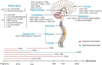
Several studies have demonstrated the effects of protein restriction in oxidative balance and mitochondrial function in the CNS (see Fig. 2). Bonatto et al.Reference Bonatto, Polydoro and Andrades 133 evaluated these parameters in the hippocampus of rats exposed to moderate protein restriction from the 1st day of the gestational period until 75 days of postnatal life and found an increase in protein oxidation but a decrease in lipid oxidation. The investigators suggested that the opposing effects of a protein-restricted diet on proteins v. lipids resulted from an overall increase in SOD activity in the protein-restricted group, wherein the elevated SOD protected lipid, but not protein, from oxidation. As the activity of CAT did not change with restricted protein, it was suggested that the higher activity of SOD without a concomitant up-regulation of CAT drives the accumulation of H2O2, followed by formation of the hydroxyl radical (∙OH),Reference Halliwell 134 considered among the most reactive of ROS. Thus hydroxyl radical formation could be the responsible for the increased protein oxidation observed in the face of increased SOD activity.Reference Jackson, Schraufstatter and Hyslop 135 , Reference Gutteridge and Wilkins 136 As evidence of this, when the protein-restricted animals were supplemented with methionine, SOD activity was reduced and the animals exhibited increased oxidative damage to their lipids.
Main findings on early protein restriction and central nervous system oxidative balance. Red lines represent protein restriction, blue lines, normoprotein diet, and the length of each approximates the time during which either feeding state was applied for each study referred to in the text. Numbers in parentheses refer to the references cited in the text. MDA, malondialdehyde; SOD, superoxide dismutase; CAT, catalase; GPx, glutathione peroxidase; GST, glutathione-S-transferase; GR, glutathione reductase; G6PDH, glucose-6-phosphate dehydrogenase; GSH, reduced glutathione; GSSG, oxidized glutathione; TAR, total antioxidant reactivity; Try, tryptophan; Tyr, tyrosine; and RCR respiratory control ratio.

Further investigations in 21-day-old rats,Reference Bonatto, Polydoro and Andrades 133 , Reference Bonatto, Polydoro and Andrades 137 showed that low-protein diet increases oxidative damage to lipids in the cerebellum and hippocampus but has no influence on oxidation, in the cortex. Evaluation of SOD and CAT activities in those brain regions, moreover, showed that SOD activity was reduced only in cerebellum with low-protein diet, and not in either the cortex or hippocampus. A possible explanation for the cerebellar damage, is that low-protein induced decrease in SOD activity enhances interaction between ∙O2 − and NO∙ to form peroxynitrite, which is capable of oxidizing lipids, proteins and thiol compounds as well as DNA.Reference Alvarez and Radi 138
Feoli et al.,Reference Feoli, Siqueira and Almeida 139 on the other hand, showed no difference in ROS production in either cerebellum, cortex or hippocampus of animals fed a low-protein diet (casein 7%) from the first gestational day until 60 days of life. Although ROS levels did not change, an increase in lipid peroxidation in the cerebellum and cortex occurred due to a decrease in SOD activity in the cerebellum, and the reduction in total antioxidant reactivity in the cortex. When the authors evaluated the content of tryptophan and tyrosine, (important neurotransmitter precursors) as a measure of damage, all three brain regions were negatively affected by low-protein diet, and the damage was closely related to the lower serotoninergic and catecholaminergic neurotransmitter concentrations.Reference Voog and Eriksson 140
Tatli et al.,Reference Tatli, Guzel and Kizil 141 by evaluating three types of induced CNS damage: (1) intrauterine growth restriction (IUGR), (2) moderate and (3) severe protein restriction in five different CNS regions (cortex, cerebellum, cervical, thoracic and lumbar cord) showed that only severe undernutrition triggered lipid oxidation in all five CNS structures at 60 days of life. Protein oxidation was shown to vary in proportion to the degree of undernutrition, with the cerebellum showing greater sensitivity to protein oxidation than the other CNS regions. In addition to increasing oxidative biomarkers, early nutritional adversity also decreased the activity of SOD and CAT in all regions analyzed.
In our laboratory, we have evaluated offspring through 100 days of age from mothers fed a low-protein diet throughout the perinatal period (gestation and lactation) and found that low-protein animals had increased oxidative damage in the brainstem. The data showed a marked decrease in antioxidant capacity, wherein enzymatic activities of SOD, CAT, GPx and glutathione-S-transferase were decreased by over 15%. The redox state was also affected by the maternal low-protein diet through a reduction in glutathione concentrations as a consequence of lower glutathione resynthesis and a decrease in NADPH supply.Reference Ferreira, Liu, Fernandes and Lagranha 142 Oxidative imbalance in certain brain regions is directly related to the occurrence of cardiovascular impairments,Reference Cardoso, Colombari and Menani 143 mainly hypertension.Reference Hirooka 23 , Reference Paravicini and Touyz 90 , Reference Chan and Chan 91 , Reference Chan and Chan 144 Thus, it is feasible that early nutritional insult is a central trigger for the development of hypertension in adulthood.
Assessing brain O2 consumption, Muzzo et al.Reference Muzzo, Gregory and Gardner 145 found that newborns from mothers fed throughout the gestational period with a diet containing only 4% protein, exhibited decreases in both brain mitochondrial protein and in O2 consumption. Animals that were reefed from the 1st until 16th postnatal day also showed a reduction in O2 consumption and phosphorylation capacity. Although the gestational period encompasses most of the period during which neurogenesis occurs,Reference Alamy and Bengelloun 146 in rats, the duration of maternal lactation represents the most important period for brain development.Reference Morgane, Mokler and Galler 131 Thus, several studies have described effects of protein restriction during other periods of brain development that may lead to lasting changes even after refeeding due to the impairment in the metabolic activity.Reference McGaughy, Amaral and Rushmore 118 , Reference Mokler, Galler and Morgane 147
In rats experiencing severe protein restriction during a period of 30 days (from the 18th to the 48th postnatal day),Reference Olorunsogo 148 brain mitochondria were shown to have alterations in Krebs cycle, isocitrate and succinate dehydrogenase, as well as in ETC enzymes, cytochrome c oxidase and ATP synthase. In addition, the mitochondrial ETC exhibited an impairment in function, such that low-protein animals were less responsive to ADP stimulation by complexes I and II substrates.
Despite the important role of the mitochondrion in several neurogenic diseases (i.e. PD, AD, ALS, etc.), very few studies have investigated the role of mitochondrial dysfunction in the central oxidative imbalance induced in protein restriction models. The antioxidant system, however, has been shown to be significantly affected by a low-protein diet, which also contributes to the disruption of mitochondrial capacity and may compromise the overall brain function.
Conclusion
In this review, we discuss the importance of mitochondrial dysfunction and oxidative stress in the development of neural disorders, and show how such diseases could be induced by nutritional insult during development. Although decreases in mitochondrial content and/or activity have been demonstrated in several studies employing nutritional manipulation either during the gestational and/or locational period, further investigations will be necessary to determine the specific mechanism causing mitochondrial dysfunction due to a protein-poor diet within a restricted developmental window. Compelling evidence has already shown that several neurogenic diseases are associated with mitochondrial disruption accompanying high-fat intake. Therefore it seems likely that a diet low in protein could also disrupt mitochondrial function sufficiently to induce neurogenic disease. Many studies to date have shown that protein restriction deeply affects central oxidative balance by decreasing antioxidant capacity. Further studies must be conducted, however, to assess the contribution of ROS generators in this oxidative disruption.
The underlying mechanisms responsible for impaired mitochondrial function in metabolic disorders induced by low-protein diet during embryonic development have not yet been elucidated. It is hoped, however, that prospective clinical investigations of mitochondrial function in healthy and diseased humans will begin to provide insight into precisely how early nutritional deficit and the occurrence of metabolic disease in adulthood are linked.
Acknowledgments
The authors are thankful to the FACEPE (Foundation to Support Science and Research from Pernambuco State – Brazil, APQ-1026.4-09/12) that provided financial fund for the laboratory.