INTRODUCTION
The leishmaniasis are a heterogeneous group of diseases that affect millions of people in tropical and subtropical areas of the world (Desjeux, 1996). Depending on the Leishmania species and on the immunological response of the human host, the disease ranges from asymptomatic infections to self-limiting cutaneous lesion(s) or fatal visceral forms. During their life-cycle, parasites alternate between 2 stages: flagellated promastigotes which develop in the midgut of the insect vector and amastigotes which multiply in the host macrophage (Alexander and Russell, 1992; Handman, 1999). At this latter stage, Leishmania parasites are sequestered and reside in the phagolysosomal vacuole, which results from the fusion of phagosomes with lysosomes (Handman, 1999; Duclos and Desjardins, 2000; Sacks, 2001; Amer and Swanson, 2002; Cunningham, 2002). Over the past decades, several molecules involved in the biology of the parasite or targeted by host-specific antibodies or cellular responses have been identified. Previous reports stressed that excreted/secreted (ES) molecules from intracellular pathogens such as Mycobacterium tuberculosis and Toxoplasma gondii contain antigens highly immunogenic and protective in vaccine models (Prigione et al. 2000; Mustafa, 2002; Daryani, Hosseni and Dalimi, 2003; Pym et al. 2003; Shams et al. 2004). Similarly, Leishmania promastigote culture filtrate proteins elicit strong immunity and protection in L. major-infected BALB/c mice (Webb et al. 1988; Mendez et al. 2002; Tonui et al. 2004). However, only few proteins were fully characterized as ES molecules. Antigens excreted/secreted by Leishmania parasites may generate peptides that can be loaded onto major histocompatibility complex (MHC) class I or class II molecules and trigger protective cellular immune responses. In addition, molecules released into the Leishmania phagolysosomal vacuole may also subvert the presentation machinery associated with the endoplasmic reticulum-mediated phagocytosis, which could represent an immune evasion strategy to avoid a cellular immune response.
In an attempt to identify new ES molecules of L. major parasites, we prepared culture supernatants of stationary-phase promastigotes incubated during 6 h in serum-free medium, at a pH and temperature that partly reproduce the phagolysosomal vacuole conditions and raised immune sera in rabbits then used them to screen an L. major cDNA expression library. A total of 33 different clones were isolated and sequenced. Gene expression analysis revealed that all tested genes are transcribed and, interestingly, some of them are stage regulated.
MATERIALS AND METHODS
Parasite cultures and preparation of L. major ES proteins (LMES) and messenger RNA
A highly virulent isolate of L. major (MHOM/TN/94/GLC94, zymodeme MON25) obtained from a human cutaneous sore was used in this study (Louzir et al. 1998; Kebaier et al. 2001). Parasites were cultivated on NNN medium at 26 °C and then progressively adapted to RPMI 1640 medium (Sigma, St Louis, Mo.) containing 2 mML-glutamine, 100 U/ml penicillin, 100 μg/ml streptomycin, and 10% heat-inactivated foetal calf serum (complete medium). Promastigotes collected at the logarithmic-growth phase culture were adjusted to 106 parasites/ml in a constant volume and further incubated at 26 °C. The stationary phase was reached after 6–7 days with a parasite concentration of ~8×10⁷ parasites/ml. Stationary-phase promastigotes were used for protein labelling and preparation of L. major ES antigens (LMES). For the preparation of LMES, stationary-phase or metacyclic (metacyclics were purified from stationary-phase promastigotes using Ficoll gradient 5–20% centrifugation) promastigotes were washed 3 times in PBS, re-suspended at 2×10⁷ parasites/ml in serum-free RPMI medium under classical promastigote culture conditions (pH 7·2, 26 °C) or at pH 5·5 and 35 °C, to partially reproduce phagolysosomal vacuole conditions, and were then incubated for 6 h. Parasite viability was assessed by the Trypan blue dye exclusion test (Barankiewicz, Dosch and Cohen, 1988). Supernatants of parasite cultures containing LMES were then concentrated 10-, 20- and 200-fold using, respectively, Centricon YM-10 tubes (Millipore, Molsheim, France), Vivaspin 6 ml concentrator (Vivascience Hannover, Germany), Centricon Plus 80 (Millipore) according to the manufacturer's instructions. The protein content of LMES was quantified using the Lowry method.
For gene expression analysis, 3 parasite growth conditions were used: procyclics, metacyclics and stationary-phase parasites incubated at acidic pH and 35 °C (AP, for acidic promastigotes). Procyclic promastigotes were collected at the logarithmic-growth phase culture. For differentiation into metacyclics, a fresh subculture was grown and a growth curve was plotted to ascertain when the cells entered stationary phase (between day 6 and day 7). Metacyclics were then purified from stationary-phase culture using density-gradient centrifugation as previously described (Spath and Beverley, 2001). AP correspond to parasites cultivated using the same conditions as for LMES preparation except that the parasites were cultivated in complete medium.
Generation of rabbit anti-LMES sera
Two rabbits were injected intramuscularly with 250 μg of the LMES antigens emulsified (vol/vol) in incomplete Freund's adjuvant (Sigma) and boosted similarly 15 days later. The second booster dose was administered 30 days later by intradermal injections in 8 different sites. Rabbits were bled starting 10 days after the final injection. Sera were separated and stored at −80 °C until use.
Radio-isotope labelling of Leishmania ES proteins
Leishmania proteins were radio-isotope labelled after incubation of stationary-phase promatigotes (1×10⁸ cells) in 5 ml of methionine-free medium (35 °C, pH 5·5 or 26 °C, pH 7·2) containing 50 μCi of [35S] methionine (specific activity>1000 Ci/mmol; Amersham) for 6 h. The supernatants, collected after a centrifugation at 4000 g for 20 min at 4 °C, was added with an anti-protease cocktail (2 μg of leupeptin per ml, 2 μg of antipain per ml, 2 μg of pepstatin per ml, 2 μg of chymostatin per ml, 16 μg of aprotinin per ml) (Boehringer Mannheim, Germany) and concentrated 10-fold. The proteins were separated by sodium dodecyl sulfate–polyacrylamide gel electrophoresis (SDS–PAGE) and revealed by autoradiography (Kodak, Biomax MR film).
Immunoscreening of an L. major promastigote cDNA library
A λ ZAPII (Stratagene, La Jolla, Calif.) cDNA expression library (Ben Achour et al. 2002) was screened using anti-LMES antiserum according to the manufacturer's instructions. Approximately 5×10⁶ plaques were screened. Filters were first incubated with the anti-LMES antibodies diluted to 1[ratio ]500 in TBS-T (20 mM Tris-HCl, pH 7·5, 150 mM NaCl, 0·05% (v/v) Tween 20) supplemented with 5% (w/v) non-fat dried milk at room temperature for 2 h then with a peroxidase-conjugated goat anti-rabbit IgG (Amersham) diluted to 1[ratio ]2000 in TBS-T for 1 h at room temperature. Finally, filters were developed using the diaminobenzidine tetrahydrochloride protocol (Sigma). Positive clones were subjected to secondary and tertiary screenings and clones isolated from the tertiary screening were subjected to pBK-CMV phagemid vector excision from the ZAP Express vector using the manufacturer's helper phage excision protocol. Recombinant pBK-CMV phagemid vector containing cDNAs of interest were then sequenced.
Bioinformatics analysis
Searches for nucleotide and deduced amino acid sequences homologous to the isolated genes and proteins were performed by BLAST at NCBI and EBI (http://www.ncbi.nlm.nih.gov/Tools/index.html, http://www.ebi.ac.uk). The same searches were carried out against the L. major databases at L. major OmniBlast Server from the Sanger Institute (http://www.sanger.ac.uk/cgi-bin/blast/submitblast/l_major/omni). Potential open reading frames (ORFs) of positive clones were identified using ‘traduction multiple’ (http://www.infobiogen.fr/) and functional domains of predicted proteins were identified using SMART (http://smart.embl-heidelberg.de), Scanprosite (http://au.expasy.org) and Motif Scan at (http://myhits.isb-sib.ch/cgi-bin/motif_scan). SignalP server (http://www.cbs.dtu.dk/services/SignalP) SecretomeP server (http://www.cbs.dtu.dk/services/SecretomeP-1.0) And TMHMM Server v. 2.0 (http://www.cbs.dtu.dk/services/TMHMM/) were used to characterize potential signal peptides, non-classical and leaderless protein secretion and transmembrane helice domains, respectively.
Expression of recombinant proteins in E. coli and Western blot analysis
XLI-Blue E. coli harbouring recombinant plasmids pBK-CMV containing cDNAs of interest were grown in LB medium, induced with 1 mM isopropyl-D-thiogalactopyranoside (IPTG) for 4 h and lysed.
Western blot experiments were performed to analyse the reactivity of the recombinant proteins synthesized in E. coli or concentrated LMES with specific antiserum. Proteins in l×-SDS sample buffer were boiled for 10 min, separated by 18% SDS–PAGE and electro-transferred to nitrocellulose membranes. Filters were blocked with PBS 0·1% Tween20 (PBS-T) containing 5% non-fat dried milk at room temperature for 1 h then incubated with specific antiserum diluted 1[ratio ]1000 in blocking solution at 4 °C overnight. Filters were washed 3 times with PBS-T, incubated with goat-anti-rabbit secondary antibody coupled to peroxidase for 1 h at room-temperature, washed 3 times with PBS-T, and revealed by peroxidase activity.
RNA isolation and cDNA synthesis
Total RNA was extracted from 109L. major parasite stocks (from the different culture conditions) by homogenization in 1 ml of TRIzol reagent (Gibco-BRL Life Technologies, INC) according to the manufacturer's instructions. Any possible genomic DNA contamination was eliminated by treatment with deoxyribonuclease I (Dnase I, Boehringer Mannheim, Germany). RNA was quantitated using a spectrophotometer. Examination of purified total RNA by gel electrophoresis revealed prominent 18S, 24Sα and 24Sβ ribosomal bands indicating that the RNA was not degraded. Five μg of total RNA was reverse transcribed in a reaction mixture containing l×RT-buffer (Gibco BRL), 1·7 UI/ μl RNasin (Promega), 250 μM each dNTP, 50 μg/ml of random hexamers (Promega), and 20 UI/μl of M-MLV RT (Murine-Moloney Leukemia) virus reverse transcriptase, Gibco BRL) for 1 h at 42 °C. The cDNA was heated at 95 °C for 10 min to inactivate the reverse transcriptase.
Gene expression analysis using quantitative real-time PCR
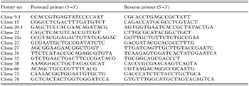
In order to quantify mRNA for L. major gene-encoding ES proteins, we performed reverse transcription and real-time RT-PCR using the SYBR Green I technique based on measuring PCR products in the logarithmic phase of the reaction by determining the CT (Heid et al. 1996), CT being the threshold cycle at which the fluorescence emission reaches the log phase of product accumulation Primers, 2 forward and 2 reverse, were designed for each sequence, including endogenous controls, using Primer express (Version 1.5, PE Applied Biosystems, Foster City, CA). The optimal upstream and downstream primer sequences of the different genes are shown in Table 1.
Table 1. Combinations of forward and reverse primers used for unknown ES gene quantification in RT-PCR reactions (All primers were desalted when HPLC purified (Genset France).)

All experiments were performed using SYBR Green I Universal PCR MasterMix (PE Applied Biosystems). PCR reactions were performed using the ABI PRISM 7700 sequence detection system (with version 1.9.1 software, PE Applied Biosystems) in 40 cycles of denaturation at 95 °C for 15 sec and annealing extension at 60 °C for 1 min. Data were normalized by referring to the expression of an endogenous control (rRNA; Accession number: CC144545) that has been chosen among 3 genes recently described as equally expressed between procyclic, metacyclic and amastigote stages of L. major using DNA microarrays and Northern blot experiments (Akopyants et al. 2004). For each sequence, a standard curve was generated by diluting a DNA stock. Threshold cycle values were used to calculate a linear regression line. The slope of standard curves has determined the PCR efficiency (EFF) in conformity with EFF=10[−1/slope]−1. All the optimized PCR have an efficiency ranging from 90 to 98%.
The results were first expressed by the calculation of the ΔCT which is the difference in threshold cycles for target mRNA and rRNA and then by the determination of the ΔΔCT which corresponds to the difference between the ΔCT of target gene in metacyclic or in AP stages and the ΔCT obtained with procyclic promastigotes used as the reference stage. For each target mRNA, results were finally expressed as a fold difference in metacyclic or in AP values compared with those found in procyclic by calculating 2−ΔΔCT.
RESULTS
Identification of Leishmania major ES molecules
In order to identify proteins that are possibly released by Leishmania parasites into the phagolysosome vacuole of host macrophages, stationary-phase promastigotes were exposed to conditions that partially reproduce the macrophages vacuole environment (pH 5·5, 35 °C): Promastigotes from L. major were first cultured in complete medium (pH 7·2, 26 °C) until the stationary-phase was reached. Parasites were then grown at pH 5·5 and at 35 °C for 6 h in the presence of [35S] methionine. Control cultures were maintained at pH 7·2 and at 26 °C. A short incubation period of only 6 h was used in order to avoid excessive cell death and protein release from dead parasites. Indeed, parasite viability assays showed the same percentage (3%) of parasite death in cultures grown in RPMI at pH 7·2 or at pH 5·5.
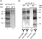
Radio-isotope labelled proteins released into culture media were concentrated 10-fold and analysed by SDS–PAGE (Fig. 1A). As expected, several proteins were detected but, interestingly, each culture condition generated a specific protein pattern: At pH 7·2 and 26 °C, few proteins were observed with 3 major bands migrating at molecular weights of 80 kDa, 66 kDa and 55 kDa (Fig. 1A, lane 2). In contrast, at pH 5·5 and 35 °C, several proteins were detected ranging from 15 kDa to 80 kDa (Fig. 1A, lane 1). In addition, 2 proteins with molecular weights of approximately 55 kDa and 30 kDa appeared to be highly induced at these culture conditions.

Fig. 1. Analysis of ES proteins of Leishmania major incubated at different culture conditions and detection of ES proteins from acidic culture conditions by Western blot experiments. (A) Total radio-isotope labelled proteins released by Leishmania parasites incubated in normal culture conditions (pH 7·2 and 26 °C) (lane 2) or in conditions that partially mimic macrophage vacuole environment (pH 5·5 and 35 °C) (lane 1) were concentrated and analysed by 18% SDS–PAGE followed by autoradiography. (B) ES proteins from AP concentrated 20-fold (lane 1) or 200-fold (lanes 3 and 4) or from metacyclics incubated at pH 5·5 and 37 °C concentrated 20-fold (lane 2) were analysed by SDS–PAGE, electroblotted on a nitrocellulose membrane and incubated with the anti-LMES (lanes 1, 2 and 4) or the pre-immune serum (lane 3).
Characterization of Leishmania major ES antigens
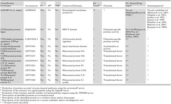
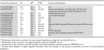
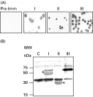
In order to characterize L. major ES proteins, we generated a specific rabbit antiserum directed against these components. ES antigens were obtained in the same culture conditions as for [35S] methionine-labelling except that culture medium did not contain FCS. The antiserum was then tested by Western blot to analyse its reactivity to concentrated ES proteins. Pre-immune serum was used as negative control. As shown in Fig. 1B, only the immune serum strongly reacts with the majority of proteins previously detected in the culture supernatant (Fig. 1B, lanes 1 and 4). In addition, no differences were observed between ES protein patterns from stationary-phase and purified metacyclic promastigotes incubated in conditions that partially reproduce the macrophages vacuole environment (Fig. 1B, lanes 1 and 2). This result may indicate that Leishmania parasites from these stages excrete or secrete a set of identical proteins. The immune anti-LMES serum was then used to screen a cDNA expression library from L. major promastigotes. From a screen of approximately 5×10⁶ plaques, 52 immuno-reactive clones were isolated and sequenced (Fig. 2). Sequence analysis revealed that some of them were identical and therefore a total of 33 different clones were identified. Sequence search for homology of the isolated clones with known sequences carried out using bioinformatic programs Blast from NCBI and EBI and L. major OmniBlast servers for both nucleotide and peptide sequences revealed that 20 cDNA clones displayed significant homologies with known genes from Leishmania or other species and the remaining 13 clones correspond to new genes. In an attempt to characterize the potential functions of the positive clones, we have identified their ORFs and analysed their deduced amino acid sequences. These analyses allowed the classification of the predicted amino-acid sequences in 3 different groups as follows. The first group (9 proteins) represents proteins which have been previously described as excreted/secreted in Leishmania and/or other micro-organisms (Table 2). The second group (11 proteins) displays proteins that have been already characterized in Leishmania and/or in other species (Table 3). However, it is unknown whether they are excreted/secreted. Finally, the third group (13 proteins) contains unknown proteins (Table 4).


Fig. 2. Identification of cDNA clones encoding potential ES proteins by immunoscreening. (A) A Leishmania major cDNA expression library was screened using the anti-LMES or the pre-immune antiserum as control. An example of positive clones belonging to groups I (see Table 2), II (see Table 3) and III (see Table 4) after 3 rounds of screening are shown in this figure. (B) Western blot analysis was used to confirm the recognition of recombinant proteins corresponding to the positive clones by the anti-LMES. Total lysate of XL1-blue E. coli strain harbouring the recombinant plasmid pBK-CMV containing cDNAs from the different groups of positive clones (I, II and III) induced with 1 mM IPTG were analysed by immunoblot using the anti-LMES. An asterisk (*) shows the recombinant proteins. Non-transformed BL21 were used as negative control (C).
Interestingly, using specific programs we showed that 21 of the isolated proteins were predicted to contain a single peptide or to be secreted via non-classical pathways (devoid of signal peptide). Among these proteins, 13 are predicted to be secreted via non-classical manners and the others via the ER/Golgi network.
LmPDI is a L. major secreted protein
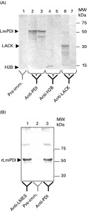
In a previous study (Ben Achour et al. 2002), we have identified a member of the Protein Disulfide Isomerase family (LmPDI) as a protein expressed at high levels by L. major isolates showing high virulence in BALB/c mice (Kebaier et al. 2001). In the present study, we could demonstrate that LmPDI is also an ES parasite antigen (Table 2). Indeed, affinity-purified antibodies to LmPDI (Ben Achour et al. 2002) strongly react with a band of the expected size (55 kDa) in the 200-fold concentrated culture supernatant of L. major parasites (Fig. 3A, lane 3) or in parasite lysates (Fig. 3A, lane 2) incubated at pH 5·5 and 35 °C. On the contrary, and as expected, the nuclear Leishmania histone H2B and the cytosolic Leishmania LACK (used here as negative controls) are detected only in parasite lysates using specific polyclonal antibodies raised against the L. major H2B and LACK respectively (Fig. 3A, lanes 4, 5, 6 and 7). In addition, we have shown that the anti-LMES reacts strongly with the recombinant LmPDI (Fig. 3B, lane 3). This result constitutes additional proof of the specificity of LmPDI secretion.

Fig. 3. LmPDI is an ES protein. (A) Western blot analysis shows that the native LmPDI released in the concentrated culture medium (lane 3) or present in the Leishmania lysate (lane 2) is highly recognized by the anti-LmPDI. As expected, the pre-immune antibody did not react with the LmPDI present in the Leishmania lysate (lane 1). On the other hand, the nuclear protein H2B (lane 5) and the cytosolic protein LACK (lane 7) are not detected in the culture medium but as expected in the L. major lysate (lane 4 and 6) using specific polyclonal anti-H2B or anti-LACK. (B) The purified recombinant LmPDI synthesized in BL21 E. coli analysed by immunoblot experiments reacts with the anti-LMES (lane 1) and with the anti-LmPDI (lane 3) but not with the pre-immune antibody (lane 2).
Gene expression analysis
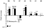
In order to better characterize unknown genes (n=13) encoding potential L. major ES proteins, we have analysed their expression patterns in procyclic, metacyclic and stationary-phase L. major parasites incubated for 6 h at pH 5·5 and 35 °C (AP). mRNA quantification was performed using reverse transcription and real-time PCR. The relative amounts of target gene mRNAs were first expressed as a ratio of target mRNA to rRNA, used as an endogenous control, and then expressed as a fold difference in metacyclic or in AP relative to procyclic promastigote. Interestingly, 5 clones seemed to be clearly stage regulated (Fig. 4). Indeed, clones 22, 66 and 72, were at least 3 times upregulated in AP in comparison with the procyclic stage. Moreover clones 22 and 72 were also upregulated in the metacyclic stage (2·6 and 3·93 times more, respectively). Unlike, clones 22 and 37, clones 66 and 72 displayed predicted functional domains. Indeed, they contain specific zinc-binding domains (Table 4) that are probably involved in protein-protein interactions. The other clones did not display significant gene expression variations. Finally, our data suggest that these new genes are indeed expressed by Leishmania parasites and, interestingly, at least for 2 of them, their relative expression (mRNA) was higher in culture conditions that we used to partially reproduce the phagolysosomal vacuole.

Fig. 4. Expression profile of unknown ES genes using quantitative real time RT-PCR in different Leishmania major stages and culture conditions. Real-time PCR experiments were conducted on cDNAs prepared by reverse transcription of total RNAs from the different L. major stages; procyclic, metacyclic and AP using specific primers to unknown ES genes. The fold increase corresponding to log10(2−ΔΔCT), which indicates the fold change in gene expression relative to the procyclic stage was calculated as described in the Material and Methods section. Genes 22, 37, 66 and 72 are stage regulated. On the contrary, the other tested genes are equally expressed between parasite stages (expression of genes 15, 31 and 38 is shown as example).
DISCUSSION
In the present study, we characterized a set of 33 Leishmania genes coding for proteins that are probably released by the parasite after 6 h incubation at a pH and temperature that partially reproduce the phagolysosomal vacuole conditions. Among these, 13 correspond to new genes. Interestingly, 21 isolated proteins are predicted to be exported in the extracellular medium through classical (via the ER, Golgi apparatus and flagellar pocket secretory system) or non-classical secretory pathways. These proteins are thus likely secreted by the parasite in the phagolysosomal compartment and may constitute virulence factors and, due to their location, may serve as an interesting target for the host cellular immune responses.
Several approaches have been used to characterize new Leishmania genes that are important for the biology of the parasite or/can be used as vaccine candidates. These include serological expression cloning, stage-specific cDNA probes, epitope-tagged expression library and biochemistry and other methods prior to cloning and sequencing. Here, we targeted the excreted/secreted proteins of L. major. Previous studies indicate the Leishmania promastigote culture filtrate proteins elicit strong immunity in humans, mice and dogs (Webb et al. 1988; Tonui et al. 2004; Lemesre et al. 2005; Rosa et al. 2005). Since the description of Bates, Gottlieb and Dwyer (1988) of around 40 glycoproteins that could be secreted by L. donovani promastigotes, only few ES proteins were extensively characterized (Symons et al. 1994; Wiese et al. 1995; Yahiaoui, Taibi and Ouassi, 1996; Webb et al. 1998; Sereno et al. 2005). In addition, these proteins were described in L. donovani and some molecules, such as the acid phosphatases, are not produced by L. major (Lovelace and Gottlieb, 1986).
The ES parasite proteins could play crucial roles in the biology or virulence of Leishmania parasites, which are very well adapted to survive in the harsh conditions of the host macrophage phagolysosomal compartment. They could also be degraded into peptides, which, upon association with MHC class II molecules and presentation to CD4+T cells, trigger host cellular immune responses. In addition, antigen-presenting cells such as macrophages and dendritic cells have been shown to be able to capture, process and present, in an MHC class I restricted manner, various exogenous antigens, including those derived from intracellular pathogens like Leishmania parasites (Houde et al. 2003). ES parasite proteins may probably generate such peptides for presentation to CD8+T cells and serve as a target for the cytotoxic immune response (Bertholet et al. 2005). Moreover, we recently demonstrated that Leishmania-specific cytotoxic immune responses are developed by individuals living in areas of L. major transmission (Bousoffara et al. 2004) and play a crucial role in resistance to re-infection (Louzir et al. 2005). We believe that ES proteins may constitute vaccine candidates. Finally, these molecules may constitute interesting new drug targets.
Our approach was based on the use of a specific antiserum raised against culture supernatants of L. major parasites to screen an expression library. The antiserum was obtained by immunizing rabbits with culture supernatants of stationary-phase promastigotes incubated in serum-free medium for only 6 h under pH and temperature conditions that partially reproduce the parasitophorous vacuole. This short incubation period was chosen in order to identify proteins that are potentially released by the parasite immediately after its internalization in host macrophages and to avoid excessive cell death and protein release from dead parasites. Our approach differs from previous studies targeting ES proteins in which authors used classical promastigote culture conditions (Webb et al. 1998; Tonui et al. 2004; Rosa et al. 2005) with the exception of Lemesre et al. (2005) who used chemically defined medium for the culture of L. infantum parasites at acidic pH conditions. Interestingly our culture conditions (35 °C, pH 5·5) revealed the presence of several proteins including 2 prominent bands of approximately 55 kDa and 30 kDa that were highly induced compared to the protein pattern generated by classical promastigote culture conditions. The screening of a cDNA expression library with antiserum raised against ES proteins allowed us to identify 33 different Leishmania sequences that are listed in Tables 2–4. It is interesting to stress that several of them present strong homologies or are identical to sequences coding for proteins already reported as ES proteins in Leishmania and/or in other organisms (9 proteins); a result that strengthens our approach to identify new proteins secreted by the parasite. In addition, using bioinformatical tools, we have shown that a majority (64%) of the isolated proteins either contain a predicted signal sequence and may therefore be exported via the ER and Golgi apparatus (25%) or are predicted to be secreted via non-classical pathways independent from the ER-Golgi network (39%). The non-classical secretory pathways have not yet been fully identified (Tanudji, Heri and Chuck, 2002; Bendtsen et al. 2004). The proteins described in the present study were indirectly identified using their capacity to induce specific antibodies in rabbits immunized with concentrated culture supernatants. The confirmation of this excreted/secreted characteristic requires Western blot analysis of culture supernatants using specific antibodies raised against each one of them or alternatively, a proteomic approach.
Some of ES proteins identified in the present study could constitute virulence factors. This was the case for a Protein Disulfide Isomerase (LmPDI) that we previously identified (Ben Achour et al. 2002). Here, we show that LmPDI is also an ES parasite antigen. Due to the presence of a signal peptide in the protein, LmPDI is probably secreted through the ER/Golgi network. Interestingly, PDIs from other micro-organisms have also been described as extracellular proteins (Turano et al. 2002; Geldhof et al. 2003; Vercauteren et al. 2003). Intracellular PDIs are essential for assisting unfolded or incorrectly folded proteins to attain their native state in the ER (Ferrari and Soling, 1999; Wilkinson and Gilbert, 2004). However, the role of extracellular PDIs is presently unknown.
Another interesting virulence factor identified in the present study is the eukaryotic elongation factor 1-alpha (EFlα). EF1α is a multifunctional protein that is essentially involved in protein biosynthesis (Ryazanov, Rudkin and Spirin, 1991; Condeelis, 1995; Browne and Proud, 2002). In Leishmania donovani, EFIα is involved in the survival of the parasite in infected macrophages and therefore was proposed as a virulence factor (Nandan et al. 2002; Nandan et al. 2003; Nandan and Reiner, 2005). It was recently shown that Leishmania EFlα activates macrophage Src homology domain containing tyrosine phosphatase (SHP-1) leading to cell deactivation (Nandan and Reiner, 2005; Nandan et al. 2002). In infected macrophages, the activation of SHP-1 may occur through a specific interaction probably with Leishmania excreted/secreted EF1α. Indeed, the presence of Leishmania EF1α in the cytosol of infected macrophages was also shown (Nandan and Reiner, 2005; Nandan et al. 2002). However, this protein does not display any signal sequence and therefore seems to belong to the group of proteins secreted via non-classical pathways.
The Leishmania spermidine synthase and cathepsin L identified in this work are also involved in the pathogenicity of the parasite (Mottram et al. 1996; Matlashewski, 2001; Roberts et al. 2001; Kaiser et al. 2003). Indeed, the spermidine synthase is essentially involved in the polyamine biosynthetic pathway (Kaiser et al. 2003). In Leishmania, it plays a crucial role in cell proliferation, cell differentiation, and biosynthesis of macromolecules (Kaiser et al. 2003). However, the mechanism by which this protein is released in the extracellular medium is not yet elucidated. On the other hand, cathepsin L, a member of the papaïn superfamily, may play an important role in the intracellular survival of the parasite, and act as a modulator of host immune responses (Mottram et al. 1996; Alexander, Coombs and Mottram, 1998; Matlashewski, 2001; Buxbaum et al. 2003; Denise et al. 2003). The presence of a signal peptide in the cathepsin L may suggest its export into the extracellular medium via the ER and Golgi apparatus. Moreover, the secretion of cysteine proteases outside the parasite may involve the flagellar pocket, a highly specialized area in secretory and endocytic activities (Landfear and Ignatushchenko, 2001). Indeed, it was shown that cysteine proteases traffic through the flagellar pocket on their way through the secretory system to the lysosome (Brooks et al. 2000). Once in the flagellar pocket, some enzyme may be released from the parasite. Interestingly, in the parasite Faciola hepatica, cathepsin L protease is also excreted/secreted and involved in the virulence of the parasite (Collins et al. 2004). However, the function of extracellular F. hepatica or L. major cathepsin L is not yet characterized. Cathepsin L is also proposed as a vaccine candidate (Rafati et al. 2001a,b; Mottram Coombs and Alexander, 2004; Zadeh-Vakili et al. 2004): this protein induces a strong T-helper1 cytokine type response in human, dog and an experimental mouse model. In mice, this response is associated with a partial protection against a L. major challenge. In F. hepatica, cathepsin L was also described as a good vaccine candidate (Dalton et al. 2003; Harmsen et al. 2004).
Other ES proteins described in this work could also constitute good vaccine candidates. Indeed, one of the Leishmania proteins already identified as excreted/secreted (group 1) is the Promastigote Surface Antigen-2 (PSA-2) (Symons et al. 1994; Webb et al. 1998). This protein is involved in parasite attachment and invasion of host macrophages via interactions with the complement receptor 3 (CR3) (Kedzierski et al. 2004). Previous studies indicated that the PSA-2 complex proteins are strong inducers of Th1 immune response in humans (Handman et al. 1995b; Kemp et al. 1998) and protection against virulent challenge in the mouse experimental infection (Handman et al. 1995b). PSA-2 is a family of glycosylinositol phospholipid-anchored polypeptides which are differentially expressed during the parasite life cycle (Handman et al. 1995a; Jimenez-Ruiz et al. 1998): some members of this family are essentially expressed by Leishmania amastigotes suggesting that they may exert their function during the intercellular stage of the parasite. The export of PSA-2 in the extracellular environment occurs most probably via their signal sequence through the ER/Golgi pathway.
We have also identified one member of the heat shock proteins 70 family, a well-known group of proteins with chaperone activity that are involved in protein folding and transport (Garrido et al. 2003; Mayer and Bukau, 2005). Leishmania hsp 70 gene is involved in parasite survival and proliferation in mammalian host cells (Garlapati, Dahan and Shapira, 1999). Interestingly, Hsp-70 were described as ES proteins in several species (Cao et al. 1998; Pockley, Shepherd and Corton, 1998; Liao et al. 2003; Hunter-Lavin et al. 2004). In Leishmania, Hsp-70 could be released in the culture medium via its signal peptide through the ER/Golgi apparatus. Recent studies suggest that Leishmania Hsp-70 may constitute a candidate vaccine (Rico et al. 1999; Campbell, Diao and Soong, 2003). Another immunogenic protein that we identified as ES is the Kinetoplast Membrane Protein-11 (KMP-11). Leishmania KMP-11 is a surface glycoprotein that is tightly associated with lipophosphoglycan (LPG) and contributes to its stability (Jardim et al. 1995). The absence of a signal peptide in its sequence and its prediction as a secreted protein via a non-classical mechanism suggest that KMP-11 is secreted independently of the ER-Golgi pathway. KMP-11 was proposed by different authors to be a vaccine candidate. Indeed, it elicits potent lymphoproliferative and antibody responses in leishmaniasis patients or experimentally infected mice (Jardim et al. 1995; Jensen et al. 1998; Requena et al. 2000; Ramirez et al. 2001; de Carvalho et al. 2003; Delgado et al. 2003). Interestingly, a strong protective effect was observed in mice vaccinated with Langerhans cells pulsed with KMP-11 (Berberich et al. 2003).
Among the identified proteins, a high percentage (26·5%) represented ribosomal proteins. Two kinds of ribosomal protein families have been detected in the culture medium: those associated with the large subunit of the ribosome (L) and those associated with the small subunit (S). All these proteins are well conserved among eukaryotic and prokaryotic species. It was shown that different ribosomal proteins are released into the culture medium of different pathogens including Leishmania (Ouaissi et al. 2004). Moreover, some ribosomal proteins were proposed as good vaccine candidates (Kurar and Splitter, 1997; Pontes et al. 2003) including the Leishmania ribosomal protein S4 (Probst et al. 2001). Sequence analysis of the Leishmania ribosomal proteins does not reveal any signal peptide and thus it is not clear by which mechanism they might be secreted.
Interestingly, we have identified a high proportion of proteins (35·3%) involved in protein synthesis and/or associated with the proteasome. This fact reflects the over-expression of this group of genes in response to low pH and temperature changes in order to protect the parasite against the drastic culture conditions.
Finally, 13 proteins described here as potentially secreted by the parasite do not correspond to any proteins described in sequence libraries. Interestingly, 9 of them contain signal peptide sequences or are predicted to be secreted via non-classical pathways. Moreover, 6 of them displayed specific conserved functional domains. Among these proteins, 2 clones, 15 and 22 show respectively, similarities with specific domains of Trp repressor and transcription factor IIB (TFIIB) which are involved in the regulation of gene expression. Clones 66 and 72 contain 2 different zinc-binding domains: a C3HC4 type zinc-finger (clone 66) and a DUF701 (clone 72). In general, these kinds of domains are involved in protein-protein interactions or in DNA binding and are found in regulatory proteins. Finally, clone 20.2 includes specific S-adenosyl-methionine transferase domains and clone 31 contains domains characteristic of the chaperone prefoldin/GimC. We can therefore hypothesize that these new proteins may be involved in the modulation of macrophage gene expression and/or the host immune response and/or interference with some cellular processes. Gene expression analyses using quantitative real-time PCR have shown that all tested genes (n=13) were transcribed. In addition, 3 of them (clones 22, 66 and 72) are upregulated in stationary-phase parasites incubated at acidic pH and at 35 °C and 1 clone (clone 37) is downregulated in metacyclic and at acidic and high temperature conditions. Clones 22, 66 and 72 encode 385, 479 and 221 amino acids proteins respectively and, since they contain predicted specific functional domains, they may play important biological functions in the mammalian infective parasite stage. Additional studies are in progress to characterize these new proteins.
We gratefully acknowledge Dr Yosser Ben Achour-Chenik for manuscript reading and helpful discussions, Ines El Akhal-Naouar for providing the anti-LACK antibodies and Meherzia Ben Fadhel for sequencing.