Turner’s syndrome is the most common genetic disorder in females, affecting approximately 1 in 2,500 live births. There are currently 50,000 to 75,000 affected individuals in United States of America alone.Reference Saenger1 Aortic dilation, with potential for dissection, is a major source of premature mortality in these individuals.Reference Lin, Lippe and Rosenfeld2–Reference Gravholt, Landin-Wilhelmsen and Stockholm4 Risk factors for aortic dilation and dissection in general population are systemic hypertension, bicuspid aortic valve, and coarctation of aorta, all of which are also common in patients having Turner’s syndrome.Reference Sybert and McCauley5 The bicuspid aortic valve, for example, is reported in almost one-third of individuals with Turner’s syndrome, as compared to a prevalence of 1 to 2% in the general population, and is strongly associated with aortic dilation.Reference Sachdev, Matura, Sidenko, Ho, Arai, Rosing and Bondy6–Reference Fedak, de Sa, Verma and Nilli8 Aortic dilation is also reported individuals with Turner’s syndrome who do not have cardiovascular risk factors, suggestive of possible generalized aortopathy, as in those with Marfan’s syndrome.Reference Ostberg, Donald, Halcox, Storry, McCarthy and Conway9, Reference Hirata, Triposkiadis, Sparks, Wooley, Bowen and Boudoulas10 The spectrum of aortic dilation is widely variable in the setting of Turner’s syndrome, which is responsible for lack of specific guidelines for echocardiographic follow-up, and stratification of risk. Abnormalities of the connective tissues, seen with variable expression in patients with Turner’s syndrome, can contribute to abnormal elastic properties of the aorta, leading to aortic dilation. In this study, we have evaluated aortic distensibility as a measure of aortic elasticity, the dimensions of the aortic root as a measure of aortic dilation, and determined their relationship with age, karyotype, and hormonal therapy.
Population studied
Our group comprised 24 individuals with Turner’s syndrome, their ages ranging between 1.7 and 43.5 yrs. All of them underwent a detailed history and physical examination. Our control group included 24 age-matched normotensive individuals with structurally normal hearts referred for evaluation of cardiac murmurs or chest pain.
Echocardiography
Cross-sectional echocardiography, with M-mode and Doppler, was done on all patients and their controls. Standard echocardiographic views included those in the parasternal long axis to measure the diameters of aortic root at the level of the basal attachments of the leaflets, the sinuses of Valsalva, the sinutubular junction, and the ascending aorta in systole, those in the parasternal short axis to assess aortic valvar morphology and left ventricular mass, and those in the suprasternal long axis to exclude coarctation and measure the diameters of aortic arch and isthmus. A measurement of aortic root dimensions at the level of the sinuses of Valsalva was considered abnormal when above two standard deviations indexed to body surface area.Reference Roman, Devereux, Kramer-Fox and O’Loughlin11
Doppler ultrasound was used to estimate the degree of stenosis and/or regurgitation in the setting of aortic valves with 2 leaflets, and recurrence of obstruction in cases of previously repaired coarctation of aorta.
Aortic distensibility was calculated using changes in aortic diameter at the level of sinuses of Valsalva during systole and diastole in centimeters, and the pressures measured in the brachial artery using a cuff with the patient recumbent by the formula:Reference Hirata, Triposkiadis, Sparks, Wooley, Bowen and Boudoulas10, Reference Stefanadis, Wooly, Bush, Kolibash and Boudoulas12
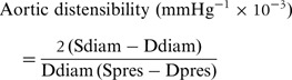
where Sdiam and Ddiam are the diameters of the aorta, and Spres and Dpres are the measured pressures in systole and diastole.
Statistical analysis
Values were expressed as mean values plus and minus standard deviations. Analysis of variance by Bonferroni adjusted multiple t-test was used for comparing data between groups. The relationship between two variables was evaluated by linear regression analysis and by calculation of Pearson correlation coeficients. A p value of less than 0.05 was considered statistically significant.
Results
Aortic dilation
Aortic dilation was found in 11 of the 24 patients (46%), with 6 of these individuals being aged from 10 to 20 years. The maximal dilation was seen at the level of the sinuses of Valsalva. Such dilation was the most common cardiovascular anomaly, with 6 of the patients (25%) having bicuspid aortic valves, 4 having aortic coarctation (17%), and 2 with systemic hypertension (8%).
Aortic dilation and cardiovascular risk factors
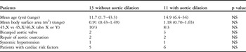
Aortic dilation was seen almost equally in patients with and without cardiovascular risk factors (Table 1), with 5 individuals without, and 6 with cardiovascular risk factors having aortic dilation. This included a girl, aged 12 years, with a bicuspid aortic valve who had a mild gradient of 20 mmHg. This did not change over 8 years of follow-up, and her entire aortic root was dilated up to 32 mm. About half of individuals with risk factors had a normal aortic root, suggestive of variable expression of this entity.
Clinical data of the patients with and without aortic dilation.

Aortic dilation and aortic distensibility
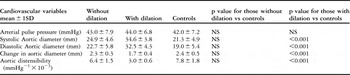
Changes in aortic diameters between diastole to systole were significantly less in those with aortic dilation when compared to both individuals without aortic dilation and normal control subjects (p < 0.001). Though pulse pressure was not significantly different between the patients and their controls, aortic distensibility was significantly less in those with aortic dilation when compared to those without aortic dilation and the controls (p < 0.001) (Table 2). There was an inverse correlation between aortic dimensions at the sinuses of Valsalva and calculated aortic distensibility (Table 2). There were two girls, aged 6 and 10 years, who had bicuspid aortic valves with normal aortic diameters and reduced distensibility.
Pulse pressures, aortic diameters at the level of the sinuses of Valsalva, and aortic distensibility in subjects with and without aortic dilation, and in the control group.

Aortic dilation and hormonal treatment
Of our 24 patients, 15 were receiving recombinant human growth hormone, 2 having completed their treatment. Of the 7 subjects without exposure to growth hormone, 3 had aortic dilation, as did 8 of the 17 subjects who had been exposed to the hormone. No relationship was found by paired t-test between the dose or duration of treatment with growth hormone and aortic dilation or corrected left ventricular mass.
Aortic dilation and karyotype
Of the patients, 18 had a single 45,X cell line. Mosaic subjects were found in 3 patients, specifically 45,X/46,X (abnY), and 3 were 45,X/46,X (abnX). Of the 18 subjects with a 45,X karyotype, 8 had aortic dilations, as did half the 6 subjects with mosaic karyotypes. So, no significant relation was found between the karyotype and aortic dilation (Table 1).
Discussion
Cardiovascular risk factors for aortic dilation and dissection include a bicuspid aortic valve, systemic hypertension, and coarctation of the aorta. As in the general population, presence of a bicuspid aortic valve independently predicts a larger proximal aorta in individuals with Turner’s syndrome.Reference Lopez, Arheart and Colan7 Such valves, or systemic hypertension, have been reported in up to nine-tenths of individuals with Turner’s syndrome suffering aortic dissection.Reference Lopez, Arheart and Colan7, Reference Carlson and Silberbach13 Patients lacking cardiovascular risk factors account for one-tenth of instances of aortic dissection, as revealed by a study including 85 cases reported between 1961 and 2006.Reference Carlson and Silberbach13 Accumulating data suggests that the Turner phenotype includes a generalized vasculopathy characterized by arterial dilation, thickening of the aortic walls, and abnormal propogation of the pulse wave.Reference Ostberg, Donald, Halcox, Storry, McCarthy and Conway9 Turner’s syndrome by itself, therefore, is considered an independent predictor of a larger proximal aorta.Reference Lopez, Arheart and Colan7
Aortic dilation
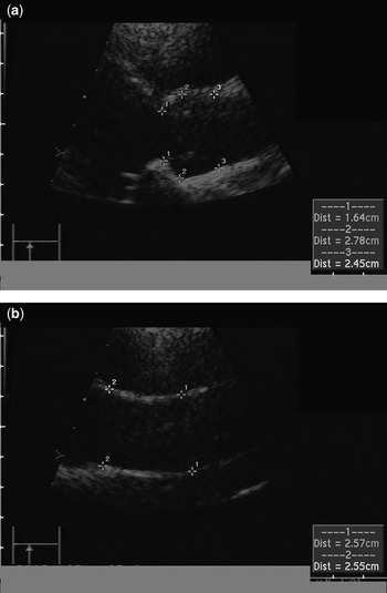
Dilation of the aortic root was the most common cardiovascular anomaly, found in almost half our cohort. Half of the individuals with cardiovascular risk factors, however, had aortic roots of normal dimensions, suggestive of its variable expression in this study. Just over half of the individuals with aortic dilation were aged from 10 to 20 years age, while 5 patients lacking risk factors had aortic dilation (Fig. 1). The dose and duration of growth hormone had no impact on either aortic dilation or corrected left ventricular mass. We also failed to find any significant relationship between the karyotype and aortic dilation.
Parasternal long axis views showing a dilated aortic root in girl of 10 years with Turner’s syndrome but without risk factors. The root measures 27.8 mm at the level of the sinuses of Valsalva, the upper normal limit for a body surface area of 0.92 m2 being 22.5 mm.

Aortic dissection
Aortic dissection is relatively rare, occurring in about 1.4% of individuals with Turner’s syndrome, with these patients presenting at a younger age as compared to the general female population.Reference Gravholt, Landin-Wilhelmsen and Stockholm4 An estimated incidence of dissection is 36 per 100,000 Turner syndrome years, as compared with an incidence of 6 per 100,000 in general population. The clinical profile of girls and women with Turner’s syndrome at risk of aortic dissection and rupture is not well described due to lack of data of its natural history. The predictive value of the size of the aortic root alone for dissection is uncertain in these patients. Aortic dissection and rupture are known to occur in patients with aortas of significantly smaller diameters when compared to those with Marfan’s syndrome.Reference Lopez, Arheart and Colan7, Reference Carlson and Silberbach13, Reference Loeys, Schwarze and Holm14 Deaths were reported in 2 adults, aged 24 and 44 years, with aortic dissection and their aortic diameters were 3.8 and 2.9 cm, respectively.Reference Lopez, Arheart and Colan7 As we have shown, aortic dilation is the most common anomaly seen in individuals with Turner’s syndrome, but its extent is variable, in those both with and without cardiovascular risk factors. Guidelines of aortic size as a marker for aortic dissection in the general population, based upon natural history data of patients with Marfan’s syndrome, therefore, cannot be applied for serial echocardiographic monitoring or surgical intervention for individuals with Turner’s syndrome.
Aortic elasticity
Connective tissue disorders associated with aortic root dilation include Marfan’s syndrome, involving a mutation of Fibrillin, Ehlers-Danlos syndrome with a mutation of collagen, and Loeys-Dietz syndrome, with mutations of transforming growth factor-B receptor, although they have a different natural history. Aortic dissection or rupture occurs in patients with aortas of smaller size in the setting of Loeys-Dietz than in Marfan’s or Ehlers-Danlos syndromes.Reference Loeys, Schwarze and Holm14 A connective tissue defect of a variable expression is seen in those with Turner’s syndrome. Histological evidence of cystic medial necrosis in aortic tissue taken from patients of aortic dissection with bicuspid aortic valves, Marfan’s syndrome, and Turner’s syndrome suggests a common aetiology, despite genetically diverse backgrounds.Reference Carlson and Silberbach13 An intrinsic abnormality of elastic property of aortic wall could be a predictive risk factor for aortic dilation and dissection in the setting of Turner’s syndrome, which can be measured echocardiographically as aortic distensibility. The predictive value of aortic distensibility in individuals with Turner’s syndrome, however, has not thus far, to the best of our knowledge, been evaluated.
Aortic distensibility
The aortic distensibility is a measure of aortic elasticity, which depends on elastic integrity of the aortic wall, and is also affected by haemodynamic stresses, such as aortic valvar disease, coarctation and systemic arterial hypertension, as well as sex hormones and advancing age.Reference Hirata, Triposkiadis, Sparks, Wooley, Bowen and Boudoulas10, Reference Carlson and Silberbach13 Aortic distensibility depends on change of aortic diameters rather than individual absolute values and pulse pressures. We used pressures in the brachial artery to estimate aortic pulse pressure, as these values have a good correlation with direct aortic pressures.Reference Stefanadis, Stratos, Boudoulas, Kourouklis and Toutouzas15 We showed an inverse relationship between ascending aortic diameters and aortic distensibility (Table 2). We found that 2 patients with bicuspid aortic valves had aortic roots of normal dimensions with reduced distensibility. The abnormal aortic elastic properties in these young individuals could be due to underlying connective tissue defect, and may be detected by measuring the serial aortic distensibility before an eventual aortic dilation. Hence, serial measurement of aortic distensibility may help to identify young individuals at risk of dissection at relatively smaller aortic diameters.
The combination of aortic diameters and aortic distensibility is a better echocardiographic tool rather than dimensions alone with which to follow individuals with Turner’s syndrome. The observations about aortic distensibility from our study, despite its cross-sectional nature and the relative small size of our cohort, are distinct. They now need to be validated by long term follow up of a larger group.
Conclusion
Aortic dilation is seen in individuals with Turner’s syndrome, both with and without risk factors. Aortic dilation in those without risk factors is accompanied by decreased aortic distensibility, suggestive of an intrinsic abnormality in elastic property of the ascending aorta. Individuals with aortic dimensions in excess of the 95th percentile with decreased distensibility should be followed closely, both clinically and echocardiographically.