

Introduction
The acanthocephalan Pseudocorynosoma constrictum (Van Cleave, 1918) is one of the most abundant species of the family Polymorphidae in North America. Like all acanthocephalans, this specie has an indirect life cycle involving two hosts. Successful transmission occurs when the definitive host, a waterfowl (e.g. the migratory blue-winged teal Anas discors) ingests a mature cystacanth of P. constrictum residing in the hemocoel of the amphipod Hyalella azteca (Duclos et al., Reference Duclos, Danner and Nickol2006). This species attaches to the small intestine of the definitive host, which, in birds, is located in the ventral peritoneal sac and occupies the caudal part of the body cavity and is the main section for food digestion and nutrient absorption (Sisson & Grossman, Reference Sisson and Grossman2002; Pawlina & Ross, Reference Pawlina and Ross2015). Different studies report that young acanthocephalans are usually found in the jejunum, and when they reach sexual maturity they migrate to the ileum, where adult worms are usually found (Aznar et al., Reference Aznar, Cappozzo, Taddeo, Montero and Raga2004).
Little is understood about clinical signs in birds infected with acanthocephalans, but, in some cases, infections of low intensity seem to have serious adverse effects on an infected animal; thus, acanthocephalan infection at any intensity should be considered to have pathogenic potential (Richardson & Nickol, Reference Richardson, Nickol, Atkinson, Thomas and Hunter2008). In this study, we used light microscopy to describe the histopathology of the small intestine of individuals of A. discors, associated with P. constrictum.
Material and methods
We processed 17 dead individuals of A. discors obtained by local hunters in the wetland Chimaliapan (19°11′20″N, 99°29′30″W), located in San Pedro Tultepec, State of Mexico, Mexico, between February 2016 and April 2017. The birds were identified taxonomically using Preston (Reference Preston1998) and Van Perlo (Reference Van Perlo2006) field guides.
Between two and three hours after bird hunt, the individuals were taken to the laboratory where the small intestine was removed and examined using a stereomicroscope. Pseudocorynosoma constrictum was found in the ileum of ten of these teals (prevalence 72.7%; intensity 1–33 specimens, mean intensity 11.75). For the acanthocephalan identification, a total of 12 individuals were carefully extracted with entire proboscis using micro-dissecting needles. The proboscis and the presence of genital spines – that are considered in the genera Pseudocorynosoma and Corynosoma – were used as taxonomic identification criteria (Aznar et al., Reference Aznar, Pérez-Ponce de León and Raga2006). The acanthocephalans were kept in distilled water and refrigerated at 4°C for 24 h to correct eversion of the proboscis; then, they were fixed, stained and mounted on permanent slides using helminthological techniques, as described in Salgado-Maldonado (Reference Salgado-Maldonado2009), and we used light microscopy to identify the individuals using keys by Yamaguti (Reference Yamaguti1963) and McDonald (Reference McDonald1988). Specimens of P. constrictum collected in the current study were deposited in the Colección Nacional de Helmintos (CNHE), Instituto de Biología, Universidad Nacional Autónoma de México, Mexico City, Mexico, and the voucher numbers are pending at the time of this publication.
To perform the histopathological analysis, ileum segments with attached P. constrictum were fixed in Bouin's solution for 24 h to maintain the structural and molecular composition of the tissue (Shaikh et al., Reference Shaikh, Ursani, Naz, Dhiloo and Solangi2016). After this time, the fixative was changed for 70% ethanol every 24 h until Bouin's solution was completely eliminated. All segments of the ileum were then embedded in Paraplast X-TRA® (Leica Biosystems, Richmond, IL, USA) (Rodríguez-Antolín et al., Reference Rodríguez-Antolín, Xelhuanzi, García-Lorenzana, Cuevas, Hudson and Martinez-Gómez2009); the resulting blocks were cut with a microtome into longitudinal, transverse and sagittal tissue sections of c. 6 μm. Each section was placed in a 45°C water bath containing gelatin until the tissue and paraplast were completely smooth. Then, the sections were removed from the water bath using gelatin-coated glass slides, the excess of water was drained and the slides were placed on a paper towel with the tissue section facing down. Afterwards, slides were stained using haematoxylin–eosin and were observed using a light-microscope in order to describe the different types of tissue and lesions.
Results and discussion
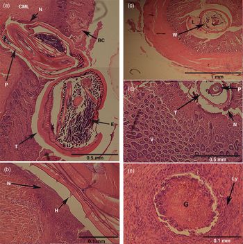
Pseudocorynosoma constrictum inserts the entire proboscis into the inner muscle layer (fig. 1a) and remains held in the tissue with hooks (fig. 1b), which allows the parasite to be completely attached to the gut while its body remains free in the lumen to absorb the nutrients that the host provides. The mucosal surface also shows damage caused by the spines of the presoma (fig. 1c). Lesions around the area where the proboscis inserts consist of destruction of villi, degeneration of intestinal crypts, presence of numerous necrotic cells in the adjacent tissue, lamina propria infiltrated by heterophils and monocytes, inflammatory reaction in the surrounding and, sometimes, the presence of erythrocytes and free granulocytes in the lumen or around the lesion, which is indicative of bleeding (figs. 1a–d). This damage could be related to proteolytic enzymes that some species of acanthocephalan produce; those enzymes degrade peptides in the tissue surface, which allows the worm to penetrate rapidly and deeply into the intestinal tissue (Polzer & Taraschewski, Reference Polzer and Taraschewski1994).
Histopathology of Pseudocorynosoma constrictum in the ileum of Anas discors. (a) The proboscis (P) of P. constrictum reaching the circular muscle layer (CML), surrounded by necrotic tissue (N); blood cells (BC) are observed next to the villi and eggs (E) within the worm trunk (T). (b) Higher magnification (40×) showing hooks (H) and abundant necrotic tissue (N). (c) Worm body (W) obstructing much of the intestinal lumen (L). (d) Section at the mucosa level, showing villi (V) around the proboscis (P) and trunk (T) with extensive necrotic damage (N). (e) Granuloma (G) surrounded by a lymphocyte collar (Ly).

Previous studies indicate than not all species of acanthocephalans produce the same type of damage or penetrate the same gut layer. For example, in vertebrate hosts like fish (Dezfuli et al., Reference Dezfuli, Pironi, Domeneghini and Bosi2002), amphibians (Heckmann et al., Reference Heckmann, Amin, Tepe, Dusen and Oguz2011), marine mammals (Amin et al., Reference Amin, Heckmann, Halajian and El-Naggar2011, Reference Amin, Heckmann, Halajian, El-Naggar and Tavakol2013) and terrestrial mammals (Choi et al., Reference Choi, Lee, Go, Park, Chai and Seo2010; Heckmann et al., Reference Heckmann, Amin, Halajian and El-Naggar2013), the complete proboscis penetrates only the mucosa, causing damage in villi, crypts and connective tissue. However, in the marine bird Leucophaeus modestus, the proboscis of Profilicolis altmani reaches the muscularis of the jejunum and ileum (Gonzales-Viera et al., Reference Gonzales-Viera, Luján-Vega, Chavera-Castillo, Cárdenas-Callirgos and Tantaleán2009), which agrees with the scope of injuries associated with P. constrictum in this analysis; these lesions are possibly facilitated by the shape and length of the proboscis of this species.
The extent of damage that helminths cause in the intestine is related to the intensity of the infection and depth of parasite penetration in the host tissues (Dezfuli et al., Reference Dezfuli, Pironi, Domeneghini and Bosi2002). The penetration of the proboscis of P. constrictum in the muscularis and the aforementioned damage could alter the homeostasis in the host. It has been shown that the isotonicity of luminal contents in the presence of structural damage on the mucosa is difficult to maintain, because intestinal epithelium promotes the strong exchange of water and electrolytes with the body's extracellular fluids (Nighot & Nighot, Reference Nighot and Nighot2018). In addition, enterocyte alteration can cause intestinal secretion – reabsorption function can quickly promote severe body fluid depletion and affect the absorption of salts by channels (Nighot & Nighot, Reference Nighot and Nighot2018).
Amin et al. (Reference Amin, Heckmann, Halajian and El-Naggar2011) observed marks at the mucosa level that suggest the migration pathways of individuals of Corynosoma strumosum along the intestine of the seal Pusa caspica. These kinds of marks were not observed in this study; instead, we observed that the mucosa had several granulomas not associated to an embedded proboscis (fig. 1e), which could be the result of migratory movement of the worm to another site or could perhaps be the result of scars from worms that had died. The granulomas seem to be the result of new tissue covering the lesions resulting from the host's immune reaction; although other histopathological analyses have demonstrated the presence of this type of lesions (Gonzales-Viera et al., Reference Gonzales-Viera, Luján-Vega, Chavera-Castillo, Cárdenas-Callirgos and Tantaleán2009; Choi et al., Reference Choi, Lee, Go, Park, Chai and Seo2010), no details are presented.
We found two cases of high infection with more than 30 individuals each. We observed that most parasites crossed the gut layers completely, perforating the entire ileum wall, which not only compromises the structural and functional design of the organ but also represents a new route for secondary infections. The high number of parasites severely infecting the hosts may cause malabsorption of nutrients by the host as a result of mucosal reduction due to the host–parasite nutritional competition (Bush et al., Reference Bush, Fernández, Esch and Seed2001). Similarly, the increased damages in the intestinal tissue due to a higher number of parasites could alter the homeostasis in the host (Nighot & Nighot, Reference Nighot and Nighot2018). We are not aware of any report of similar damages.
Finally, we did not observe any evidence of the encapsulation of the parasite with connective tissue as a response of the host's immune system, which allows the acanthocephalans to continue migrating along the intestine (Amin et al., Reference Amin, Heckmann, Halajian, El-Naggar and Tavakol2013).
In conclusion, the insertion of the proboscis and hooks of P. constrictum into the muscular layer of the small intestine of the blue-winged teal causes inflammatory reactions, haemorrhaging, necrosis and the destruction of villi and crypts, which could alter the physiological functioning of the host's alimentary canal, which, in turn, can affect secretion, absorption and homeostasis. Furthermore, the existence of a large number of worms causes the occlusion of several lumens and sometimes a total perforation of the intestine.
Acknowledgements
We are grateful to Dr Raul Fajardo for allowing us to perform histological analyses in the Centro de Investigación y Estudios Avanzados en Salud Animal (CIESA) of UAEMex, and to Ms Berenit Mendoza Garfias for her valuable support in the use of the electron microscopy of the Instituto de Biología in the Universidad Nacional Autónoma de México (UNAM). Finally, we would like to thank Héctor Vázquez and Tonia De Bellis for commenting on previous versions of the manuscript and for their revision of the text.
Financial support
This study was supported in part by the Consejo Nacional de Ciencia y Tecnología (CONACyT) through a scholarship to Carmen Caballero-Viñas to complete her doctoral thesis, of which this work is a result (grant number 252123).
Conflicts of interest
None.
Ethical standards
Our study received the field permits and the approval of the ethics committee from Universidad Autó noma del Estado de Mé xico (0110104).