Intramammary infection (mastitis) is common in dairy cows, major consequences of which are lower milk yield and shortened productive life causing significant economic losses (Rajala-Schultz et al. Reference Rajala-Schultz, Gröhn, McCulloch and Guard1999; Petrovski et al. Reference Petrovski, Trajcev and Buneski2006; Halasa et al. Reference Halasa, Huijps, Østerås and Hegeveen2007). In mastitis treatment, intramammary infusion is the most common route of applying a drug. This delivers a high drug concentration directly into the infected quarter. Each drug has a prescribed period of withhold of milk from sale to avoid antimicrobials in the milk supply. Violative residues following intramammary treatment of mastitis may arise following inappropriate treatment records, inability to identify and withhold milk from treated cows, too short a withhold period (WP) and off-label, and combination usage (Oliver et al. Reference Oliver, Maki and Dowlen1990; McEwen et al. Reference McEwen, Black and Meek1992; FDA, 2015). Antimicrobial residues in milk are of concern because of the risk of developing resistant microorganisms, allergic reactions in milk consumers, and inhibition of starter cultures used in the dairy food industry (Seymour et al. Reference Seymour, Jones and McGilliard1988; McEwen et al. Reference McEwen, Black and Meek1991).
Some studies have reported prolonged excretion of antimicrobials in milk after treating cows for mastitis. However, to our best knowledge, reports describing excretion of antimicrobial residues in milk from individual quarters in cows treated with combined antimicrobial therapy do not exist. The pharmacokinetic properties of a drug applied through either the intramammary or systemic route is greatly affected by the pH of the medium and the dissociation constant (pKa) of the drug, as well as the ability of protein binding (Ziv, Reference Ziv1975). Differences occur in the physico-chemical characteristics between the healthy milk and milk from mastitic udder, with most of the pharmacokinetic studies being performed in healthy cows. Studies performed in mastitic cows showed slower drug elimination. Poelarends et al. (Reference Poelarends, Hogeveen and te Giffel2001) noted no significant difference of WP after treating cows for mastitis with combination therapy and single intramammary therapy. However, the mean excretion time was longer for combination therapy. Slower elimination of azithromycin from milk of mastitic quarters was noted after treating bovine subclinical mastitis via the intramuscular route (Lucas et al. Reference Lucas, Errecalde and Mestorino2009). Different pharmacokinetic properties of norfloxacin were noted after intramuscular treatment of lactating cows suffering from clinical and subclinical mastitis (Gips & Soback, Reference Gips and Soback1999). Change in elimination kinetics are not restricted to antimicrobials only. Elimination kinetics of other drugs are also affected. For example, prolonged clearance from the body of mastitic cows have been reported after the systemic use of non-steroidal anti-inflammatories (Lohuis et al. Reference Lohuis, van Werven, Brand, Van Miert, Rohde, Ludwig, Heizmann and Rehm1991; Kissell et al. Reference Kissell, Leavens, Baynes, Riviere and Sminth2015). These studies suggest that antimicrobial drugs in therapy of mastitis in dairy cows pose risk of antimicrobial residues in milk after the prescribed WP. Treatment of mastitis with a combination of antimicrobial drugs is frequently practiced in Slovenia and elsewhere (du Preez, Reference du Preez1988; Oliveira & Ruegg, Reference Oliveira and Ruegg2014).
To protect public health and prevent any adverse effects, Maximum Residue Limits (MRL) for pharmacologically active substances are required in the European Union (EU) by Commission Regulation (EU) 37/2010 (European Commission, 2010). The WP, indicated in the instructions’ label for a medicine should be long enough for any residues in animal products intended for human consumption to fall below the MRL. Investigations concerning the WP are encouraged by Directive 2001/82/EC (EC, 2001), but persistence of antimicrobial residues in milk after the WP in dairy cows is a problem reported for decades (Johnson et al. Reference Johnson, Martin, Baker and Parsons1977; Oliver et al. Reference Oliver, Duby, Prange and Tritschler1984; Seymour et al. Reference Seymour, Jones and McGilliard1988; McEwen et al. Reference McEwen, Black and Meek1992; Podpečan et al. Reference Podpečan, Pengov, Zrimšek, Sekulovski and Kirbiš2014).
Bulk tank milk occasionally tests positive to antimicrobials, but far less frequently than in the past. Slow and prolonged excretion of antimicrobials in milk after mastitis treatment has been reported in a few published studies but has not received much attention (McEwen et al. Reference McEwen, Black and Meek1991, Reference McEwen, Black and Meek1992; FDA, 2015). If the excretion of antimicrobials is slower in mastitic quarters treated with combination of antimicrobial drugs, bulk tank milk may be contaminated with antimicrobial residues even when the prescribed WP has been respected. The purpose of this study was to examine, on commercial farms, if treating clinical mastitis with a combination of certain antimicrobial drugs (extra-label use) requires a longer withhold period.
Materials and methods
Sampling
Quarter milk samples were collected on detection from 35 lactating Holstein Friesian cows presenting clinical mastitis, from two commercial dairy farms (one with 200 and the other with 180 dairy cows) in Slovenia. Each farm had daily veterinary attendance. Cows had access to drinking water ad libitum and were fed twice daily after milking with a total mixed ration (based on home produced forages: grass silage, maize silage, hay and straw; concentrates and vitamin-mineral supplement) according to National Research Council (2001) recommendations. Both dairy farms had a free-stall housing system. On both farms cows were milked twice daily, in a milking parlour, and were producing an average 8800 and 9000 kg liquid milk per lactation respectively.
Cows enroled in the study had no concomitant diseases diagnosed. The veterinarian chose all treatments for the mastitis. A similar method was used on both farms with minor differences according to cow. All cows were treated by the intramammary route with a preparation containing tetracycline, neomycin, bacitracin and prednisolone (Mastijet Fort®, Intervet International, Boxmeer, the Netherlands). Additionally, the cows received other antimicrobial drugs (details in online Supplementary Table S1). Post treatment milk samples were taken from the mastitic quarters and from untreated healthy quarters in the same cow. Milk samples were collected aseptically in sterile 10 ml test tubes and kept at 4–8 °C until bacteriological analysis. Identification of the bacteria responsible for the infection was carried out within 24 h. For the analysis of drug residues, milk samples were collected from treated and healthy quarters at two milkings before, and two milkings after the prescribed WP. The samples were collected in 25 ml containers and stored at 0 °C until analysis. Nine milk samples were collected per cow, making a total of 315 samples from 35 cows.
The attending veterinarians completed a questionnaire, providing data about the cow; the prescribed WP, age, lactation number, number of infected udder quarters, basic clinical signs (fever, quarter oedema, decreased milk production, abnormal milk); and treatments applied.
Analytical methods
Identification of bacteria
Each milk sample was plated onto blood agar (Trypticase soy agar with 5% sheep blood) and incubated at 37 °C for 18 to 24 h. Mastitis-causing pathogens were identified by Matrix-Assisted Laser Desorption Ionization-Time of Flight mass spectrometry (MALDI-TOF, Bruker Daltonics, Bremen, Germany). A fresh colony was smeared onto a ground steel MALDI target plate in a thin film and covered with 1·5 µl of a saturated solution of α-cyano-4-hydroxy-cinnamic acid in 50% acetonitrile-2·5% trifluoroacetic acid. The mass spectrum was evaluated in the linear positive mode (Microflex; Bruker Daltonics, Bremen, Germany). The mass spectra (ranging from 2000 to 20 000 Da) were compared with the reference spectra of the integrated database provided by the manufacturer, using MALDI Biotyper software (Bruker Daltonics, Bremen, Germany).
All samples taken from the healthy untreated mammary quarters before and after the prescribed WP were analysed by a microbiological method using five-plates so-called ‘the STAR protocol’ (Gaudin et al. Reference Gaudin, Maris, Fuselier, Ribouchon, Cadieu and Rault2004). The samples taken after the WP from treated mastitic quarters were analysed by the STAR protocol, Delvotest® SP-NT (DSM, Delft, the Netherlands) and TwinsensorBT KIT020 (Unisensor, S.A., Angleur, Belgium). If a positive sample was detected, all milk samples taken from that quarter, including those taken before the prescribed WP, were analysed by the appropriate quantitative method (quinolones with HPLC-fluorescence detector, tetracyclines with LC-MS/MS, and penicillins with UPLC-MS/MS). The performances of screening tests and confirmative methods are presented in the online Supplementary Table S2.
Antimicrobial analysis
Amoxicillin trihydrate, penicillin G potassium salt, oxacillin sodium salt monohydrate, cefquinome sulphate, cephalexin, tetracycline hydrochloride, neomycin trisulfate salt hydrate, gentamicin sulphate salt, kanamycin A disulfate salt hydrate, dihydrostreptomycin sesquisulfate, streptomycin sesquisulfate hydrate, novobiocin sodium salt, enrofloxacin and ciprofloxacin were sourced from Sigma-Aldrich (St. Louis, MO, USA).
Sample analysis and validation were carried out using TwinSensorBT Kit020 (Unisensor, S.A., Angleur, Belgium) as described in the manufacturer's manual and as described by Perme et al. (Reference Perme, Bizjak, Gačnik and Kirbiš2010), respectively. The sensitivity limits for amoxicillin, penicillin, cefquinome, cefalexin and tetracycline are listed in the Supplement file. The dipsticks were read visually and by ReadSensor (Unisensor, S.A., Germany). In each set of samples, one positive and one negative control were added.
Milk samples were analysed using the Delvotest® SP-NT (DSM, Delft, The Netherlands) according to the manufacturer's instructions. In each analysis run, positive and negative controls were included.
The STAR protocol, a five-plate microbiological test for detection of antimicrobial residues in milk, was used with Bacillus cereus ATCC 11778, Escherichia coli ATCC 10536, Staphylococcus epidermidis ATCC 12228, Kocuria rhizophila ATCC 9341 (KWIK-STIK, Microbiologics, MN, USA), and Bacillus subtilis B.G.A. spore suspension (Merck, Darmstadt, Germany). For tetracycline, samples were prepared as described by Raspor Lainšček et al. (Reference Raspor Lainšček, Biasizzo, Henigman, Dolenc and Kirbiš2014). Details of the preparation are listed in the Supplement file.
Enrofloxacin and ciprofloxacin were analysed using HPLC-fluorescence detector following the procedure of Kirbiš et al. (Reference Kirbiš, Marinšek and Flajs2005) with minor modifications (Supplement file). The method was validated according to the European Commission Decision 2002/657/EC (EC, 2002).
For tetracycline analysis with LC-MS/MS we prepared milk samples following the method of MacNeil et al. (Reference MacNeil, Martz, Korsrud, Salisbury, Oka, Epstein and Barnes1996) with minor changes (Supplement file), and measurements were made according to Založnik et al. (Reference Založnik, Bajc and Gačnik2007). The method was validated according to the European Commission Decision 2002/657/EC (EC, 2002).
Amoxicillin and procaine benzylpenicillin were identified, and quantified following the method of Pezdir et al. (Reference Pezdir, Gačnik and Dolenc2012), with minor changes using UPLC-MS/MS (Supplement file). The method was validated according to the European Commission Decision 2002/657/EC (EC, 2002).
Statistical analysis
Statistical analysis comprised significance testing (P ≤ 0·05) of the difference between tetracycline positive and tetracycline negative milk samples using a two-way ANOVA (IBM SPSS Statistics, Version 22, 2013). To equalise sample number between the two groups, 15 samples were selected randomly from the tetracycline negative group. Bacterial infection, combination antimicrobial therapy, treatment interval and duration, and cow status (parity, fever, udder health, and milk production) were evaluated for statistical significance.
Results and discussion
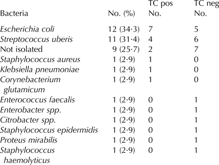
In 9 (25·7%) of 35 cows no bacteria were isolated (Table 1), the upper range of culture negative results (Guterbock et al. Reference Guterbock, Van Eenennaam, Anderson, Gardner, Cullor and Holmberg1993). A total of 31 colonies were isolated and identified from 26 milk samples. These comprised 11 bacterial species.
Bacterial found in milk samples collected from mastitic quarters in correlation to tetracycline (TC) residues.

TC pos – milk from quarters positive for tetracycline residues after the WP.
TC neg – milk from quarters negative for tetracycline residues after the WP.
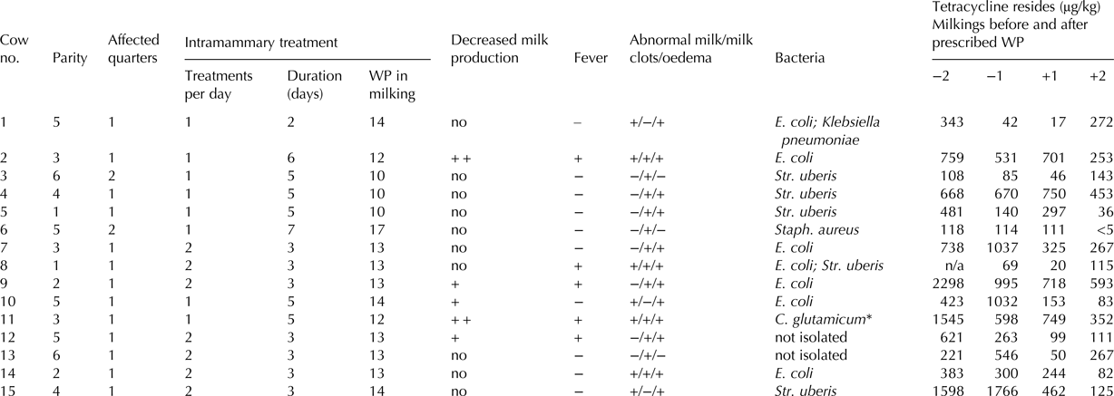
All milk samples from healthy untreated quarters were free of antimicrobials before and after the WP. In 15 (42·9%) cows the samples from the treated quarters contained tetracycline above the MRL (100 µg/kg) after the prescribed WP (Table 2). In four cases (cows 2, 4, 9, and 11) the tetracycline concentration was at least seven times the MRL. In two cows (2 and 11) milk production was highly depressed (>50%) accompanied by fever, abnormal milk with clots and quarter oedema (Table 2). In four samples (26·7%), the levels of tetracycline detected were below the MRL in the first milking intended for consumption, while, at the next milking, the concentration of tetracycline was above the MRL. A similar finding has been reported by McEwen et al. (Reference McEwen, Black and Meek1992). Due to dilution with milk from the other three untreated quarters, whole udder milk was below the MRL. Serraino et al. (Reference Serraino, Giacometti, Marchetti, Zambrini, Zanirato, Fustini and Rosmini2013) have reported tetracycline residues above the MRL in 0·34% of the bulk tank milk samples.
Clinical signs (abnormal milk, milk clots, oedema, decreased milk production, fever), parity, number of affected quarters, treatment frequency and duration, bacterial identification and tetracycline concentration in milk of cows treated for clinical mastitis exceeding the MRL value (100 µg/kg) for tetracycline after the prescribed WP.

* Corynebacterium. glutamicum is probably a result of contamination or misidentification as it is not known as a mastitis-causing species.
n/a, not available.
−2 – two milkings before WP.
−1 – One milking before WP.
+1 – one milking after WP.
+2 – two milkings after WP.
++ very decreased.
The label instructions for the intramammary tetracycline product advises a WP of 4 d (8 milkings), but because of the off-label use, the farm veterinarians prescribed a longer WP (Table 2). Numerically, more of the cows having tetracycline residues in milk after the WP (7/15, 46·7%) had been treated every 12 h than were the negative cows (6/20, 30·0%). Treatment duration and interval (12 or 24 h) did not differ significantly between the tetracycline positive and tetracycline negative cows (P > 0·05).
Cow description and health observations, including parity, decreased milk production, abnormal milk and fever, differed significantly between tetracycline positive and tetracycline negative cows (P < 0·05). Older cows were slower to eliminate tetracycline. The average parity of cows exceeding the tetracycline MRL after the WP was 3·7 (median 4). In cows not exceeding the tetracycline MRL, the average parity was 2·6 (median 2) (Table 2). Fever and decreased milk production occurred in 5 (33·3%) tetracycline positive cows, while, in tetracycline negative group 6 (30·0%) cows were pyrexic and 5 (25·0%) cows had a decreased milk production.
Four tetracycline positive cows and one tetracycline negative cow displayed abnormal milk colour, quarter oedema and milk clots (Table 2). All tetracycline positive samples contained residues of aminoglycosides. Aminoglycosides were detected in 21 (61·8%) samples after the prescribed WP. Neomycin was administered to all cows as part of the tetracycline preparation, along with dihydrostreptomycin (4 cases), gentamicin (2 cases) and kanamycin (1 case). In only two cases (cows 10 and 15) was the MRL value for neomycin (1500 µg/kg) exceeded (3741 and 2884 µg/kg).
Enrofloxacin and its metabolite ciprofloxacin were not found in samples taken two milkings before the WP by HPLC with a fluorescence detector. Enrofloxacin was not detected in any of the other samples analysed with the STAR protocol. Enrofloxacin was always administered parenterally and had a WP of 3 d, hence its absence.
β lactams were detected in milk samples from 12 cows (34·3%), taken after the WP. Eleven samples also contained tetracycline residues above the MRL (Table 2), but procaine benzylpenicillin and amoxicillin were below the MRL (4 µg/kg). Cefquinome was detected in cow no. 2 and 10 with the Twinsensor after the WP. The estimated concentration of cefquinome in cow no. 2, detected with Twinsensor, but not with STAR-protocol, was <40 µg/kg, in cow no. 10, it was 60 µg/kg. In both cases the MRL of 20 µg/kg was exceeded. Cephalexin exceeded the MRL (100 µg/kg) in the first milking after the WP (171 µg/kg) in cow no. 16.
According to the European Medicinal Agency the harmonised method in the EU to determine a WP is the ‘Time to Safe Concentration’ (TTSC). Using healthy dairy cows, It determines the time necessary for the concentration to drop below, and remain below, the MRL (European Medicines Agency, 2000). The WP is evaluated by the manufacturer of the drug preparation. However, the pharmacokinetics of antimicrobials administered to cows with mastitis differ from those of healthy animals (Cagnardi et al. Reference Cagnardi, Villa, Gallo, Locatelli, Carli, Moroni and Zonca2010). During mastitis the pH of milk often increases. An antimicrobial may pass through the ‘leaky’ epithelial barrier into milk, most probably by passive diffusion. Only unbound, nonionized and lipid-soluble drugs pass through the udder's epithelium into the plasma (Ziv & Sulman, Reference Ziv and Sulman1975; Gehring & Smith, Reference Gehring and Smith2006). The prolonged excretion of residues in milk, as shown in our results, can be partly attributed to the physical and chemical changes in the mastitic mammary gland.
Conclusions
Residues of tetracycline had prolonged excretion in mastitic cows, particularly in older cows with decreased milk production and abnormal milk. If milk from several post treatment cows, when combination treatment is used, is consigned for sale along with milk from untreated cows then milk quality and safety may be at risk. Withhold periods only apply to individual cows. When several cows have been treated the veterinarian should prescribe a WP safety margin.
Supplementary material
The supplementary material for this article can be found at https://doi.org/10.1017/S0022029918000389
The research has been founded with support from the European Commission (Erasmus Mundus, BASILEUS project) and the Slovenian Research Agency (research programme P4-0092). The authors thank Prof. de Almeida (Lisbon, Portugal) for critical reading of the manuscript.