A subgroup of patients diagnosed prenatally at mid-gestation with aortic stenosis and certain echocardiographic features progress to hypoplastic left heart syndrome at late gestation.Reference Danford and Cronican 1 – Reference Makikallio, Mc Elhinney and Levine 3 Hypoplastic left heart syndrome is a congenital heart defect that occurs in ∼1 of 2800–6200 live births. It is lethal if left untreated, and it accounts for 23% of cardiac deaths diagnosed during the 1st week of life and 15% within the first month of life.Reference Tweddell, Hoffman, Ghanayem, Frommelt, Mussatto and Berger 4 In spite of significant improvement in survival rates over the past decades after stage I Norwood and hybrid procedures, mortality and morbidity remain high.Reference Mahle, Spray, Wernovsky, Gaynor and Clark 5 , Reference Tweddell, Hoffman and Mussatto 6 Neurologic damage attributed to a lack of anterograde flow through the aorta during foetal life has also been described in this population.Reference Rogers, Msall and Buck 7 , Reference Mc Elhinney, Benson and Brown 8
It has been reported that prevention of hypoplastic left heart syndrome is possible in selected patients by relieving aortic stenosis in utero by foetal aortic valvuloplasty.Reference Tworetzky, Wilkins-Haug and Jennings 9 – Reference Artz, Wertaschnigg, Veit, Klement, Gitter and Tulzer 12 The purpose of this study is to show our experience in percutaneous ultrasound-guided balloon valvuloplasty in foetuses with severe aortic stenosis at risk of progression to hypoplastic left heart syndrome.
Materials and methods
We reviewed foetal echocardiograms and medical records of patients with prenatal diagnosis of severe aortic stenosis considered at risk of progression to hypoplastic left heart syndrome who underwent prenatal valvuloplasty at the Fetal Medicine Unit in Hospital Italiano of Buenos Aires from May, 2005 to December, 2010.
Foetal echocardiograms were performed in all cases by three paediatric cardiologists. Foetal echocardiograms to guide aortic valvuloplasties were performed by one foetal medicine specialist.
Patient selection for foetal aortic valvuloplasty
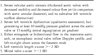
Patients were selected as candidates for foetal aortic valvuloplasty when the following criteria were met: severe aortic stenosis at risk of progression to hypoplastic left heart syndrome (echocardiographic criteria listed in Table 1), single pregnancy, no associated major malformations, normal karyotype, and no maternal contraindications.
Echocardiographic criteria for selection of patients for foetal aortic valvuloplasty.

z-Scores for foetal cardiac measurements were calculated relative to estimated gestational age based upon the data reported by Schneider et al,Reference Schneider, McCrindle, Carvalho, Hornberger, McCarthy and Daubeney 13 and for neonatal cardiac measurements upon data reported by Daubeney et al.Reference Daubeney, Blackstone, Weintraub, Slavik, Scanlon and Webber 14
This study was approved by the Institutional Review Board of the Hospital Italiano de Buenos Aires. Patients undergoing the procedure were previously counselled by members of our Fetal Medicine Unit regarding the intervention and its potential harms and benefits for both mother and foetus.
Foetal intervention
The interventions were performed under ultrasonographic guidance by maternal–foetal medicine specialists with experience in foetal therapy, and by a team of paediatric cardiologists with experience in cardiac catheterisation.
After maternal epidural anaesthesia, foetal position was evaluated and, if necessary, external foetal version was performed in order to achieve an optimal foetal position: left side of the foetal chest wall anteriorly oriented, without limbs between the left ventricle apex and the uterine wall, and with the left outflow tract parallel to the intended cannula course. Once positioning was achieved, atropine (20 μg/kg), vecuronium (0.2 mg/kg), and fentanyl (10 μg/kg) were administered intramuscularly to the foetus for paralysis and anesthesia.
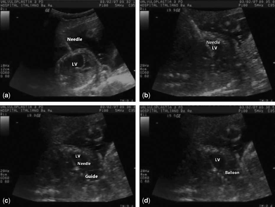
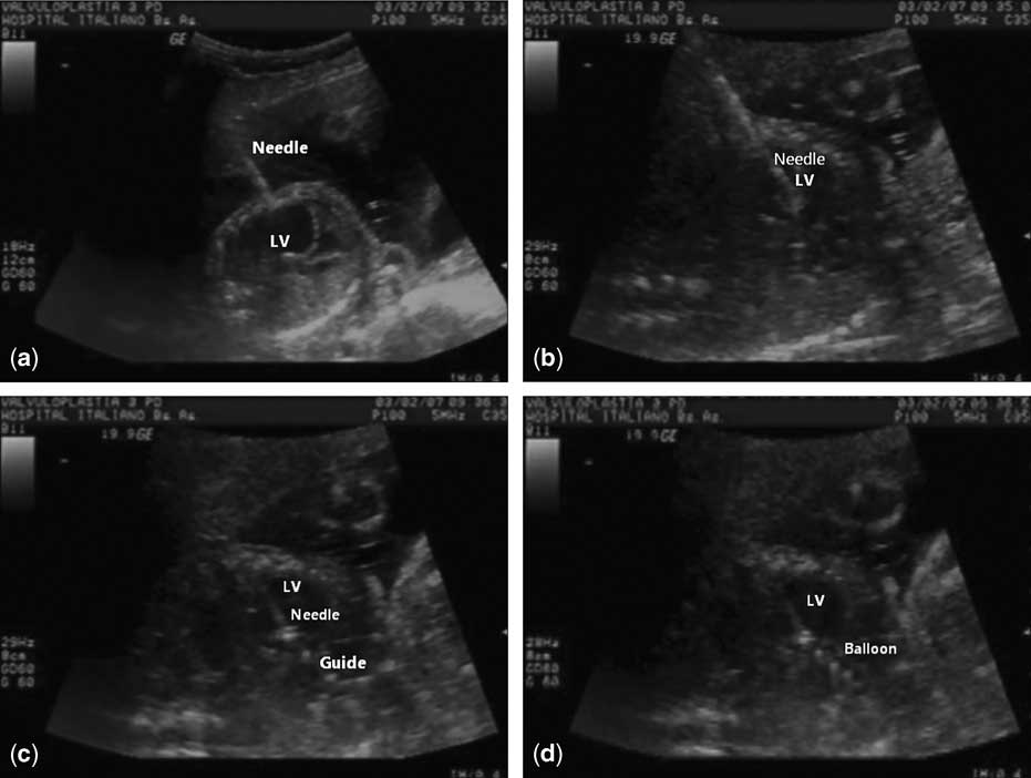
A 12 cm-long, 18 gauge, in the first three procedures, or 19 gauge, in the last two cases, cannula and stylet needle were introduced through the maternal abdomen and the uterine wall into the amniotic cavity and then guided through the foetal chest wall into the foetal left ventricle. Once inside the left ventricle, the cannula was moved to the aortic valve. The stylet was then removed and a 0.014-inch coronary guidewire with a coronary balloon was progressed across the aortic valve. The balloon was then inflated to a diameter between 0.9 and 1.0 times the size of the aortic valve annulus, gauging the pressure to allow precise estimates of inflation diameters (Fig 1). Finally, after aortic valvuloplasty, both the cannula and the guidewire with the balloon were removed all together in order to reduce the chance of tearing up the balloon and leaving a portion inside. Intervention time period was defined by the interval between the introduction of the cannula and stylet needle through the maternal abdomen and their withdrawal. It did not include foetal positioning time.
( a ) Foetal aortic valvuloplasty: a needle through the maternal abdomen towards the left ventricle, ( b ) the needle inside the left ventricle, ( c ) the guidewire across the aortic valve, and ( d ) balloon inflation. LV = left ventricle.
Statistical analysis
Continuous variables are presented as either mean ± standard deviation or as median with ranges. Categorical data are presented in number (n) and percentages.
Results
Foetal echocardiographic evaluation
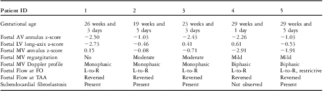
In the study period, five foetuses were selected for foetal aortic valvuloplasty. Median maternal age was 28 years (range: 23–32 years). Median gestational age at initial evaluation was 23 weeks (range: 19 weeks and 5 days to 29 weeks and 5 days). All the cases had severe aortic stenosis with dysplastic thickened and rigid valves, and no subvalvar obstruction. The mean aortic valve z-score was −1.85 (±0.67). The mean left ventricle inlet z-score was −0.54 (±1.19). In the first patient, there was a delay between the first assessment and the procedure, and thus when the valvuloplasty was performed the left ventricle z-score had gone down to −2.73. The mean mitral valve z-score was −1.09 (±1.16). All foetuses presented with severely decreased left ventricular contractility, and all but one had subendocardial fibroelastosis. None of the patients presented with pericardial effusion, tricuspid regurgitation, or other cardiac malformation. Echocardiographic features are detailed in Table 2.
Foetal echocardiographic evaluation before foetal aortic valvuloplasty.

AV = aortic valve; FO = foramen ovale; L-to-R = left-to-right; LV = left ventricle; MV = mitral valve; TAA = transverse aortic arch
Foetal intervention
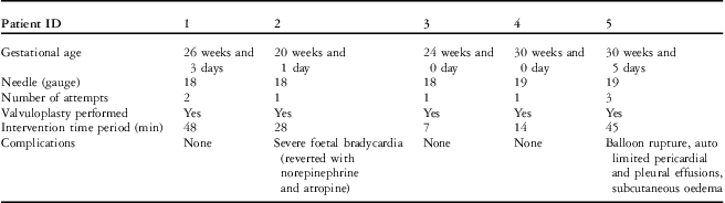
The median gestational age at valvuloplasty was 26 weeks and 3 days (range: 20 weeks and 1 day to 30 weeks and 5 days). In all the cases, external foetal version was performed and achieving an optimal foetal position was the most time-consuming part of the procedure. The median intervention time period was 28 minutes (range: 7–48 min). In all foetuses, the aortic valve was crossed and the balloon was inflated (Table 3). In one foetus (case 3), reversed blood flow in the aortic transverse arch became antegrade after valvuloplasty. None of the intervened foetuses presented aortic regurgitation. Severe foetal complications occurred in two of five cases. There was one foetus that presented extreme bradycardia while the cannula was still inside the left ventricle, reverting with intracardiac epinephrine and atropine administration. In one case, there was a balloon rupture, and pericardial and pleural effusion, both recovering spontaneously after a few days. There were no foetal losses or premature delivery due to the procedure. There were no maternal complications.
Foetal aortic valvuloplasties: procedural features.

Outcome
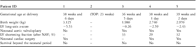
In one case, the couple decided to terminate the pregnancy 3 weeks after foetal valvuloplasty. Another patient was born at 36 weeks and 5 days with intrauterine growth restriction, and the other three were born at term (38–39 weeks) with appropriate weight for gestational age (mean of 2945 g (±158); Table 4).
Postnatal features of patients who underwent foetal aortic valvuloplasty.

LV = left ventricle; NAV = neonatal aortic valvuloplasty; TOP = termination of pregnancy
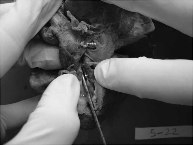
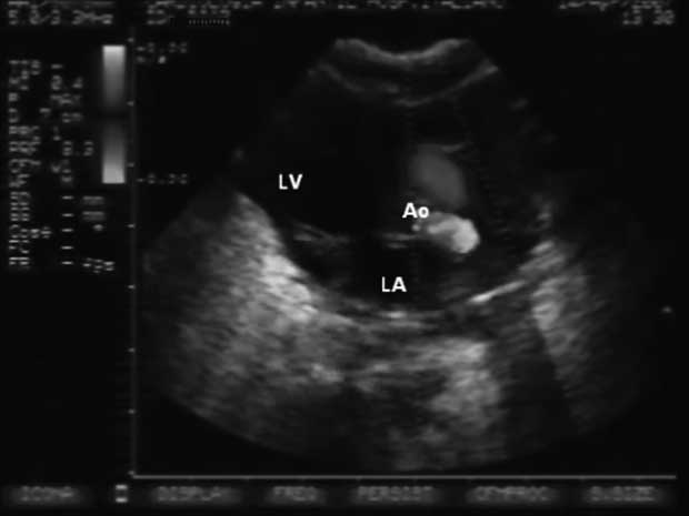
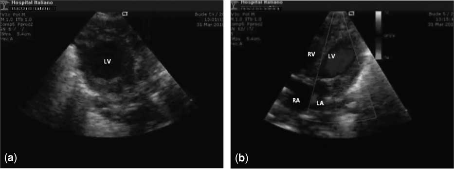
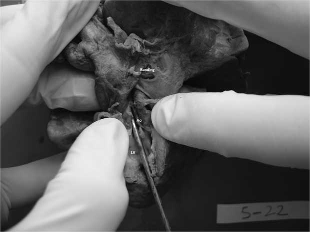
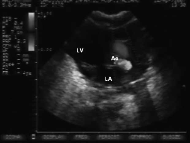
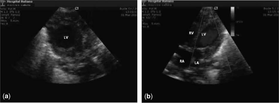
There was one patient who was born with a hypoplastic and hypocontractile left ventricle (long-axis z-score: −5.53), dying on the 4th day of life after a hybrid procedure (Fig 2). Another baby was born with a normal sized left ventricle (left ventricle long-axis z-score: −0.26) and poor contractility, requiring neonatal aortic valvuloplasty, which significantly improved left ventricular contractility (Fig 3). That patient evolved with respiratory failure and haemodynamic decompensation refractory to treatment with inotropes and alveolar surfactant. The last two cases were born with small borderline left ventricles (long-axis z-score −2.59 and −2.43) and slightly improved contractility (Fig 4, Table 4). They underwent neonatal aortic valvuloplasty with further contractility recovery. Diastolic function remained impaired in both neonates, with a restrictive foramen ovale and severe pulmonary hypertension. Accordingly, they were submitted to cardiac surgery at 3 and 2 weeks of age, as a bridge to eventual biventricular reconversion, but both died as a consequence of pulmonary complications.
The heart of the first patient to undergo foetal aortic valvuloplasty, who died after a hybrid procedure, with a hypoplastic left ventricle and a patent aortic valve. Ao = aorta; LV = left ventricle.
Postnatal long-axis echocardiographic view of a patient who had foetal aortic valvuloplasty performed, showing flow across the aortic valve and a normal sized left ventricle. AO = aorta; LA = left atrium; LV = left ventricle.
Postnatal short-axis ( a ) and long-axis ( b ) echocardiographic views of a patient who underwent foetal aortic valvuloplasty, showing the left ventricle's borderline. LA = left atrium; LV = left ventricle; RA = right atrium; RV = right ventricle.
Discussion
In the present series, percutaneous foetal aortic valvuloplasty could be performed in the five selected cases, with no associated foetal mortality or maternal morbidity. Except for one case in which the parents decided termination of pregnancy, all other cases were born after 36 weeks. There was one foetus that evolved to hypoplastic left heart syndrome, and although left heart structures’ growth was observed in the other three cases postnatal outcome was poor, without survival.
In utero aortic valvuloplasty, in order to prevent progression to hypoplastic left heart syndrome in selected patients, has been performed during the last 20 years in a few centres around the world. They initially showed limited technical success and high rate of complications.Reference Maxwell, Allan and Tynan 15 , Reference Kohl, Sharland and Allan 16 In the last few years, further experience and technical improvement have allowed better patient selection and more efficient procedures, as well as better postnatal patient management.Reference Mc Elhinney, Marshall and Wilkins-Haug 11 , Reference Artz, Wertaschnigg, Veit, Klement, Gitter and Tulzer 12 , Reference Wilkins-Haug, Tworetzky, Benson, Marshall, Jennings and Lock 17 – Reference Mizrahi-Arnaud, Tworetzky and Bulich 19 In the two largest series reported, intervention was attempted in 70 and 23 patients, respectively, with technical success in 52 (74%) and 15 (65%), and from these with neonatal biventricular heart at birth observed in 15 (29%) and 10 (67%) patients.Reference Mc Elhinney, Marshall and Wilkins-Haug 11 , Reference Artz, Wertaschnigg, Veit, Klement, Gitter and Tulzer 12
In our hospital, the criteria for patient selection for foetal aortic valvuloplasty were established on the basis of anatomic and physiological criteria initially reported by Tworetzky et alReference Tworetzky, Wilkins-Haug and Jennings 9 and Huhta et al.Reference Huhta, Quintero, Suh and Bader 10 There have been changes over the years as a result of the experience obtained by the two centres with the largest series of cases, especially regarding anatomical criteria, as reported by Tworetzky et alReference Mc Elhinney, Marshall and Wilkins-Haug 11 and Artz et al.Reference Artz, Wertaschnigg, Veit, Klement, Gitter and Tulzer 12 In aortic stenosis and evolving hypoplastic left heart syndrome, there is depressed left ventricle function (qualitatively observed) and inverted flow across the foramen ovale and in the aortic arch. The potential for the intervention's technical success and postnatal biventricular outcome is defined mainly by the dimensions of the left heart structures – left ventricle, aortic valve annulus, and mitral valve annulus – at the time of intervention. Left ventricle long-axis z-score and high left ventricle pressure have been independently associated with higher probability of biventricular outcome from the time of birth.Reference Mc Elhinney, Marshall and Wilkins-Haug 11 , Reference Artz, Wertaschnigg, Veit, Klement, Gitter and Tulzer 12 The guidelines for patient selection have been updated in order to improve identification of patients in whom foetal intervention will result in improved left heart growth and postnatal survival with a biventricular circulation.Reference Mc Elhinney, Marshall and Wilkins-Haug 11 The five cases we report fit the original broader criteria, but not all the new ones, which could partly explain unfavorable postnatal outcome.
Although in the five cases we were able to cross the aortic valve and inflate the balloon, we found it difficult to evaluate with precision the immediate effectiveness of the procedure. Given cardiac prenatal physiology, and associated myocardial dysfunction in our patients, we found it difficult to assess with certainty whether the aortic valve was sufficiently dilated in order to achieve the amount of antegrade blood flow necessary to allow enough growth of left heart structures for a biventricular outcome after birth. Antegrade aortic blood flow may not reflect accurately valve permeability in the setting of severely depressed left ventricular systolic function. In one foetus, flow at the transverse aortic arch became antegrade after valvuloplasty. In another case, visualisation was limited by the development of pericardial effusion. The use of a pressure guidewire for intraprocedural foetal haemodynamic monitoring has been recently reported, and it may contribute to improve assessment during valvuloplasty and outcome prediction.Reference Goldstein, Fifer and Armstrong 20
We used a balloon to aortic valve ratio of 0.9:1 to 1:1, and none of our patients had new aortic regurgitation after foetal valvuloplasty. It has been reported that foetal aortic valves can be dilated safely with larger balloons than are usually used for postnatal dilation, with spontaneous improvement in post-dilatation aortic regurgitation.Reference Mc Elhinney, Marshall and Wilkins-Haug 11 , Reference Artz, Wertaschnigg, Veit, Klement, Gitter and Tulzer 12 In fact, larger balloons, with ratios of 1.2:1, are more commonly being used.Reference Artz, Wertaschnigg, Veit, Klement, Gitter and Tulzer 12
In this series, there was no foetal mortality or maternal morbidity, and regarding foetal morbidity one case presented bradycardia requiring intracardiac medication, and in another one there was balloon rupture with autolimited pericardial and pleural effusion. In the largest series, ∼12% (8/70 and 3/24) of foetuses in whom valvuloplasty was attempted did not reach a viable birth – extreme prematurity or stillbirth.Reference Mc Elhinney, Marshall and Wilkins-Haug 11 , Reference Artz, Wertaschnigg, Veit, Klement, Gitter and Tulzer 12 Few maternal complications have been reported.Reference Wilkins-Haug, Tworetzky, Benson, Marshall, Jennings and Lock 17 In 40% (28/70) of foetuses, there were haemodynamic changes and/or haemopericardium requiring medication and/or pericardial drainage.Reference Mc Elhinney, Marshall and Wilkins-Haug 11 , Reference Mizrahi-Arnaud, Tworetzky and Bulich 19 In contrast to what has been happening to foetoscopy and open foetal surgery, whose success has been hampered by high rates of preterm rupture of membranes, premature delivery, and maternal complications, foetal cardiac interventionism today seems to be acceptably safe for both mother and foetus.Reference Mc Elhinney, Marshall and Wilkins-Haug 11 , Reference Artz, Wertaschnigg, Veit, Klement, Gitter and Tulzer 12 , Reference Wilkins-Haug, Tworetzky, Benson, Marshall, Jennings and Lock 17 , Reference Mizrahi-Arnaud, Tworetzky and Bulich 19 , Reference Bruner, Tulipan and Paschall 21 – Reference Deprest, Gratacos and Nicolaides 23
Progress in prenatal therapy needs to go along with progress in postnatal therapy. The three patients who did not evolve to hypoplastic left heart syndrome required intensive neonatal care and neonatal cardiac interventionism; cardiac surgery was performed in two. The three patients died within their first month of life. These results are poor, but given the small size of our sample gathered over a long period of time they are within what would be expected according to the literature. New therapeutic approaches should be developed for this new type of aortic stenosis that underwent foetal aortic valvuloplasty, with a different prenatal natural history and pathophysiological features. Recent studies have reported unusual surgical strategies such as resection of subendocardial fibroelastosis, and early Ross operation after foetal aortic valvuloplasty with promising results.Reference Brown, Boshoff, Eyskens and Gewillig 24 – Reference Moon-Grady, Moore and Azakie 26
Foetal aortic valvuloplasty, with promising results in the largest series, still remains an experimental therapy, actually reduced to a highly select group of patients. Further research regarding this complex disease's physiopathology, especially regarding foetal haemodynamics, myocardial damage, and heart function, will possibly allow us to better understand its natural history and genetic variants, to incorporate new aspects useful in identifying salvageable left ventricles at initial evaluation, and to improve postnatal management and optimisation of therapeutic strategies. Efforts are being made in carrying out multi-centre collaborative studies in order to clear up the risk/benefit balance of foetal cardiac interventionism.Reference Oepkes, Moon-Grady, Wilkins-Haug, Tworetzky, Arzt and Devlieger 27 In this context, we believe that reporting our small series of patients actually contributes to such effort.
Conclusions
Foetal aortic valvuloplasty is a procedure with acceptable rate of foetal and maternal complications when performed by experienced hands. It could have the potential of significantly improving prognosis in a selected subgroup of patients with severe aortic stenosis, by preventing progression to hypoplastic left heart syndrome. The challenge remains to further increase our knowledge regarding this complex disease both prenatally and postnatally in order to continue to enlighten its understanding and management, as well as the role of foetal aortic valvuloplasty.
Acknowledgements
We would like to thank all of our fetal interventionism team members, nurses and physicians, for their effort and dedication.