Cardiac Echinococcosis is often caused by Echinococcus granulosus and, while the clinical presentation can vary, it often has an asymptomatic course until a rupture occurs. The cyst can involve the liver (75%), lungs (15%), and other organs (10%). A cardiac hydatid cyst involves the left ventricle, particularly the myocardium (55–75%), the right ventricle (15–25%), and the right or left atrium (10–14%).Reference Von Sinner 1 A cardiac hydatid cyst may manifest with chest pain, dyspnoea, cough, and haemoptysis, and it may cause a right or left ventricular outlet obstruction, valvular insufficiency, myocardial contraction problems, rhythm abnormalities, for example non-specific changes or ST-T changes resembling ischaemia on an electrocardiogram, or a complete branch block, and cardiac failure. Ruptures of such cysts are associated with a high mortality rate caused by anaphylactic shock, intracardiac perforation, and pulmonary, cerebral, or coronary embolism.Reference Teggi 2 Once a diagnosis is made using imaging techniques, for example ultrasound and CT, and serologic tests, the treatment involves surgical removal of the cyst.Reference Brunetti, Kern and Vuitton 3 Differential diagnoses should include cardiac tumors, parietal thrombosis, and mitral valvular thrombosis in patients with a biological valvular implant, and in those with valvular problems.
Case report
A 13-year-old male presented to his paediatrician with chest pain for which he underwent a chest X-ray that revealed enlargement of the heart. The patient was referred to a paediatric cardiologist. Echocardiography revealed a cardiac mass within the pericardium adjacent to the right ventricle suspected to be teratoma, and the patient was referred to our clinic with this pre-diagnosis.
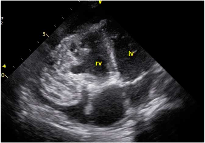
During the physical examination, the patient was conscious and cooperative. His body temperature was 38°C, his respiratory rate was 25/minute, and his respiratory sounds were normal. The patient’s pulse rate was 115/minute and his blood pressure was 100/60 mmHg. The patient’s liver and spleen were non-palpable, and Traube’s space was resonant on percussion of the abdomen. The results of a complete blood count showed total leukocyte count, 13.3/µl; haemoglobin, 10.7 g/dl; platelet count, 250,000/µl; erythrocyte sedimentation rate, 75 mm/hour; C-reactive protein-positive, 15.3 mg/dl; prothrombin time, 11 seconds; prothrombin activity, 105%; activated partial thromboplastin time, 31 seconds; and international normalised ratio. Electrocardiography showed sinus rhythm. Echocardiography showed a thick and trabeculated right ventricular free wall. A well-defined cystic mass lesion surrounded by fibrous tissue, with variable shapes during systole and diastole, was noted adjacent to the wall. A cardiac tumour was suspected; thus, the patient underwent cardiac MRI, and an aneurysmatic structure on the right ventricular free wall was reported. An indirect hemagglutination test for a hydatid cyst was requested, and the result was positive (1/1280). Whole abdominal, cranial, and thoracic CT scans revealed no additional lesions, and the patient underwent surgery. The pericardial effusion was evacuated carefully, and the pericardial cavity was irrigated with 1% povidone–iodine and 18% saline. The cyst was excised and the aneurysmatic structure was repaired. No additional problems occurred during follow-up. The patient began therapy with albendazole 15 mg/kg/day and was discharged after 1 week. During 1 year of follow-up, no recurrence was observed.
Discussion
A cardiac hydatid cyst was described for the first time by Williams in 1836, and the first successful surgical intervention was carried out by Long in 1932.Reference Ameli, Mobarhan and Nouraii 4 Such cysts are often detected incidentally and may be an intramyocardial, pericardial, paracardial, or intracavitary lesion. An intracardiac rupture results in an embolism to the lungs and other organs through the systemic circulation, and an intrapericardial rupture results in acute pericarditis, cardiac tamponade, and, eventually, constrictive pericarditis.
On echocardiography, the cyst may appear as a solitary echolucent lesion with internal trabeculations. It may be solid, and multiple cysts may be present. A cardiac hydatid cyst is usually the primary lesion. It has a haematogenous origin and involves the left heart rather than the right heart specifically, the left ventricular free wall (55–60%), the interventricular septum (5–9%), the right atrium (3–4%), the left atrium (8%), the pulmonary artery (7%), and the pericardium (8%); the right ventricle is involved in only 15% of cases.Reference Fiengo, Bucci and Giannotti 5
A cyst on the ventricular wall extends to the epicardium or endocardium. Subendocardial cysts show intracavitary enlargement, whereas subepicardial cysts extend to the pericardium. Intrapericardial ruptures occur in 10% of cardiac hydatid cysts.
There are two types of pericardial infestation. The primary type occurs with haematogenous spread from the artery that supplies the pericardium and involves the pericardium. This type is extremely rare. The secondary involvement category can be divided into two types: type A, in which the cardiac hydatid cyst is perforated in the pericardial cavity, and type B, which occurs when a hydatid cyst affecting the lungs, mediastinum, abdominal organs, and other neighbouring organs ruptures into the pericardial cavity. A hydatid cyst that ruptures into the pericardium can result in a severe inflammatory reaction, pericardial effusion, and intense pericardial fibrin formation.Reference De Martini, Nador and Binda 6
It is sometimes difficult to differentiate rupturing of a hydatid cyst into the pericardium from primary or secondary metastatic tumors. The differential diagnosis of a solid mass lesion involving the apex of the heart includes fibroma, rhabdomyoma, rhabdomyosarcoma, lipoma, teratoma, and thrombotic apical pseudoaneurysm. Transthoracic echocardiography has a critical role in the differentiation of uninfected hydatid cysts on the cyst wall and heterogeneous cyst material.Reference Heyat, Mokhtari, Hajallioo and Shakibi 7 The differentiation of an infected cyst from solid tumors poses challenges, although the definitive diagnosis is based on a pathological examination.
The standard diagnostic approach for cardiac hydatidosis involves a combination of imaging techniques and serological analysis. Serological tests such as indirect hemagglutination test and enzyme-linked immunosorbent assay can assist in the diagnosis of cardiac hydatid cyst infection, but since they have a sensitivity of only 80%, false negative results should be considered. The sensitivity of these screening tests is relatively high but varies depending on the affected organ. Usually the diagnosis starts with the clinical suspicion of the disease.Reference Tabesh, Tafti and Ameri 8
The role of CT and MRI in the differential diagnosis of cardiac echinococcosis is important. CT is most useful for depicting the peripheral calcifications surrounding established echinococcal cysts, and MRI imaging is most helpful for identifying echinococcosis of the central nervous system. CT is superior to other imaging modalities in observing intra-cystic gas, minute calcifications, and in anatomical mapping.Reference Nemes, Geleijnse and van Geuns 9 Cysts may be identified as single or multiple, and uni- or multi-locular. Pathognomonic findings are the presence of a single cyst with a wall, daughter cysts surrounded by a capsule with peripheral calcifications, and membrane detachment. Separation of the laminated membrane from the pericyst (connective tissue layer) produces a split wall appearance. MRI depicts the exact anatomic location, and nature of internal and external structures. The appearance is usually of a characteristic oval lesion, which is hypointense on T1-weighted images, and hyperintense on T2-weighted images. A typical finding on T2-weighted images is a hypointense peripheral ring, representing the pericyst, which is a dense fibrous capsule from reactive host tissue. Cysts may be single or multiple, uni- or multi-loculated, and thin- or thick-walled. More specific signs include calcification of the cyst wall, presence of daughter cysts, and membrane detachment.Reference Dursun, Terzibasioglu and Yilmaz 10
The treatment of cardiac hydatid cysts involves surgical removal of the cyst. Under cardioplegic arrest the cyst is punctured with a wide aspiration needle connected to the suction device. After the aspiration, without removing the needle, 10% hypertonic saline is injected into the cystic cavity for sterilisation. The endocyst and the remaining daughter cysts are then removed. The residual cavity is closed either with continuous or multiple interrupted prolene suturesReference Tanyeli, Dereli, Mercan, Gormus and Yuksek 11 , Reference Sabzi and Faraji 12 (Fig 1).
Aneurysmatic structure on the right ventricular free wall.

Medical therapy with 10–15 mg/kg of albendazole and 40–50 mg/kg of mebendazole before surgery must be avoided, as this may cause thinning of the cyst wall in patients with a high risk of cyst rupture. Post-operative medical therapy decreases recurrence significantly and should therefore be appliedReference Brunettia, Kernb P and Vuittonc 13 in case of possible cyst rupture and dissemination of daughter cysts during the operation and to prevent recurrence of the cysts. There is a lack of consensus on the ideal duration of therapy. One article recommended albendazole 400 mg twice a day for a period of 6 months.Reference Altas, Sarikaya, Sacli, Yerlikhan and Kirali 14
Given the information in this case report, hydatid cysts should not be overlooked in the differential diagnosis of pericardial tumors.
Acknowledgements
We would like to thank the doctors and nurses working in our hospital.
Financial Support
This research received no specific grant from any funding agency, commercial or not-for-profit sectors. The authors have indicated they have no financial relationships relevant to this article to disclose.
Conflicts of Interest
The authors have indicated they have no potential conflicts of interest to disclose.