Persistent left superior caval vein is the most common venous anomaly in the thorax – one-twentieth or more of patients with congenital malformed hearts – and most frequently it is found draining into the coronary sinus with a right superior caval vein also draining into the morphologically right atrium.Reference Bartram, Van Praagh, Levine, Hines, Bensky and Van Praagh1 Only rarely is the right-sided vein absent, causing an isolated persistent left superior caval vein. Most reported cases of isolated persistent left superior caval vein have been incidentally discovered during a central catheterisation or a pacemaker implantation,Reference Schummer, Schummer and Frober2 and the multidetector computed tomography findings of isolated persistent left superior caval vein, to our knowledge, have been discussed only in two cases.Reference Heye, Wengenroth, Schipp, Johannes Dengler, Grenacher and Werner Kauffmann3, Reference Xiong and Shi4 Here, we report a case of isolated persistent left superior caval vein, which was responsible for the termination of an attempted endocardial insertion of cardiac pacemaker lead wires and conversion to a subxiphoid approach.
Case report
An 81-year-old man on stroke rehabilitation was referred to our radiology department for further evaluation of cough and fever. In addition to hypertension, he had a history of atrial fibrillation and a pacemaker with the lead wire inserted using a subxiphoid approach – 15 years ago – at another hospital. According to the transfer note, an endocardial insertion of pacemaker somehow ended in failure.
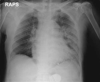
Chest X-ray revealed pneumonic infiltrations in the left lower lobe (Fig 1). It also showed mild cardiomegaly and a pacemaker located in the left upper quadrant of the abdomen from which terminal electrodes originate and enter at the cardiac apex into the cardiac shadow.
Patient with persistent left superior caval vein 81-year-old male – with complete absence of the right superior caval vein. Anterior–posterior chest radiograph shows pneumonia in the left lower lobe. Also noted is a pacemaker located in the left upper quadrant of the abdomen from which terminal electrodes originate and enter at the cardiac apex into the cardiac shadow (RAPS, right side of an anterior–posterior supine).

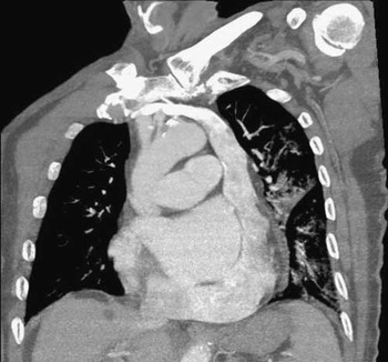
In addition to confirming the presence of pneumonic infiltrations in the left lower lobe, multidetector computed tomography also revealed a large aberrant vessel draining from the right and left brachiocephalic veins, which ran inferiorly passed the left side of the aortic arch and drained into the coronary sinus (Fig 2). The coronary sinus was somewhat dilated but otherwise normal, curving around the heart base and into the right atrium. Owing to the fact that there was no direct communication between the right brachiocephalic vein and the right atrium, the diagnosis of isolated persistent left superior caval vein could be made. Computed tomography showed no associated cardiac or visceral anomalies.
Patient with persistent left superior caval vein – 81-year-old male – with complete absence of the right superior caval vein. An oblique reconstructed multidetector computed tomography shows an aberrant vessel draining from the right and left brachiocephalic veins, running inferiorly passed the left side of the aortic arch and flowing into the coronary sinus. No evidence of right superior caval vein can be seen.

His outside medical records were obtained later, and revealed that it was the anomalous venous course that led to the decision to terminate the endovascular approach and perform the implantation of the generator using a subxiphoid approach with the pacemaker placed in the posterior fascial layer of the rectus abdominis muscle. During the endovascular implantation attempt, a catheter was inserted through the right subclavian vein, but instead of flowing into the right atrium the venous drainage of the right subclavian vein traversed to the left side and eventually drained into the coronary sinus and the right atrium.
Discussion
A persistent left superior caval vein is a structure that normally obliterates during embryonic development. Around the 24th day of gestation, the venous blood of the upper half of the body drains into the primitive heart through the left and right anterior cardinal veins. Although the caudal part of the left anterior cardinal vein usually resolves into the ligament of Marshall in most cases, they remain to function as a normal vessel in 0.2 to 8% of population, which results in bilateral superior caval veins. Rarely, the right, instead of the left, anterior cardinal vein obliterates to create an isolated persistent left superior caval vein.Reference Heye, Wengenroth, Schipp, Johannes Dengler, Grenacher and Werner Kauffmann3
Despite the fact that a persistent left superior caval vein itself does not have any haemodynamic significance, it may be associated with many congenital heart diseases, such as atrial septal defect, biscuspid aortic valves or aortic coarctation in Turner syndrome, left isomerism, atresia of the coronary sinus orifice, and many more. The persistent left superior caval vein was also known to be associated with anatomical and architectural abnormalities of the normal pacemaker and conduction tissues, causing arrhythmia in a small number of cases.Reference Bartram, Van Praagh, Levine, Hines, Bensky and Van Praagh1, Reference Lenox, Hashida, Anderson and Hubbard5
More importantly, persistent left superior caval vein has been blamed for troubles when inserting venous catheters, pacemakers or cannulating the heart for cardiopulmonary bypass.Reference Girerd, Gressard, Berthezene and Lantelme6, Reference Konvicka and Villamaria7 Serious complications including, angina, arrhythmia, and even cardiac arrest have been reported during catheterisation of the persistent left superior caval vein. Despite most experienced practitioners being aware of bilateral persistent left superior caval vein, the rare isolated persistent left superior caval vein can cause much confusion as in our case. The operator in the current case did notice the drainage of the left superior caval vein flow into the coronary sinus; however, it is likely that the operator recognised it as an isolated persistent superior caval vein and was unaware of the fact that it is not a contraindication for implanting a pacemaker using an endocardial approach.Reference Girerd, Gressard, Berthezene and Lantelme6 Permanent pacemaker leads have been successfully advanced into the right ventricle through persistent left superior caval vein as early as 1971–1972 with good long-term results.Reference Ratliff, Yousufuddin and Lieving8 Thus, when fully informed, a cardiothoracic interventionist may be able to successfully implant pacemakers in patients with persistent left superior caval vein through the left superior caval vein – the endocardial approach – and avoid the alternative subxiphoid approach, which is known to be more painful and have higher complication rates.Reference Jaroszewski, Altemose and Scott9
As the number of catheterisation and pacemaker implantation increases, the clinical significance of vascular anomaly may also rise. However, it is difficult to diagnose an isolated persistent left superior caval vein with a simple X-ray film unless there is a central catheter or a pacemaker placed through the subclavian or internal jugular vein. The echocardiogram can show enlarged coronary sinus draining into the right atrium, and a contrast echocardiography using agitated saline injected into the antecubital vein can show a specific finding of a contrast echo flowing into the right atrium from only the coronary sinus.Reference Alhaj, Pizzarello, Bergmann, Garcia and Trofimovsky10, Reference Konecky, Freedberg, McCauley and Kronzon11 However, cross-sectional imaging with computed tomography can help recognise the detailed anatomy and should be considered before an interventional procedure, if persistent left superior caval vein is suspected in the plain film or if there is a history of troubled endovascular procedure. With the ability to display a normal or abnormal course of the central veins on a single frame, multidetector computed tomography provides accurate anatomical information necessary for a successful catheterisation.