Introduction
American paleobotanists have long been criticized by their European contemporaries for a perceived negligence in documenting Pennsylvanian-aged macrofloral assemblages preserved as adpressions (i.e., compression-impression floras). For example, after a reconnaissance fieldtrip to the USA in 1933, W.J. Jongmans summarized the status of Carboniferous paleobotany in America in an unflattering quote (Jongmans and Gothan, Reference Jongmans and Gothan1934, p. 17, translated from German), “If one considers then how poor most of the American illustrations are, how relatively few works are supplied with illustrations—most papers only contain lists with many names, which provide us no clue on which to base a judgment,—how confused the nomenclature is, because of parallel efforts by rather than collaboration between the American and European paleobotanists—, then it becomes clear to anyone that any attempt [of comparison] without one’s own consideration [of the evidence] will be very difficult if not impossible.” Thereafter, Jongmans (Reference Jongmans1937a, p. 364–366) and Bode (Reference Bode1958, p. 217, 218) expressed discontent about correlating between American and European successions, and pointed to the large size of (and distance between) Pennsylvanian basins in the USA, the use of parochial stratigraphic nomenclature in different states, the reliance of American paleobotanists on outdated literature that did not take European taxa into account, and the scarcity of publications with well-illustrated macrofloras. In particular, it was felt that the lack of consultation with the European literature had caused the introduction of many ‘new species’, creating the illusion that Pennsylvanian vegetation preserved on either side of the Atlantic Ocean differed more than expected.
Since the mid-20th century, the breakdown of language barriers, better international access to publications, and collaboration between American and European paleobotanists has seen the resolution of many taxonomic issues, as W.C. Darrah foretold in a footnote in Jongmans (Reference Jongmans1937b, p. 394): “The number of [P]aleozoic plants with wide distributions in both Europe and America will increase rather than diminish with careful study.” Furthermore, publications that involve North American adpression assemblages grew substantially after the 1920s (Pfefferkorn and Gillespie, Reference Pfefferkorn and Gillespie1980a). Nonetheless, the idea persists that Pennsylvanian macrofloral assemblages lack documentation in the USA (e.g., Cleal et al., Reference Cleal, Opluštil, Thomas, Tenchov, Abbink, Bek, Dimitrova, Drábková, Hartkopf-Fröder, Van Hoof, Kędzior, Jarzembowski, Jasper, Libertín, McLean, Oliwkiewicz-Miklasinska, Pšenička, Ptak, Schneider, Schultka, Šimůnek, Uhl, Waksmundzka, Van Waveren and Zodrow2009, p. 184), thus hindering comparisons and biostratigraphic correlations of strata across the Euramerican paleocontinent. A task of this paper is to demonstrate that such claims are exaggerated.
This contribution focuses on the description and illustration of a Middle Pennsylvanian (middle Moscovian) macrofloral assemblage collected from wetland deposits above the Minshall Coal (lowermost Staunton Formation) of Indiana, situated on the eastern margin of the Illinois Basin. Plant remains were collected at a single locality from four distinct beds, each containing a compositionally unique suite of fossil-taxa (sensu Cleal and Thomas, Reference Cleal and Thomas2010), although all specimens are treated as belonging to one macrofloral assemblage to simplify biostratigraphic comparisons. The strata accumulated close to the Atokan-Desmoinesian Stage boundary, which has been correlated with the Bolsovian-Asturian Substage boundary of Western Europe (Peppers, Reference Peppers1996; Blake et al., Reference Blake, Cross, Eble, Gillespie and Pfefferkorn2002). The microfloral assemblage from the clastic beds and underlying Minshall Coal (uppermost Brazil Formation) also is tabulated, but only touched on in biostratigraphic context. Comprehensive synonymy lists are compiled for fossil-taxa in the macrofloral assemblage based on comparison with a suite of North American literature, which were singled out because they contain well-illustrated records of Middle Pennsylvanian plant remains, with a focus on the Illinois Basin. Homotaxial biostratigraphic comparisons with American and European biozonation schemes are presented, and uncertainties about the placement and correlation of the Atokan-Desmoinesian and Bolsovian-Asturian boundaries are discussed. Evaluating the congruence of these boundaries is timely because they involve an interval that is being increasingly recognized as one of significant environmental change throughout Euramerica (cf., Cecil et al., Reference Cecil, Stanton, Neuzil, Dulong, Ruppert and Pierce1985; Bertier et al., Reference Bertier, Swennen, Lagrou, Laenen and Kemps2008; Van Hoof et al., Reference Van Hoof, Falcon-Lang, Hartkopf-Fröder and Kerp2013). Furthermore, nearly all macrofloral taxa identified also are present in contemporaneous deposits of Europe, and the paleobiogeographic and paleoecologic ramifications of this finding are considered in the context of Pennsylvanian wetland ecosystems in tropical Euramerica.
Geologic setting
Illinois Basin
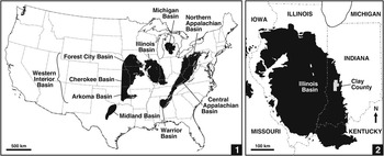
The Illinois Basin (also called the Eastern Interior Basin) is an interior cratonic basin (Leighton et al., Reference Leighton, Kolata, Oltz and Eidel1991), one of several large depocenters in the USA that contain coal-bearing strata of predominantly Pennsylvanian age (Fig. 1.1). In its present configuration, the basin underlies much of Illinois and parts of western Indiana and western Kentucky, with small outliers in Iowa and Missouri (Fig. 1.2). The Illinois Basin contains a largely siliciclastic succession of Morrowan to Virgilian (Bashkirian to Gzhelian, upper Namurian to upper Stephanian) strata (Nelson et al., Reference Nelson, Trask, Jacobson, Damberger, Williamson and Williams1991, Reference Nelson, Greb and Weibel2013), which accumulated on a low-gradient slope that dipped toward a shallow epeiric sea (Watney et al., Reference Watney, Wong and French1989; Greb et al., Reference Greb, Andrews, Eble, DiMichele, Cecil and Hower2003). Glacioeustatic fluctuations from the west resulted in cyclic marine transgressions across the continental platform (Wanless and Shepard, Reference Wanless and Shepard1935, Reference Wanless and Shepard1936; Heckel, Reference Heckel1986; Langenheim and Nelson, Reference Langenheim and Nelson1992; Algeo and Heckel, Reference Algeo and Heckel2008), and in accord with its paleogeographic position, depositional environments in the Illinois Basin tend to be intermediate between the marine-dominated rocks of the Western Interior (Midcontinent) Basin and mainly deltaic to terrestrial strata of the Appalachian Basin to the east (Fig. 1.1; Heckel, Reference Heckel1977, Reference Heckel1980; Nelson et al., Reference Nelson, Greb and Weibel2013). These basins developed in a paleoequatorial setting in the west-central part of Euramerica (Witzke, Reference Witzke1990), and were separated from eastern parts of the paleocontinent (present-day Europe) by uplands of the Laurentian Shield and, in part, by the Appalachian-Variscan Mountains (Blakey, Reference Blakey2013).
Basins in contiguous USA that contain coal-bearing strata of Pennsylvanian age. (1) Western Interior, Illinois, Appalachian, and Michigan basins, and their components. (2) Illinois Basin, and position of Clay County, Indiana on eastern margin of basin.

Study locality
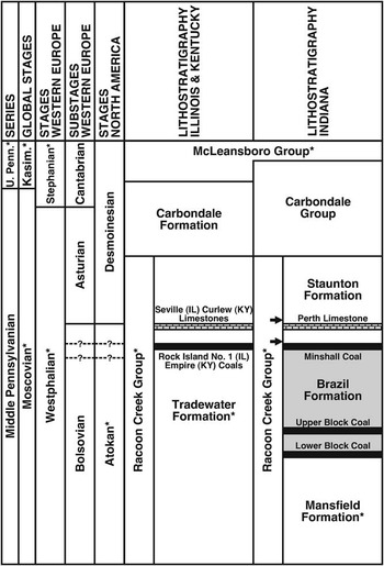
The study area is situated in Clay Country, west-central Indiana, on the eastern margin of the Illinois Basin (Fig. 1.1), where the thin and laterally discontinuous Lower Block, Upper Block, and Minshall coals of the upper Atokan Brazil Formation (Fig. 2) have been exploited due to their low to medium sulfur and ash content (Mastalerz et al., Reference Mastalerz, Padgett and Eble2000, Reference Mastalerz, Ames and Padgett2003; Kvale et al., Reference Kvale, Mastalerz, Furer, Engelhardt, Rexroad and Eble2004). The Brazil Formation, defined as being from the base of the Lower Block Coal to the top of the Minshall Coal (Hutchison, Reference Hutchison1976), is equivalent to the middle part of the Tradewater Formation of Illinois and Kentucky (The Tri-State Committee on Correlation of the Pennsylvanian System in the Illinois Basin, 2001). In Indiana, strata above the Minshall Coal are assigned to the Staunton Formation, which includes the Perth Limestone, the top of which defines the Atokan-Desmoinesian Stage boundary based on palynology (Peppers, Reference Peppers1996). The Minshall Coal and overlying Perth Limestone correlate with the Rock Island (No. 1) Coal and Seville Limestone of northwestern Illinois and the Empire Coal and Curlew Limestone of southwestern Kentucky (Fig. 2; Tri-State Committee, 2001; Nelson et al., Reference Nelson, Greb and Weibel2013).
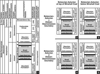
Stratigraphy of Middle Pennsylvanian of Illinois Basin (as currently understood), showing correlation between chronostratigraphic and lithostratigraphic units (* indicates incomplete unit); compiled from Davydov et al. (Reference Davydov, Crowley, Schmitz and Poletaev2010, fig. 2), Falcon-Lang et al. (Reference Falcon-Lang, Heckel, DiMichele, Blake, Easterday, Eble, Elrick, Gastaldo, Greb, Martino, Nelson, Pfefferkorn, Phillips and Rosscoe2011, fig. 1), and Nelson et al. (Reference Nelson, Greb and Weibel2013, fig. 2). Brazil Formation (shaded) incorporates strata from base of Lower Block Coal to top of Minshall Coal. Macrofloral assemblage collected from lowermost Staunton Formation (arrows). Atokan-Desmoinesian Stage boundary and Bolsovian-Asturian Substage boundary shown as coeval and equivalent to top of Perth, Seville, and Curlew limestones, after Peppers (Reference Peppers1996). Dashed lines denote possible positions of one or both boundaries based on biostratigraphic data presented herein (see Fig. 7; Biostratigraphy and age section).

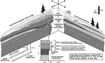
The macrofloral assemblage was collected from an open-pit mine north of Indiana Route 46, the now inaccessible Ashboro Pit (SW 1/4, SE 1/4, Section 17, Township 11N, Range 6W) operated by Log Cabin Coal Company. Plant fossils were collected during a reconnaissance in 1980, when the Lower Block and Upper Block coals were being mined, but the pit was idle and partly filled with water during a 1982 visit, leaving the upper highwall inaccessible. The west- and north-facing highwalls of the L-shaped pit are reproduced schematically in Figure 3. About 15 m of the Brazil and overlying Staunton formations were exposed, the lithostratigraphic boundary being the top of the Minshall Coal.
Schematic drawing of succession exposed in Ashboro Pit (now inaccessible open-pit mine of Log Cabin Coal Company; SW 1/4, SE 1/4, Section 17, Township 11N, Range 6W), north of Indiana Route 46, Clay County, Indiana, during visits in June 1980 and July 1982. Brazil Formation incorporates strata from base of Lower Block Coal (under water) to top of Minshall Coal, and overlying strata belong to Staunton Formation. Top of Perth Limestone corresponds to Atokan-Desmoinesian boundary (Peppers, Reference Peppers1996). Macrofloral assemblage collected from roof-shales above Minshall Coal (Units 1 to 3, marked with *) on west-facing highwall, and presumably from claystone-filled hollow (Unit 4, marked with ?) on north-facing highwall. Note that Unit 4 is at stratigraphic position above the Perth Limestone. All sampled beds belong to USNM locality 38354.

Correlation of limestone and coal members of the Brazil and Staunton formations is challenging due to their lenticular nature and variable lithology, but marker beds in the Ashboro Pit are identified with confidence (Fig. 3). The Lower and Upper Block coals are distinctive in this area, consisting of hard, dull-banded or ‘splint’ coal that separates into large cubic blocks along widely spaced joint planes. The type area of these coals is 15 km north of the locality, whereas the Minshall Coal and Perth Limestone have type sections five to 15 km farther north. No other marine unit in the Brazil or lower Staunton formations in this part of Indiana is likely to be confused with the Perth Limestone.
Most macrofloral remains were collected from an ~35 cm-thick package of ‘roof-shales’ immediately above the Minshall Coal on the west-facing highwall (Units 1 to 3; Fig. 3), whereas some adpressions presumably are from a bed (Unit 4) on the north-facing highwall at a stratigraphic position above the Perth Limestone. All sampled beds belong to USNM Locality 38354. Although the strata belong to the lowermost Staunton Formation, as currently understood and depicted in Figure 2, Units 1 to 3 are assigned to the uppermost Atokan and Unit 4 to the lowermost Desmoinesian. Based on new data presented here, the age of the fossiliferous strata and position of the Atokan-Desmoinesian and Bolsovian-Asturian boundaries will be discussed in the Biostratigraphy and age section.
Fossiliferous units
A light-gray, rooted paleosol (‘underclay’) underlies the 15 cm-thick Minshall Coal (Fig. 3), which is relatively uniform in thickness, but undulatory due to irregularities on the paleosol surface. Following the ‘low-stand coal’ model (cf., Cecil, Reference Cecil1990; Cecil et al., Reference Cecil, Dulong, West, Stamm, Wardlaw and Edgar,.T.2003, Reference Cecil, DiMichele and Elrick2014; DiMichele, Reference DiMichele2014), the Minshall Coal is interpreted to record peat buildup due to paludification and base-level rise under the (per)humid (everwet) conditions that attended a sea-level lowstand at maximum glaciation.
The ~35cm-thick package of fossiliferous roof-shales above the Minshall Coal is divided into three distinct beds based on lithology and plant-fossil content (Units 1 to 3; Fig. 3). Gradational with the underlying coal, Unit 1 (~10 cm thick) is a medium- to dark-gray, fissile, highly carbonaceous shale that contains abundant but fragmentary and poorly preserved plant remains. Spirorbiform worm-tubes (microconchids) are affixed to some fossil leaves, and inarticulate brachiopods occur sporadically. Fine root structures are present at the top of the bed. Unit 1 is interpreted to record deposition in the shallow, dysaerobic, and at least hyposaline waters of a tidally influenced mudflat or estuary, which developed above the precursor Minshall Coal during initial stages of transgression as deglaciation commenced (cf., Archer et al., Reference Archer, Feldman, Kvale and Lanier1994, Reference Archer, Elrick, Nelson and DiMichele2016; Mastalerz et al., Reference Mastalerz, Stankiewicz, Salmon, Kvale and Millard1997). Accordingly, plant remains mainly represent the remnants of drowned peat-forming vegetation. Roots at the top of the unit indicate upward shallowing, emergence, and development of a ‘clastic swamp’ (sensu Gastaldo, Reference Gastaldo1987; Demko and Gastaldo, Reference Demko and Gastaldo1992). The time required for establishment and colonization of the clastic swamp implies a temporary lull in sea level rise, possibly even a minor regressive event. Elsewhere in Clay County, the Minshall Coal consists of two distinct ‘benches’ separated by a clastic unit (up to 2.7 m thick) with roots at the top. A similar pattern occurs farther south in Daviess County, where a persistent claystone (~5 cm thick) exists near the middle of the Minshall Coal (incorrectly called the Buffaloville Coal by mine operators). As such, clastic swamp deposits at the top of Unit 1 may coincide with, and/or are a lateral equivalent of, the upper bench (Clay County) or upper part (Daviess County) of the Minshall Coal.
Unit 2 (~10 cm thick) is gradational with Unit 1, and comprises medium-gray, fissile, and well-indurated shale. Adpressed plant fossils are abundant and well preserved. Unit 2 is interpreted to record renewed transgression and further accumulation of tidally influenced mudflat or estuarine deposits. Macrofloral remains in this bed are thought to represent litter shed from clastic swamp vegetation on the periphery of the mudflat or estuary.
Unit 3 (~15 cm thick) is gradational with Unit 2, and consists of maroon, very well-indurated mudstone that contains siderite nodules. Macrofloral remains are well preserved but only moderately abundant. Unit 3 presumably resulted from additional base level rise due to transgression, with the presence of siderite nodules reflecting a change in depositional and/or diagenetic conditions on the mudflat or estuary. Adpressed plant fossils probably were derived from wetland vegetation in local clastic swamp habitats.
Unit 4 (10s of cm thick) consists of pinkish light-gray, well-laminated claystone that contains sparse but well-preserved plant adpressions. The most probable provenance is the fill of a hollow (Fig. 3) that was eroded down to the level of the Minshall Coal during the onset of regression, a consequence of polar ice buildup. If this stratigraphic level is correct, Unit 4 may record the rapid filling of an abandoned channel, either in a fluvial or tidally influenced deltaic setting. Plant debris probably was shed from clastic wetland plant communities alongside the abandoned channel.
Quantification and general characteristics of floral assemblages
Macrofloral assemblage
Most plant remains are preserved as coalified compressions or impressions (i.e., adpressions; Shute and Cleal, Reference Shute and Cleal1987), although a small number of specimens are ‘naturally macerated’ (cf., Zodrow and Mastalerz, Reference Zodrow and Mastalerz2009). Proximate analysis of a sample of the underlying Minshall Coal yields a vitrinite reflectance (%Romax) of 0.57, a coal rank of high-volatile C bituminous (Teichmüller and Teichmüller, Reference Teichmüller and Teichmüller1982). Such a low rank indicates that phytoleims (coalified compressions) probably contain epidermal cuticles, although no attempt was made to procure cuticles.
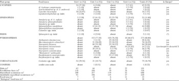
Table 1 presents a quantitative breakdown of the rock slabs and fossil-taxa in Units 1 to 4, and for the macrofloral assemblage as a whole. Of the 191 slabs collected, 326 slab faces bear macrofloral remains (excluding barren faces and counterparts). The quantification technique used is a variant of that described by Pfefferkorn et al. (Reference Pfefferkorn, Mustafa and Hass1975): each slab face was treated as a quadrat, and a fossil-taxon was recorded as ‘present’ regardless of the number of specimens on the face. Through this process, 688 macrofloral occurrences were recorded on the 326 fossiliferous slab faces. Excluding seeds and some axes, which could not be tied to a particular fossil-plant taxon with confidence (termed ‘unidentifiable’ macrofloral occurrences in Table 1), there were 449 ‘identifiable’ macrofloral occurrences.
Fossil-taxa in macrofloral assemblage, from Units 1 to 4 above Minshall Coal (Fig. 3), segregated into plant groupings. Right-hand column indicates if fossil-taxon present in Europe. Symbols: (a) total slabs from unit; (b) slab faces with macrofloral remains (i.e., excludes barren faces and counterparts); (c) identifiable macrofloral occurrences + ‘unidentifiable’ macrofloral occurrences (e.g., axes and seeds); (d) occurrence indicates presence (in any quantity) on slab face; (e) ‘biological’ fossil-taxa + ‘non-biological’ fossil-taxa (Cyperites bicarinatus Lindley and Hutton, Reference Lindley and Hutton1832, Lepidostrobophyllum Hirmer, Reference Hirmer1927 [sp. indet.], Calamites Brongniart, Reference Brongniart1828a [spp. indet.], and putative conifer cone scale); (f) reassembled conceptualization of once-living, whole fossil plant. See text for details.

Of the 22 fossil-taxa identified (Table 1), 18 can be considered ‘biological’ taxa (i.e., a reassembled conceptualization of a whole and once-living fossil plant; sensu Cleal and Thomas, Reference Cleal and Thomas2010), whereas only four ‘non-biological’ taxa were recorded (Cyperites bicarinatus Lindley and Hutton, Reference Lindley and Hutton1832, Lepidostrobophyllum Hirmer, Reference Hirmer1927 [sp. indet.], Calamites Brongniart, Reference Brongniart1828a [spp. indet.], and a putative conifer cone scale). It is noted, however, that each fossiliferous bed contains a low-diversity assemblage, ranging from six to 12 fossil-taxa and four to nine biological taxa. When relative abundances for the entire macrofloral assemblage are considered, pteridosperms are the predominant (60.8%) and most diverse plant group (nine biological fossil-species). The sphenopsids (11.8%) are relatively diverse (five biological species), whereas all cordaitalean remains (16.7%) are included in Cordaites Unger, Reference Unger1850 (spp. indet). The lycopsids (9.4%) are represented by two biological species, and fern and putative conifer remains are very rare.
Microfloral assemblage
Samples of the Minshall Coal and four fossiliferous clastic units (~150 g of rock/coal crushed and sieved through –20 mesh) were oxidized using Schulze’s Solution (nitric acid saturated with potassium chlorate) to liberate spores and pollen, followed by digestion in 5% potassium hydroxide. The organic fraction was concentrated with zinc chloride solution, and treated with ethylene glycol monoethyl ether (2-ethoxyethanol) and ultrasonic vibration to remove fine particulate matter from the spore/pollen fraction. Slides were prepared by strew-mounting a small amount of spore/pollen residue with polyvinyl alcohol on cover glasses, which were fixed with a synthetic piccolyte resin. Line counts (250 palynomorphs per sample) were performed at 640X magnification to establish relative abundances, and additional slides were scanned at 250X to document forms of potential biostratigraphic significance that were not encountered in statistical counts. All slides, maceration residues, and unused portions of samples are stored at the Kentucky Geological Survey (KGS), University of Kentucky, Lexington, KY under KGS sample numbers 3264–3268.
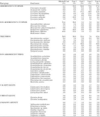
Table 2 gives a quantitative breakdown of the plant groups and fossil-taxa found in the Minshall Coal and Units 1 to 4. Forty-nine miospore species were identified, seven of unknown affinity. The beds are dominated by spores of arborescent or sub-arborescent lycopsids (Minshall Coal, Units 1 to 3) or by tree ferns (Unit 4). Non-arborescent fern and calamitalean spores are rare, as is cordaitalean pollen. Although pteridosperm foliage dominates the macrofloral assemblage, it is emphasized that no samples contain pteridosperm pollen, despite targeted searches, even in maceration residues of the ≤250 µm size fraction. Thus, although pteridosperm pollen typically is very large (100–600 µm; Taylor, Reference Taylor1978) and may be excluded by traditional preparation techniques (Peppers and Pfefferkorn, Reference Peppers and Pfefferkorn1970), some grains should have been found if present. Because this paper focuses on the macrofloral assemblage, the microfloral data are only used to provide complementary biostratigraphic control.
Fossil-taxa in microfloral assemblage, from Minshall Coal and Units 1 to 4 above coal (Fig. 3), segregated into plant groupings/growth forms.

Biostratigraphy and age
The Minshall Coal and overlying Perth Limestone of western Indiana (Fig. 2), along with their correlates in southern Indiana (‘Buffaloville’ Coal [see below], Curlew Limestone), western Kentucky (Empire Coal, Curlew Limestone), and western Illinois (Rock Island [No. 1] Coal, Seville Limestone), have figured prominently in the discussion surrounding placement of the contentious Atokan-Desmoinesian Stage boundary in the Illinois Basin (e.g., Shaver, Reference Shaver1984; Peppers, Reference Peppers1996; Heckel, Reference Heckel2013; Nelson et al., Reference Nelson, Greb and Weibel2013). Because a precisely defined Atokan stratotype has yet to be realized (Sutherland and Manger, Reference Sutherland and Manger1984), in large part because the poorly fossiliferous Atoka Formation (defined in the Arkoma Basin, Oklahoma; Fig. 1.1) is depositionally and tectonically complex, the position of the Atokan-Desmoinesian boundary in the Illinois Basin has shifted depending on the fossil group used for biostratigraphic control. For example, marine faunas in the limestones have indicated either an early Desmoinesian age (ostracodes and fusulinids: Shaver and Smith, Reference Shaver and Smith1974, Shaver, Reference Shaver1984; conodonts: Rexroad et al., Reference Rexroad, Brown, Devera and Suman1998; fusulinids: Wahlman, Reference Wahlman2013) or a late Atokan age (fusulinid foraminifera: Douglass, Reference Douglass1987; conodonts: Barrick et al., Reference Barrick, Lambert, Heckel, Rosscoe and Boardman2013). Likewise, the Atokan-Desmoinesian boundary was placed at the top of the Seville, Perth, and Curlew limestones based on the correlation of microfloral assemblages from coal seams (Peppers, Reference Peppers1996; Fig. 2). By extension of these uncertainties, the position of the Minshall and equivalent coals remains uncertain in chronostratigraphic terms, although the seams usually are placed in the uppermost Atokan (Nelson et al., Reference Nelson, Greb and Weibel2013).
Conodonts from the limestone directly overlying the Buffaloville Coal in its type area (southern Indiana) indicate that it is younger than the Perth, Curlew, and Seville limestones (personal communication, P.H. Heckel, 2014; Fig. 2). This result corroborates the palynological study of Peppers (Reference Peppers1993), who concluded that the true Buffaloville Coal is younger than the Minshall Coal.
According to Peppers (Reference Peppers1996), the Atokan-Desmoinesian Stage boundary of North America coincides with the Bolsovian-Asturian Substage boundary of Western Europe (Figs. 2, 4). If placement of this coupled boundary at the top of the Perth Limestone (and correlates) is accepted, the implication is that the underlying Minshall Coal (and correlates) are late Bolsovian in age. In the following section, the taxonomic composition of the microfloral and macrofloral assemblages from the Minshall Coal and overlying roof-shales (Units 1 to 3) are compared with established biozonation schemes to determine whether or not this assumption holds. Unit 4, which is presumed to have been at a stratigraphic position above the Perth Limestone (Fig. 3), can be assigned an earliest Asturian age by default because it lies just above the Atokan-Desmoinesian boundary (Fig. 2).
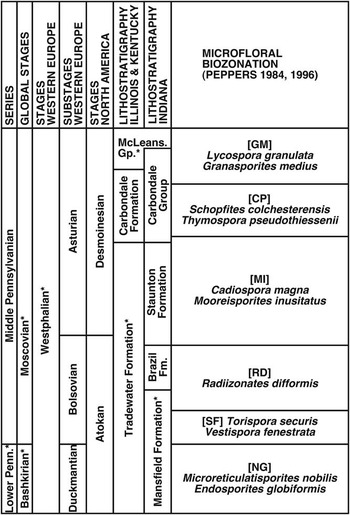
Microfloral biozonation scheme of Peppers (Reference Peppers1984, Reference Peppers1996) correlated with chronostratigraphic and lithostratigraphic units (* indicates incomplete unit). Modified from Peppers (Reference Peppers1996, pl. 1). Microfloral assemblage corresponds to upper Radiizonates difformis (RD) Biozone.

Comparison with American microfloral biozonation
Peppers (Reference Peppers1984, Reference Peppers1996) constructed a microfloral biozonation scheme to facilitate regional correlation between the Illinois Basin and other parts of Euramerica. According to this framework (Fig. 4), the Brazil Formation is in the upper (but not uppermost) Bolsovian part of the Radiizonates difformis (RD) Biozone, with the top of the Minshall Coal corresponding to the top of the biozone (Peppers, Reference Peppers1996).
The composition of the microfloral assemblage (Table 2) supports assignment of the Minshall Coal and overlying roof-shales to the Radiizonates difformis (RD) Biozone. Of particular importance is the presence of the diagnostic densospores Radiizonates difformis (Kosanke, Reference Kosanke1950) Staplin and Jansonius, Reference Staplin and Jansonius1964 and Radiizonates rotatus (Kosanke, Reference Kosanke1950) Staplin and Jansonius, Reference Staplin and Jansonius1964, which are most abundant in the coal and Unit 1. Further, none of the beds (including Unit 4) contain the namesake taxa of the succeeding Cadiospora magna-Mooreisporites inusitatus (MI) Biozone (Fig. 4). Cadiospora magna Kosanke, Reference Kosanke1950 first appeared at the Bolsovian-Asturian boundary, whereas Mooreisporites inusitatus (Kosanke, Reference Kosanke1950) Neves, Reference Neves1958 arose in the latest Bolsovian (Peppers, Reference Peppers1996). These factors collectively point to a late Bolsovian age for the Minshall Coal and overlying clastic beds.
Nonetheless, a microfloral assemblage from a clastic unit directly beneath the paleosol of the Minshall Coal adds uncertainty to this late Bolsovian assignment (these data, incorporated into another publication, are introduced here because they have important ramifications on the age of overlying strata). Most noteworthy is the statistically significant abundance of Thymospora pseudothiessenii (Kosanke, Reference Kosanke1950) Wilson and Venkatachala, Reference Wilson and Venkatachala1963. The spore is considered a reference for basal Asturian, although it first appears in uppermost Bolsovian strata (Peppers, Reference Peppers1996; Lesnikowska and Willard, Reference Lesnikowska and Willard1997) and reaches its epibole in the middle Asturian Schopfites colchesterensis-Thymospora pseudothiessenii (CP) Biozone (Fig. 4). Equally intriguing is the fact that Savitrisporites nux (Butterworth and Williams, Reference Butterworth and Williams1958) Smith and Butterworth, Reference Smith and Butterworth1967 occurs (albeit very rarely) beneath the Minshall Coal, but not in the coal itself. Because this spore disappears just below the Bolsovian-Asturian boundary (Peppers, Reference Peppers1996), its absence in the coal does not in itself imply an Asturian age for the Minshall, but it does signify peat accumulation very close to the substage boundary. It is crucial to note that the scheme of Peppers (Reference Peppers1984, Reference Peppers1996) is almost exclusively based on palynomorphs from coal, and many taxon ranges would be extended if clastic lithologies had been incorporated. These additional microfloral data signify that an earliest Asturian age for the Minshall Coal cannot be ruled out.
Comparison with American macrofloral biozonation
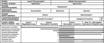
Moore et al. (Reference Moore, Wanless, Weller, Steele Williams, Read, Bell, Ashley, Cheney, Cline, Condra, Dott, Dunbar, Elias, Glenn, Greene, Hendricks, Jewett, Johnson, King, Knight, Levorsen, Miser, Newell, Plummer, Thompson, Tomlinson and Westheimer1944) and Read and Mamay (Reference Read and Mamay1964) segregated upper Paleozoic strata of the USA into fifteen macrofloral zones defined by the co-occurrence of common or diagnostic taxa. Unfortunately, the absence of range charts and ambiguity of zone boundaries make this biostratigraphic framework difficult to apply. Gillespie and Pfefferkorn (Reference Gillespie and Pfefferkorn1979) discussed the temporal distribution of key taxa and charted their ranges in the Appalachian Basin. Extending this work, Blake et al. (Reference Blake, Cross, Eble, Gillespie and Pfefferkorn2002) produced an updated chart that correlates between lithostratigraphic units of the Appalachian Basin, chronostratigraphic units of midcontinental USA and Western Europe, and macrofloral zones of the USA (Read and Mamay, Reference Read and Mamay1964) and paleoequatorial Europe (Wagner, Reference Wagner1984). According to Blake et al. (Reference Blake, Cross, Eble, Gillespie and Pfefferkorn2002, fig. 1, with updates from B.M. Blake, personal communication, 2014), the Atokan-Desmoinesian boundary, which they equated to the Bolsovian-Asturian boundary following Peppers (Reference Peppers1996), is at the Upper Block (No. 5) Coal near the contact between the Kanawha and Allegheny formations (Fig. 5). This level correlates to the middle of Zone 9 (‘Neuropteris’ rarinervis) of Read and Mamay (Reference Read and Mamay1964).
Biostratigraphic ranges of key fossil-species in macrofloral assemblage correlated with chronostratigraphic units, lithostratigraphic units of Central Appalachian Basin, and American macrofloral biozonation scheme of Read and Mamay (Reference Read and Mamay1964) (* indicates incomplete unit); compiled from Darrah (Reference Darrah1969), Gillespie and Pfefferkorn (Reference Gillespie and Pfefferkorn1979), Blake et al. (Reference Blake, Cross, Eble, Gillespie and Pfefferkorn2002), and B.M. Blake (personal communication, 2014). Maximum range overlap occurs in upper Kanawha to lower Allegheny formations (late Atokan to early Desmoinesian, late Bolsovian to early Asturian), equivalent to most of Zone 9 (‘Neuropteris’ rarinervis).

Blake et al. (Reference Blake, Cross, Eble, Gillespie and Pfefferkorn2002, fig. 2) also provided a biostratigraphic range chart that shows the distribution of important fossil-taxa in the Appalachian Basin. A pragmatic approach to determining the age of the macrofloral assemblage above the Minshall Coal has been to compile the ranges of key fossil-species in the Appalachian Basin (e.g., Read and Mamay, Reference Read and Mamay1964; Darrah, Reference Darrah1969; Gillespie and Pfefferkorn, Reference Gillespie and Pfefferkorn1979; Blake et al., Reference Blake, Cross, Eble, Gillespie and Pfefferkorn2002; personal communication, B.M. Blake, 2014). The results of this exercise (Fig. 5) are somewhat ambiguous, in part because the first appearance datum (FAD) and last appearance datum (LAD) of Neuropteris semireticulata Josten, Reference Josten1962, Neuropteris ovata Hoffmann, Reference Hoffmann1826a, Reference Hoffmann1826b, and Neuropteris flexuosa Sternberg, Reference Sternberg1825 are poorly constrained. Additionally, a short hiatus may exist between the Kanawha and Allegheny formations (personal communication, B.M. Blake, 2014), further confounding age determination. Nonetheless, the compilation indicates that the macrofloral assemblage contains several species that first appear in the middle Bolsovian but extend into and are more typical of the Asturian. Maximum range overlap occurs in a late Bolsovian to earliest Asturian interval, equivalent to much of Zone 9 (‘Neuropteris’ rarinervis) and in accord with the age suggested by the microfloral assemblage.
In contrast to Europe and Atlantic Canada (see below), in the USA Neuropteris ovata occurs sparingly in middle Bolsovian assemblages (Zone 8—‘Neuropteris’ tenuifolia), is abundant in (but not characteristic of) late Bolsovian to early Asturian assemblages (Zone 9—‘Neuropteris’ rarinervis), and only becomes more common in younger strata (Jongmans, Reference Jongmans1937c; Read and Mamay, Reference Read and Mamay1964; Darrah, Reference Darrah1969; Gillespie and Pfefferkorn, Reference Gillespie and Pfefferkorn1979; Blake et al., Reference Blake, Cross, Eble, Gillespie and Pfefferkorn2002). Likewise, Read and Mamay (Reference Read and Mamay1964) noted rare occurrences of Neuropteris flexuosa in Zone 8, but indicated that the species only became abundant in the middle to late Asturian (Zone 10—Neuropteris flexuosa and Pecopteris spp.); Darrah (Reference Darrah1969) placed the FAD in the lower Allegheny Formation (lower Asturian).
That Neuropteris ovata and Neuropteris flexuosa first appear in middle to upper Bolsovian strata in the USA is supported by re-examination of specimens illustrated by Wood (Reference Wood1963), who documented assemblages from above the middle(?) Bolsovian Lower Block Coal in western Indiana (Fig. 2). Although poorly preserved and fragmentary, both species seem to be present: N. ovata was misidentified under Neuropteris obliqua? (pl. 9, fig. 10) and Odontopteris subcuneata (pl. 10, fig. 5), whereas N. flexuosa may have been both correctly identified (pl. 9, fig. 5) and referred to Neuropteris heterophylla? (pl. 9, fig. 6). However, all of these taxa are listed as being “not common” (Wood, Reference Wood1963, p. 32), indicating that they were not characteristic of the assemblages.
This analysis indicates that roof-shales above the Minshall Coal are either late Bolsovian or early Asturian in age. However, the fact that Neuropteris flexuosa is the most abundant pteridosperm in Units 2 and 3 (Table 1), and that Neuropteris ovata also is a common element in these beds, indicates that an early Asturian age is most probable.
Comparison with European macrofloral biozonation
Building on previous biostratigraphic schemes (e.g., Dix, Reference Dix1934; Bell, Reference Bell1938; Gothan and Remy, Reference Gothan and Remy1957; Corsin and Corsin, Reference Corsin and Corsin1971; Laveine, Reference Laveine1977), a comprehensive framework for Carboniferous plant fossils was assembled by Wagner (Reference Wagner1984), who compared the ranges of key taxa and incorporated preexisting floral zones to establish sixteen distinct biozones. The biozonation has been refined to improve its resolution, with several subbiozones now recognized (Cleal, Reference Cleal1991; Cleal and Thomas, Reference Cleal and Thomas1994). Although primarily based on range data from western and central Europe, Wagner (Reference Wagner1984) intended that the scheme would aid biostratigraphic correlations throughout the Euramerican paleoequatorial belt.
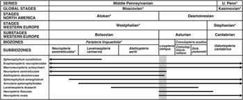
Comparison with the European biozonation (Fig. 6) is based on the compilation of taxon ranges as known in Western Europe (e.g., Laveine, Reference Laveine1987; Wagner, Reference Wagner1984; Cleal and Thomas, Reference Cleal and Thomas1994). Although some taxa are long ranging, there is minimal overlap between the LAD of Neuropteris semireticulata and Alethopteris densinervosa Wagner, Reference Wagner1968 and the FAD of Neuropteris flexuosa and members of the Neuropteris ovata complex, the latter group being considered a guide to the basal Asturian (Van Leckwijck, Reference Van Leckwijck1964; Laveine, Reference Laveine1977; Wagner, Reference Wagner1984). Accordingly, the age of the macrofloral assemblage can be tightly constrained to the early Asturian, assignable to the Linopteris obliqua Biozone. The anomalous presence of Sphenophyllum cuneifolium (Sternberg, Reference Sternberg1821) Zeiller, 1878-Reference Zeiller79a, more typical of lower Westphalian assemblages, does not override this outcome.
Biostratigraphic ranges of key fossil-species in macrofloral assemblage correlated with chronostratigraphic units and European macrofloral biozonation scheme. Compiled from Wagner (Reference Wagner1984), Laveine (Reference Laveine1987), Cleal (Reference Cleal1991), and Cleal and Thomas (Reference Cleal and Thomas1994). Range of Laveineopteris dussartii (Laveine, Reference Laveine1967) Laveine, Reference Laveine2005 included because may be synonymous with Neuropteris missouriensis Lesquereux, 1879-Reference Lesquereux80. Maximum range overlap occurs in lower Linopteris obliqua Biozone (early Asturian, early Desmoinesian).

Stratigraphic ramifications
These homotaxial comparisons yield slightly different age results for the Minshall Coal and overlying roof-shales depending on the fossil group or biostratigraphic framework used: (1) probably latest Bolsovian (American microfloras; Fig. 4); (2) latest Bolsovian or earliest Asturian (Appalachian Basin macrofloras; Fig. 5); or (3) earliest Asturian (European macrofloras; Fig. 6). Although all analyses clearly convey that both peat and clastic sediment accumulation occurred close to the Bolsovian-Asturian boundary, three crucial points need to be addressed before attempting to refine the stratigraphic resolution. First, the Minshall Coal and overlying roof-shales need not be the same age; the boundary may have been crossed in the interim between the final phases of peat accumulation and the drowning and burial of the mire during transgression. Second, significant (and perhaps undue) weight has been placed on coincidence of the Bolsovian-Asturian and Atokan-Desmoinesian boundaries, especially since they were defined in widely separated areas based on different parameters. Third, there must have been a temporal lag between the evolution of a new plant taxon and its dispersal to other parts of paleoequatorial Euramerica (cf., Laveine et al., Reference Laveine, Zhang and Lemoigne2000; Blake et al., Reference Blake, Cross, Eble, Gillespie and Pfefferkorn2002). These factors are taken into account as four stratigraphic scenarios are explored for the study section, each of which attempts to rectify the slightly disparate ages recorded in the microfloral and macrofloral assemblages.
Figure 7.1 shows the currently accepted correlation scheme in the Indiana portion of the Illinois Basin (with a focus on the Brazil and Staunton formations) and displays the relationships among chronostratigraphic, lithostratigraphic, and biostratigraphic units, at least as they have been understood prior to this analysis. The Bolsovian-Asturian Substage and Atokan-Desmoinesian Stage boundaries are equivalent and occur at the top of the Perth Limestone; the Minshall Coal belongs to the Radiizonates difformis (RD) Biozone and, by implication, the Minshall Coal and its roof-shales (Units 1 to 3) are upper Bolsovian. However, this scenario only is valid if Neuropteris ovata and Neuropteris flexuosa can occur in abundance in late Bolsovian assemblages. As noted above, both taxa make scattered appearances in middle to upper Bolsovian strata in the USA, but do not become common until the Asturian (Fig. 5; N. ovata in Zone 9, N. flexuosa in Zone 10 of Read and Mamay, Reference Read and Mamay1964).
Options for placement of Atokan-Desmoinesian Stage boundary and Bolsovian-Asturian Substage boundary based on biostratigraphic data presented herein. (1) Stratigraphy of Middle Pennsylvanian of Indiana part of Illinois Basin (as currently understood), showing correlation between chronostratigraphic and lithostratigraphic units, and American microfloral and European macrofloral biozonation schemes (* indicates incomplete unit). Compiled from Figures 2, 4, and 6. Macrofloral assemblage collected from lowermost Staunton Formation (arrows). For all four scenarios presented in 7.2–7.5, Minshall Coal retained in Radiizonates difformis (RD) Biozone and clastic units bearing macrofloral assemblage (arrows) in Linopteris obliqua Biozone. (2) Atokan-Desmoinesian and Bolsovian-Asturian boundaries remain coupled and lowered to top of Minshall Coal. (3) Atokan-Desmoinesian and Bolsovian-Asturian boundaries remain coupled and lowered to below Minshall Coal. (4) Atokan-Desmoinesian and Bolsovian-Asturian boundaries decoupled, with Atokan-Desmoinesian boundary retained at top of Perth Limestone and Bolsovian-Asturian boundary lowered to top of Minshall Coal. (5) Atokan-Desmoinesian and Bolsovian-Asturian boundaries decoupled, with Atokan-Desmoinesian boundary retained at top of Perth Limestone and Bolsovian-Asturian boundary lowered to below Minshall Coal. See discussion in text for stratigraphic ramifications of each scenario.

Introduction of the European macrofloral biozonation framework indicates that the roof-shales (Units 1 to 3) above the Minshall Coal (but not necessarily the coal itself) are no older than earliest Asturian (Fig. 6). At least four scenarios are possible (Fig. 7.2–7.5); all include the Minshall Coal in the Radiizonates difformis (RD) Biozone and its roof-shales in the Linopteris obliqua Biozone.
Scenario one
The Bolsovian-Asturian and Atokan-Desmoinesian boundaries could remain coupled and lowered to the top of the Minshall Coal (Fig. 7.2). The implications are that the precursor mire existed during the latest Bolsovian (latest Atokan), and that a chronostratigraphic boundary was crossed before transgression and deposition of the roof-shales in the earliest Asturian (earliest Desmoinesian). As a consequence, the contact between the R. difformis (RD) and Cadiospora magna-Mooreisporites inusitatus (MI) microfloral biozones would be slightly younger than the late Bolsovian (late Atokan) age proposed by Peppers (Reference Peppers1996; Fig. 7.1), and the basalmost Staunton Formation would correspond to the lower Asturian (lower Desmoinesian).
Scenario two
The coupled Bolsovian-Asturian and Atokan-Desmoinesian boundary could be situated somewhere below the Minshall Coal (Fig. 7. 3), meaning that both the peat and overlying clastics accumulated during the earliest Asturian (earliest Desmoinesian). Consequently, the upper part of the R. difformis (RD) Biozone and uppermost Brazil Formation would be younger than currently understood (Fig. 7.1).
Scenario three
The Bolsovian-Asturian and Atokan-Desmoinesian boundaries could be decoupled, with the former lowered to the top of the Minshall Coal and the latter remaining at the top of the Perth Limestone (Fig. 7.4). The ramifications mostly mirror those for point 1, except that the uppermost Brazil Formation, lowermost Staunton Formation, and lower part of the C. magna-M. inusitatus (MI) Biozone would be retained in the uppermost Atokan (although now correlative to the lowermost Asturian).
Scenario four
The decoupled Bolsovian-Asturian boundary could be placed at an even lower position below the Minshall Coal (Fig. 7.5). As in point 3, the upper Brazil, lower Staunton, and lower C. magna-M. inusitatus (MI) Biozone would remain in the upper Atokan, but would correspond to an even younger Asturian age.
All four scenarios have validity, but because this study deals with plant-fossil assemblages from one locality, no further attempt is made to recommend one over another. However, decoupling the Bolsovian-Asturian and Atokan-Desmoinesian boundaries, and lowering the former to the top of (or below) the Minshall Coal (points 3 and 4) is considered the least disruptive to the established stratigraphic framework. Similar studies at other sites in the Illinois Basin and elsewhere are necessary to rectify the position of the Bolsovian-Asturian Substage boundary of western Europe and its relationship to the Atokan-Desmoinesian Stage boundary in the USA.
Paleobiogeographic and paleoecologic implications
The temporal persistence of peat-inhabiting wetland vegetation in Pennsylvanian mires is well established, with evidence pointing to long periods of ecological stability through recurring glacioeustatic cycles, and the repeated reassembly of communities drawn from a small pool of edaphic specialists when climatic conditions were favorable (DiMichele and Phillips, Reference DiMichele and Phillips1996a, Reference DiMichele and Phillips1996b; DiMichele et al., Reference DiMichele, Pfefferkorn and Phillips1996, Reference DiMichele, Phillips and Nelson2002; Willard et al., Reference Willard, Phillips, Lesnikowska and DiMichele2007; Falcon-Lang and DiMichele, Reference Falcon-Lang and DiMichele2010). Comparable patterns of persistence and compositional recurrence also existed in vegetation from associated clastic wetland habitats (Scott, Reference Scott1978; Pfefferkorn and Thomson, Reference Pfefferkorn and Thomson1982; DiMichele et al., Reference DiMichele, Pfefferkorn and Phillips1996; Cleal et al., Reference Cleal, Uhl, Cascales-Miñana, Thomas, Bashforth, King and Zodrow2012; Tabor et al., Reference Tabor, Romanchock, Looy, Hotton, DiMichele and Chaney2013). These low-diversity ecosystems were the product of prolonged evolution under the intrinsically stressful conditions that typify wetland settings, which are effectively closed systems with low rates of speciation and turnover, and that experience little taxonomic exchange with terra-firma habitats (Knoll, Reference Knoll1985; DiMichele et al., Reference DiMichele, Phillips and Olmstead1987; Wing and DiMichele, Reference Wing and DiMichele1995; DiMichele and Phillips, Reference DiMichele and Phillips1996a; Cleal et al., Reference Cleal, Uhl, Cascales-Miñana, Thomas, Bashforth, King and Zodrow2012).
Pennsylvanian wetland communities also retained compositional conservatism at a variety of spatial scales. Over tens of meters to a few kilometers, mire vegetation was markedly heterogeneous yet encompassed a limited species pool (Pfefferkorn, Reference Pfefferkorn1979; DiMichele and Nelson, Reference DiMichele and Nelson1989; Pryor and Gastaldo, Reference Pryor and Gastaldo2000; Gastaldo et al., Reference Gastaldo, Stevanoviç-Walls, Ware and Greb2004, DiMichele et al., Reference DiMichele, Falcon-Lang, Nelson, Elrick and Ames2007, Opluštil et al., Reference Opluštil, Pšenička, Libertín, Bashforth, Šimůnek, Drábková and Dašková2009a, Reference Opluštil, Pšenička, Libertín, Bek, Dašková, Šimůnek and Drábková2009b), and clastic wetland floras displayed a comparable mosaic of low-diversity patches (Wing and DiMichele, Reference Wing and DiMichele1995; Bashforth et al., Reference Bashforth, Falcon-Lang and Gibling2010, Reference Bashforth, Drábková, Opluštil, Gibling and Falcon-Lang2011; Cleal et al., Reference Cleal, Uhl, Cascales-Miñana, Thomas, Bashforth, King and Zodrow2012). However, local-scale variability was masked at the landscape or regional scale, and disappeared into the background of the restricted wetland species pool. An expression of this homogeneity is discussed by Moore et al. (Reference Moore, Wittry and DiMichele2014), who demonstrated that two contemporaneous clastic wetland floras, separated by 1100 km on the American craton, showed convergent dominance-diversity structures and a remarkable degree of compositional similarity.
A key result of this study is confirmation that Pennsylvanian communities from clastic wetland habitats showed spatial conservatism over vast areas of tropical Euramerica. Despite promulgations about the lack of similarity between American and European adpressed macrofloral assemblages, this notion can be tempered, at least for the Middle Pennsylvanian time interval studied. Of the 18 biological fossil-taxa documented (Table 1), at least 14 species (78%; including Cordaites spp. indet.) are common in contemporaneous European assemblages. Assuming that Eusphenopteris Gothan, Reference Gothan1913 ex Simson-Scharold, Reference Simson-Scharold1934 (sp. indet.) and Sphenopteris (Brongniart, Reference Brongniart1822) Sternberg, Reference Sternberg1825 (sp. indet.) also occur in Europe, which they almost certainly do, and considering that the American form Neuropteris missouriensis Lesquereux, 1879-Reference Lesquereux80 may be conspecific with the European form Laveineopteris dussartii (Laveine, Reference Laveine1967) Laveine, Reference Laveine2005, the number of shared species could be as high as 17 (94%). Only the taxon identified as “Mariopteris” anthrapolis Langford, Reference Langford1958 seems to be unequivocally unique to American floras of this age (Table 1). Hence, it is not hyperbole to state that the macrofloral assemblage could have been collected from a lower Asturian roof-shale in Europe. It is clear, yet unsurprising, that the intrinsic stress inherent to wetland settings existed wherever such environments developed in tropical Euramerica, resulting in low-diversity communities that showed a high degree of taxonomic mutuality over the paleocontinent.
The discovery of Alethopteris densinervosa in the macrofloral assemblage, and the realization that it had previously been misidentified as Alethopteris serlii (Brongniart, 1833-Reference Brongniart34) Göppert, Reference Göppert1836 in other American floras (e.g., Basson, Reference Basson1968; Oleksyshyn, Reference Oleksyshyn1982; Moore et al., Reference Moore, Wittry and DiMichele2014), provides additional evidence for the spatial congruity in Pennsylvanian clastic wetland vegetation. Formerly only known from the Northern France Coalfield (Wagner, Reference Wagner1968; Laveine, Reference Laveine1987), and perhaps the Sydney Coalfield (Zodrow, Reference Zodrow1986; Zodrow and Cleal, Reference Zodrow and Cleal1998), the presence of a relatively obscure fossil-species such as A. densinervosa in widely separated areas from strata of similar age (early Bolsovian to middle Asturian) highlights the commonality of Middle Pennsylvanian floras in wetland deposits of Euramerica.
From a paleobiogeographic perspective, these observations are important because west-central and eastern Euramerica were separated by the expansive uplands of the Laurentian Shield to the north and, in part, by the Appalachian-Variscan Mountains (Blakey, Reference Blakey2013). The similarity of wetland communities on either side of the paleocontinent confirms that, at least during the Middle Pennsylvanian, these elevated areas did not present an insurmountable barrier to dispersal for most wetland plants (cf., White, Reference White1909; Pfefferkorn and Gillespie, Reference Pfefferkorn and Gillespie1980b; Blake et al., Reference Blake, Cross, Eble, Gillespie and Pfefferkorn2002). In fact, the mountain belt probably comprised a spatially and temporally discontinuous chain of tectonically active centers (Roscher and Schneider, Reference Roscher and Schneider2006), which rose (and eroded down) earlier in the east than in the west in zipper-like fashion (Hatcher, Reference Hatcher2002, Reference Hatcher2010), rather than a continuous belt of high-elevation that spanned the entire equatorial region of Pangea. Of course, compositional differences and endemic taxa certainly existed in various parts of Euramerica (e.g., Bell, Reference Bell1944; Pfefferkorn and Gillespie, Reference Pfefferkorn and Gillespie1980b; Gastaldo and Boersma, Reference Gastaldo and Boersma1983a, Reference Gastaldo and Boersma1983b; Cleal, Reference Cleal2002, Reference Cleal2008a, Reference Cleal2008b; Cleal et al., Reference Cleal, Opluštil, Thomas, Tenchov, Abbink, Bek, Dimitrova, Drábková, Hartkopf-Fröder, Van Hoof, Kędzior, Jarzembowski, Jasper, Libertín, McLean, Oliwkiewicz-Miklasinska, Pšenička, Ptak, Schneider, Schultka, Šimůnek, Uhl, Waksmundzka, Van Waveren and Zodrow2009, Reference Cleal, Uhl, Cascales-Miñana, Thomas, Bashforth, King and Zodrow2012; Šimůnek and Cleal, Reference Šimůnek and Cleal2011), presumably a product of regional landscape partitioning due to elevational, latitudinal, and climatic factors (Blake et al., Reference Blake, Cross, Eble, Gillespie and Pfefferkorn2002; Falcon-Lang et al., Reference Falcon-Lang, Heckel, DiMichele, Blake, Easterday, Eble, Elrick, Gastaldo, Greb, Martino, Nelson, Pfefferkorn, Phillips and Rosscoe2011). Such variables would have determined when and where speciation occurred, and must have impeded the dispersibility of wetland vegetation to some degree. For example, Neuropteris ovata and Neuropteris flexuosa make scattered appearances in middle to upper Bolsovian strata in the USA (Jongmans, Reference Jongmans1937c; Wood, Reference Wood1963; Read and Mamay, Reference Read and Mamay1964; Darrah, Reference Darrah1969; Blake et al., Reference Blake, Cross, Eble, Gillespie and Pfefferkorn2002), even though these species are key indicators to identifying the base of the Asturian in Europe and Atlantic Canada (Laveine, Reference Laveine1977; Wagner, Reference Wagner1984; Zodrow and Cleal, Reference Zodrow and Cleal1985; Cleal and Thomas, Reference Cleal and Thomas1994; Cleal, Reference Cleal2007). The paleobiogeographic ramification of these precocious appearances in the USA is that N. ovata and N. flexuosa originated in the western part of the paleocontinent before synchronously arriving in Europe. Although such paleobiogeographic vagaries may hinder the precision of homotaxial biostratigraphic comparisons between American and European macrofloral assemblages, on the whole, correlations within Euramerica should be possible so long as regional peculiarities are taken into account.
The marked lithological and biological changes that led to the recognition of the Atokan-Desmoinesian Stage boundary in the USA and the Bolsovian-Asturian Substage boundary in Western Europe may record a common climatic shift that occurred throughout equatorial regions of Euramerica (cf., Cecil et al., Reference Cecil, Stanton, Neuzil, Dulong, Ruppert and Pierce1985; Bertier et al., Reference Bertier, Swennen, Lagrou, Laenen and Kemps2008; Van Hoof et al., 2012). At the core of this environmental change was a shift to increasing seasonality of equatorial precipitation at all phases of glacioeustatic cycles (Cecil et al., Reference Cecil, Stanton, Neuzil, Dulong, Ruppert and Pierce1985; Roscher and Schneider, Reference Roscher and Schneider2006), which corresponded to a progressive decrease in grounded ice in the Southern Hemisphere (Fielding et al., Reference Fielding, Frank and Isbell2008) and the onset of a rise in global sea level (Rygel et al., Reference Rygel, Fielding, Frank and Birgenheier2008). Because the change may have begun (or at least initially was most strongly manifest) in western Pangea before rapidly spreading eastward, and given the vast equatorial area over which the modification occurred, the boundaries show a degree of temporal and spatial disjunction. As such, the expression of this major climatic shift in the rock record may present a more realistic model for the timing and effects of large-scale environmental changes than do short-term, globally contemporaneous catastrophes.
Tectonic activity also played a prominent role during this time interval in North America. In fact, the type Atokan Stage resides in the heart of the Ouachita orogenic belt (southern part of the greater Appalachian-Variscan chain), and the Ouachita orogeny culminated near the end of the Atokan (Houseknecht, Reference Houseknecht1983). Assuredly, elevation of this new mountain range also affected regional climate.
Materials and methods
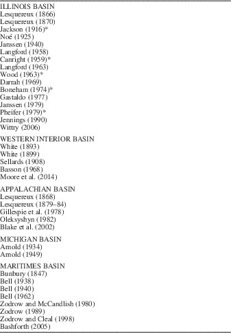
A selection of North American publications that provide detailed descriptive and illustrated records of Middle Pennsylvanian adpressions was the main source used for comparison with the macrofloral assemblage (Table 3). The list focuses on the Illinois Basin, and Indiana in particular, but includes records from the Western Interior, Appalachian, and Michigan basins of the USA, and the Maritimes Basin of Atlantic Canada. Almost all fossil-taxa in the macrofloral assemblages (Table 1) are described; the only exceptions are Calamites spp. indet. and the putative conifer cone scale. Likewise, most taxa are illustrated in Figures 8–11 (including the conifer cone scale, Fig. 11.7).
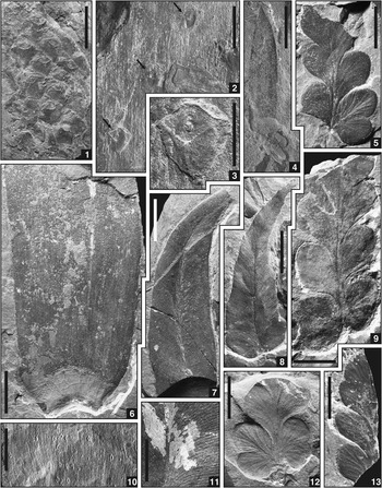
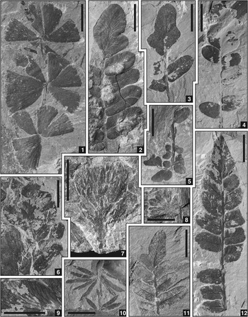
Specimens from Units 1 and 2 (Fig. 3) above Minshall Coal, Clay County, Indiana; scale bars=10 mm (1, 2, 4–10, 12, 13) or 5 mm (3, 11). (1) Synchysidendron sp. cf. S. andrewsii (Lesquereux, 1879-Reference Lesquereux80) Moore, Wittry, and DiMichele, Reference Moore, Wittry and DiMichele2014, lycopsid bark, Unit 1, USNM 559868. (2, 3) cf. Asolanus camptotaenia Wood, Reference Wood1860, lycopsid bark, with arrows (2) indicating position of leaf cushions and details of lowermost leaf cushion (3), Unit 1, USNM 559867. (4) Lepidostrobophyllum sp. indet., lycopsid cone scale, Unit 1, USNM 559870. (5), “Mariopteris” anthrapolis Langford, Reference Langford1958, lyginopteridalean pteridosperm foliage, ultimate pinna with proximal quadrate pinnules that exhibit strongly incised acroscopic margin and division into rounded lobes, passing apically into strongly decurrent and spatulate to obovate pinnules, part of 12, Unit 2, USNM 559873. (6) Cordaites sp. indet., cordaitalean leaf, with well-preserved leaf base, Unit 2, USNM 594360. (7) Macroneuropteris scheuchzeri (Hoffmann, Reference Hoffmann1826a) Cleal, Shute, and Zodrow, Reference Cleal, Shute and Zodrow1990, medullosalean pteridosperm foliage, subfalcate pinnule, Unit 2, USNM 594361. (8) Macroneuropteris scheuchzeri, subfalcate pinnule exhibiting initial stages of differentiation of pinnule base into two symmetrical lobes, eventually becoming small orbicular pinnules at base of trifoliate pinnule form, Unit 2, USNM 559875. (9) “Mariopteris” anthrapolis, Unit 2, ultimate pinna with large quadrate pinnules near base and spatulate to obovate pinnules near top, USNM 559871. (10) Cordaites sp. indet., with spirorbiform worm-tubes (microconchids) affixed to surface, Unit 1, USNM 559869. (11) Macroneuropteris scheuchzeri, exhibiting hair-like structures characteristic of taxon, Unit 2, USNM 594362. (12) “Mariopteris” anthrapolis, counterpart of 5, Unit 2, USNM 559873. (13) “Mariopteris” anthrapolis, distal end of ultimate pinna with strongly decurrent and spatulate to obovate pinnules that meld into large lobate terminal pinnule with subtly crenulate margin, Unit 2, USNM 559874.

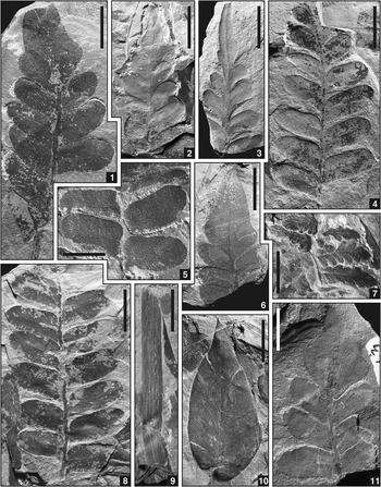
Specimens from Unit 2 (Fig. 3) above Minshall Coal, Clay County, Indiana; all scale bars=10 mm. (1) Neuropteris flexuosa Sternberg, Reference Sternberg1825, medullosalean pteridosperm foliage, pinna with large rhomboid to deltoid terminal pinnule, USNM 594364. (2) Neuropteris ovata Hoffmann, Reference Hoffmann1826a, Reference Hoffmann1826b, medullosalean pteridosperm foliage, pinna with small elongate terminal pinnule and ‘mixoneurid’ habit in distal lateral pinnules, USNM 594366. (3) Neuropteris ovata, pinna with small elongate terminal pinnule and ‘mixoneurid’ habit in distal lateral pinnules, USNM 594365. (4) Neuropteris ovata, pinna with lateral pinnules having a strongly incised acroscopic base and a pronounced basiscopic auricle, USNM 594363. (5) Neuropteris flexuosa, showing rounded acroscopic base of lateral pinnules, USNM 559872. (6) Neuropteris flexuosa, pinna with large rhomboid to deltoid terminal pinnule, USNM 594367. (7) Sphenopteris sp. indet., fern foliage of unknown affinity, USNM 594368. (8) Neuropteris ovata, pinna with lateral pinnules having a strongly incised acroscopic base and a pronounced basiscopic auricle, USNM 594359. (9) Cyperites bicarinatus Lindley and Hutton, Reference Lindley and Hutton1832, lycopsid leaf, USNM 594369. (10) Macroneuropteris scheuchzeri (Hoffmann, Reference Hoffmann1826a) Cleal, Shute, and Zodrow, Reference Cleal, Shute and Zodrow1990, medullosalean pteridosperm foliage, subtriangular pinnule with asymmetrical base, USNM 594370. (11) Neuropteris flexuosa, pinna with large rhomboid to deltoid terminal pinnule, and lateral pinnules with pronounced basiscopic auricle, USNM 594371.

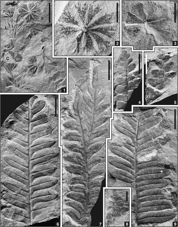
Specimens from Unit 3 (Fig. 3) above Minshall Coal, Clay County, Indiana; scale bars=10 mm (1, 4–7, 9) or 5 mm (2, 3, 8). (1) Sphenophyllum cuneifolium (Sternberg, Reference Sternberg1821) Zeiller, Reference Zeiller1879, sphenophyll foliage, with whorls of several leaf forms, including (C) cuneate ‘normal’ leaves (forma typica), (S) cuneate ‘divided’ leaves (forma saxifragaefolium), and (L) ‘deeply divided’ to ‘linear’ leaves, USNM 594376. (2) Sphenophyllum cuneifolium, whorl with cuneate ‘divided’ leaves (forma saxifragaefolium), USNM 594379. (3) Sphenophyllum cuneifolium, whorl with cuneate ‘normal’ leaves (forma typica), USNM 594377. (4) Eusphenopteris neuropteroides (Boulay, Reference Boulay1876) Novik, Reference Novik1947, lyginopteridalean pteridosperm foliage, ultimate pinna with trilobate to bilobate pinnules near base, passing upwards to quadrate or ovate pinnules and topped by small lobate terminal pinnule, USNM 594380. (5) Eusphenopteris neuropteroides, penultimate pinna, from which arises ultimate pinna with trilobate to bilobate pinnules near base and quadrate to ovate pinnules near top, USNM 594378. (6) Alethopteris densinervosa Wagner, Reference Wagner1968, medullosalean pteridosperm foliage, biconvex pinnule form, USNM 594372. (7) Alethopteris densinervosa, subtriangular pinnule form on pinnae topped by a small allantoid terminal pinnule, USNM 594375. (8) Eusphenopteris neuropteroides, strongly decurrent rounded pinnules and small terminal pinnule at distal end of ultimate pinna, USNM 594374. (9) Alethopteris densinervosa, exhibiting transition from biconvex pinnule form at top of pinna to elongate form at base, USNM 594373.

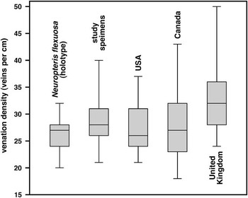
Specimens from Units 2 and 4 (Fig. 3) above Minshall Coal, Clay County, Indiana; scale bars=10 mm (2–6, 11, 12) or 5 mm (1, 7–10). (1) Sphenophyllum emarginatum (Brongniart, Reference Brongniart1822) Brongniart, Reference Brongniart1828b, sphenophyll foliage, with whorls displaying cuneate leaves, Unit 4, USNM 594381. (2) Neuropteris missouriensis Lesquereux, 1879-Reference Lesquereux80, medullosalean pteridosperm foliage, pinna with large obtusely rounded terminal pinnule, Unit 4, USNM 594388. (3) Neuropteris missouriensis, pinna with large obtusely rounded terminal pinnule, Unit 4, USNM 594387. (4) Neuropteris missouriensis, pinna with large obtusely rounded terminal pinnule and small ovate lateral pinnules, Unit 4, USNM 594390. (5) Eusphenopteris sp. indet., lyginopteridalean pteridosperm foliage, Unit 4, USNM 594385. (6, 9) Neuropteris semireticulata Josten, Reference Josten1962, medullosalean pteridosperm foliage, with details of flexuous to pseudoreticulate venation characteristic of taxon in 11.9, Unit 4, USNM 594382. (7) Possible cone scale of primitive conifer, Unit 2, USNM 558409. (8) Annularia sphenophylloides (Zenker, Reference Zenker1833) Gutbier, Reference Gutbier1837, calamitalean foliage, Unit 4, USNM 594383. (10) Annularia sp. cf. A. radiata (Brongniart, Reference Brongniart1822) Sternberg, Reference Sternberg1825, calamitalean foliage, Unit 4, USNM 594384. (11) Neuropteris missouriensis, top of pinna with small terminal and elongate lateral pinnules, somewhat atypical of taxon, and prominent compression margin on some pinnules, Unit 4, USNM 594386. (12) Neuropteris missouriensis, large pinna with smaller, less obtusely rounded terminal pinnule and lateral pinnules that display prominent compression margin, Unit 4, USNM 594389.

Main literature sources used for comparison with macrofloral assemblage, and to compile synonymy lists. All publications provide detailed descriptive and/or illustrated records of Middle Pennsylvanian plant-fossil adpressions in North America, with focus on Illinois Basin (* indicates record from Indiana).

Repositories and institutional abbreviations
Plant fossils are stored at the National Museum of Natural History (NMNH), Smithsonian Institution, Washington, DC, and illustrated specimens are reposited in the Plant Type and Illustrated Collection under USNM numbers 558409, 559867–559875, and 594359–594391. Components of the assemblage were the subject of a dissertation (Comer, Reference Comer1992) that included specimens from several stratigraphic levels and depositional facies not included in this study.
Systematic paleontology
Division Tracheophyta
Class Lycopsida
Order Isoëtales Bartling, Reference Bartling1830
Family Diaphorodendraceae DiMichele and Bateman, Reference DiMichele and Bateman1992
Genus Synchysidendron DiMichele and Bateman, Reference DiMichele and Bateman1993
Synchysidendron sp. cf. S. andrewsii (Lesquereux, 1879-Reference Lesquereux80) Moore, Wittry, and DiMichele, Reference Moore, Wittry and DiMichele2014
1879-80 Lepidodendron Andrewsii Reference LesquereuxLesquereux, p. 389, pl. 64, fig. 6.
1938 Lepidodendron dichotomum Arber var. bretonense Reference BellBell, p. 92, pl. 95, figs. 5–9, pl. 96, fig. 1, pl. 97, fig. 4.
1940 Lepidodendron dichotomum; Reference BellBell, p. 122, pl. 7, fig. 4.
1944 Lepidodendron dichotomum var. bretonense; Reference BellBell, p. 89, pl. 45, fig. 3.
1962 Lepidodendron bretonense Reference BellBell, p. 53, pl. 47, figs. 5, 6(?), pl. 48, figs. 4, 6(?).
(?)1963 Lepidodendron Sternberg sp.; Reference LangfordLangford, p. 153, figs. 712, 713.
1980 Lepidodendron bretonense; Reference Zodrow and McCandlishZodrow and McCandlish, p. 80, pl. 116, fig. 1, pl. 118, figs. 1(?), 2.
1985 Lepidodendron bretonense; Reference WnukWnuk, p. 169, pl. 2, figs. 7, 8.
2005 Diaphorodendron bretonense (Bell) Reference BashforthBashforth, p. 36, pl. 2, figs. 1–4, 7, 8.
2006 Lepidodendron andrewsii; Reference WittryWittry, p. 106, figs. 1–3 [holotype: figs. 1, 2].
2014 Synchysidendron andrewsii (Lesquereux) Reference Moore, Wittry and DiMicheleMoore, Wittry and DiMichele, p. 28, pl. 1, figs. 1–13.
Holotype
USNM 15430 (part) and USNM 15431 (counterpart), Plant Type and Illustrated Collection, National Museum of Natural History, Smithsonian Institution, Washington, from Francis Creek Shale, Carbondale Formation (middle Desmoinesian), Illinois Basin, Mazon Creek, Illinois.
Occurrence
USA and Canada: common throughout Middle Pennsylvanian (Bell, Reference Bell1938, Reference Bell1962; Bashforth, Reference Bashforth2005; DiMichele et al., Reference DiMichele, Elrick and Bateman2013a; Moore et al., Reference Moore, Wittry and DiMichele2014).
Material examined
One specimen from Unit 1.
Description
The axis fragment is 42 mm long and 23 mm wide. Leaf cushions are contiguous, crowded, asymmetrical, 7–9 mm long and 6–7 mm wide (L:W ratio ~1.3), and quadrilateral to napiform with curved to obtusely rounded lateral margins. Upper angles appear obtusely to acutely pointed, whereas lower angles appear acutely pointed (but are overlapped by the top of the subjacent cushion). Leaf scars are situated in the upper half of a cushion, slightly elevated, 1.2–1.8 mm long, 2.3–4.0 mm wide (L:W ratio ~0.5), with a broadly convex lower margin, a triangular upper margin with straight sides that converge to an obtusely pointed top, and acutely pointed lateral angles. Cicatricules are not preserved and their position and dimensions are unknown. A gently arched ridge runs from each lateral angle of the leaf scar to the lateral margin of the leaf cushion, separating the cushion into upper and lower fields that lack obvious ornamentation.
Remarks
In an adpressed state, the absence of infrafoliar parichnos scars beneath the leaf scar is the overriding character that confers assignment to Synchysidendron or Diaphorodendron DiMichele, Reference DiMichele1985 of the Diaphorodendraceae (DiMichele, Reference DiMichele1985; DiMichele and Bateman, Reference DiMichele and Bateman1992), and distinguishes it from true Lepidodendron Sternberg, Reference Sternberg1820 (sensu DiMichele, Reference DiMichele1983) of the Lepidodendraceae. Other features, such as the fleshiness of leaf cushions, the presence on large axes of flat interareas between leaf cushions, and the degree of ornamentation on the lower keel of the leaf cushion contribute to generic identification. Segregation of the Diaphorodendraceae from the Lepidodendraceae is more apparent in anatomically preserved examples, as the vegetative anatomy and reproductive organs are conspicuously different (DiMichele, Reference DiMichele1979a, Reference DiMichele1979b, Reference DiMichele1983, Reference DiMichele1985; DiMichele and Bateman, Reference DiMichele and Bateman1992). Furthermore, the two families can be differentiated by their growth habits (Wnuk, Reference Wnuk1985, DiMichele et al., Reference DiMichele, Elrick and Bateman2013a). Trunks of Synchysidendron and Diaphorodendron bear two opposite rows of large deciduous lateral branches (or branch scars) that extend into a dichotomously branched crown, whereas Lepidodendron only has a terminal dichotomous crown.
Leaf cushions of the study specimen approach equilateral dimensions (L:W ratio ~1.3), are contiguous to slightly overlapping, and markedly protrude from the stem surface, supporting assignment to Synchysidendron rather than Diaphorodendron, which tends to have relatively flat and non-protruding leaf cushions with a higher L:W ratio (Wnuk, Reference Wnuk1985; DiMichele and Bateman, Reference DiMichele and Bateman1992; DiMichele et al., Reference DiMichele, Elrick and Bateman2013a; Moore et al., Reference Moore, Wittry and DiMichele2014). The axis fragment probably was derived from a deciduous lateral branch, the most common litter elements of the Diaphorodendraceae (DiMichele et al., Reference DiMichele, Elrick and Bateman2013a).
The specimen resembles adpressed examples of Synchysidendron andrewsii, a fossil-species that was documented by Moore et al. (Reference Moore, Wittry and DiMichele2014) and is the most common form of Synchysidendron in the Middle Pennsylvanian of the USA (DiMichele et al., Reference DiMichele, Elrick and Bateman2013a). The taxon has been recorded in Atlantic Canada under Lepidodendron dichotomum var. bretonense (Bell, Reference Bell1938, Reference Bell1944), Lepidodendron bretonense (Bell, Reference Bell1962; Zodrow and McCandlish, Reference Zodrow and McCandlish1980), and incorrectly assigned to Diaphorodendron bretonense by Bashforth (Reference Bashforth2005). Anatomically preserved axes of comparable morphology have been referred to Synchysidendron dicentricum (Felix, Reference Felix1952) DiMichele and Bateman, Reference DiMichele and Bateman1993 (DiMichele, Reference DiMichele1979b; DiMichele and Bateman, Reference DiMichele and Bateman1992, Reference DiMichele and Bateman1993; DiMichele et al., Reference DiMichele, Elrick and Bateman2013a). Similar to examples of S. andrewsii in Moore et al. (Reference Moore, Wittry and DiMichele2014, pl. 1, figs. 1–13), the specimen has protuberant, napiform to quadrilateral leaf cushions, leaf scars wider than long that are situated in the upper half of the cushion, a gently arched ridge that runs between the scar and cushion margins, and the faint impression of a notch in the upper field just below the upper angle; such notches are exemplified in DiMichele et al. (Reference DiMichele, Elrick and Bateman2013a, fig. 5b). However, S. andrewsii tends to have weakly developed upper and lower keels and plications on the lower keel; absence of these features in the specimen makes referral uncertain.
Family Sigillariaceae(?) Unger (in Endlicher), Reference Unger1842
Genus Asolanus Wood, Reference Wood1860
cf. Asolanus camptotaenia Wood, Reference Wood1860
1860 Asolanus camptotænia Reference WoodWood, p. 238, pl. 4, fig. 1.
1866 Sigillaria monostigma Reference LesquereuxLesquereux, p. 449, pl. 42, figs. 1–5.
1869 Sigillaria camptotænia (Wood) Reference WoodWood, p. 342, pl. 9, fig. 3.
1870 Sigillaria (Knorria) monostigma; Reference LesquereuxLesquereux, p. 446, pl. 26, fig. 5.
1879-80 Sigillaria monostigma; Reference LesquereuxLesquereux, p. 468, pl. 73, figs. 3–6.
1899a Sigillaria (Asolanus) camptotænia; Reference WhiteWhite, p. 230, pl. 69, pl. 70, figs. 1, 3(?), 4.
1925 Sigillaria camptotaenia; Reference NoéNoé, pl. 11, figs. 1, 3.
1938 Asolanus camptotaenia; Reference BellBell, p. 102, pl. 104, figs. 1–3.
1940 Asolanus camptotaenia; Reference JanssenJanssen, p. 22, pl. 5, figs. 1, 2, 3(?), pl. 6(?).
1949 Asolanus camptotaena [sic]; Reference ArnoldArnold, p. 179, pl. 11, fig. 3.
1958 Asolanus camptotaenia; Reference LangfordLangford, p. 108, text-figs. 200–202.
1962 Asolanus camptotaenia; Reference BellBell, p. 56, pl. 48, figs. 1–3.
1968 Asolanus camptotaenia; Reference BassonBasson, p. 40, pl. 1, figs. 1, 2.
1969 Asolanus camptotaenia; Reference DarrahDarrah, p. 182, pl. 34, fig. 1.
1977 Asolanus campotaenia [sic]; Reference GastaldoGastaldo, text-fig. 21.
1978 Asolanus camptotaenia; Reference Gillespie, Clendening and PfefferkornGillespie et al., p. 50, pl. 20, figs. 1, 3.
2006 Asolanus camptotaenia; Reference WittryWittry, p. 111, fig. 1.
Holotype
ASNP 695, Paleobotany Collection, Academy of Natural Sciences of Drexel University, Philadelphia, from “Anthracite Region of Pennsylvania” (Darrah, Reference Darrah1969, p. 183).
Occurrence
USA: middle(?) Atokan to middle Virgilian, most common in upper Atokan to upper Desmoinesian (Darrah, Reference Darrah1969; Gillespie and Pfefferkorn, Reference Gillespie and Pfefferkorn1979; Oleksyshyn, Reference Oleksyshyn1982; Blake et al., Reference Blake, Cross, Eble, Gillespie and Pfefferkorn2002). UK: lower Duckmantian to lower Cantabrian (Cleal, Reference Cleal2007). Atlantic Canada: middle Asturian (Bell, Reference Bell1938).
Material examined
Two specimens from Units 1 and 4.
Description
The illustrated axis fragment is 130 mm long and 105 mm wide, and involves three preservation states that represent degrees of decortification. All preservation modes exhibit longitudinally corrugated interfoliar areas, expressed as subparallel to slightly flexuous and discontinuous ridges and furrows; corrugations are coarser on the outermost layer, and finer on the innermost layer. Leaf cushions (Fig. 8.3) are spirally arranged and widely spaced, ranging from 14–23 mm apart (measured center to center in the helix), and their morphology is only preserved in the two outermost layers. Cushions protrude slightly from the axis surface, 6–8 mm long, 5–7 mm wide (L:W ratio ~1.1), with rhomboidal to subtriangular outlines (widest at the top). Upper angles are broadly convex, lower angles are obtusely rounded, and lateral angles are obtusely rounded to pointed. Leaf scars are situated at the very top of a cushion, slightly elevated, 2.0–3.3 mm long, 3.5–5.0 mm wide (L:W ratio ~0.6), with broadly curved upper and lower margins that converge laterally into acutely rounded to pointed lateral angles. Situated in the center of a leaf scar is a single prominent cicatricule, which is circular, ~1 mm in diameter, and consists of a raised rim around a central hollow; other equivocal pits may occur above and/or below the main cicatricule. Leaf cushions appear smooth and lack obvious ornamentation, although some exhibit a faint and finely pustular texture.
Remarks
Specimens belong to one of a number of lycopsid genera characterized by widely spaced leaf scars, but identification is ambiguous because they exhibit a blend of features typical of multiple taxa. The relatively coarse longitudinal interfoliar corrugations resemble some fossil-species of Bothrodendron Lindley and Hutton, Reference Lindley and Hutton1833 (particularly Bothrodendron punctatum Lindley and Hutton, Reference Lindley and Hutton1833) and Cyclostigma Haughton, Reference Haughton1860, the latter synonymous with Pinakodendron Weiss (in Weiss and Sterzel), Reference Weiss and Sterzel1893 according to Cambier and Renier (Reference Cambier and Renier1912) and Crookall (Reference Crookall1964). However, neither of these fossil-genera have leaf cushions, leaf scars are oval to almost circular with rounded and inconspicuous lateral angles, leaf scars contain three foliar cicatricules, and a prominent ligule pit occurs above the leaf scar in Bothrodendron (absent in Cyclostigma). Because all of these morphological traits are incongruent with the material at hand, identity is unlikely.
The specimens most strongly resemble Asolanus (usually reported as Asolanus camptotaenia), which has affinities with Sigillaria Brongniart, Reference Brongniart1822 (Wood, Reference Wood1869; White, Reference White1899a). Like the study examples, this taxon has leaf cushions with a convex upper angle and subtriangular lower angle, leaf scars that are rhomboidal to lenticular with sharp lateral angles, and a centrally positioned cicatricule that is circular and ring-like (Crookall, Reference Crookall1964). Asolanus camptotaenia exhibits a wide range of morphologies depending on preservation state and position on the original plant. However, in its most recognizable form, the species has unique interfoliar ornamentation that consists of straight to slightly flexuous, parallel striations that extend perpendicular from the four edges of the leaf scar, resulting in a rhomboidal pattern on the stem surface. This is inconsistent with the longitudinally corrugated surface on the specimen, so confident assignment to A. camptotaenia is not possible.
Incertae sedis
Genus Cyperites Lindley and Hutton, Reference Lindley and Hutton1832
Cyperites bicarinatus Lindley and Hutton, Reference Lindley and Hutton1832 emend. Rex, Reference Rex1983
1832 Cyperites bicarinata Reference Lindley and HuttonLindley and Hutton, p. 123, pl. 43, figs. 1, 2.
1944 Lepidophyllum Brongniart sp.; Reference BellBell, p. 96, pl. 59, fig. 2.
(?)1959 Lepidophyllum longifolium Brongniart; Reference CanrightCanright, pl. 1, fig. 9.
1963 Lepidophyllum bicarinatum (Lindley and Hutton) Reference LangfordLangford, p. 159, text-fig. 726.
1963 Lepidophyllum longifolium; Reference LangfordLangford, p. 159, text-fig. 728.
1963 Lepidophyllum sp.; Reference LangfordLangford, p. 159, text-fig. 729.
1963 Lepidophyllum sp.; Reference WoodWood, p. 37, pl. 2, fig. 2.
1967 Lepidophyllum longifolium; Reference TidwellTidwell, p. 22, pl. 1, fig. 3.
1974 Lepidophyllum longifolium; Reference BonehamBoneham, p. 97, pl. 1, fig. 6.
1977 Lepidophylloides Snigirevakaya sp.; Reference GastaldoGastaldo, text-fig. 18.
1978 Lepidophylloides sp.; Reference Gillespie, Clendening and PfefferkornGillespie et al., p. 45, pl. 12, fig. 1, pl. 13, fig. 2.
1979 Lepidophyllum longifolium; Reference JanssenJanssen, p. 62, text-fig. 44.
1979 Cyperites bicarinatus; Reference PheiferPheifer, pl. 1, figs. 29, 30.
(?)1980 Sigillariophyllum sp. Grand’Eury; Reference Zodrow and McCandlishZodrow and McCandlish, p. 89, pl. 144.
1982 Cyperites bicarinatus; Reference OleksyshynOleksyshyn, p. 29, text-fig. 9b.
1983 Cyperites bicarinatus Lindley and Hutton emend. Reference RexRex, p. 81, pl. 1, pl. 4 [neotype: pl. 4, fig. 1].
1992 Cyperites sp.; Reference Tidwell, Jennings and BuesTidwell et al., p. 1018, text-fig. 4.4.
2005 Cyperites bicarinatus; Reference BashforthBashforth, p. 36, pl. 1, figs. 1, 2.
2006 Cyperites sp.; Reference WittryWittry, p. 103, figs. 1, 3–5.
Type material
Syntypes (lost?) from Leebotwood Coal Pit, Halesowen Formation (upper Asturian or lower Cantabrian), Shropshire, UK. Neotype (V.61563), Natural History Museum, London, from roof-shale of No. 10 Coal Seam, Kilmersdon Tip, Farrington Formation (upper Asturian), Radstock Coalfield, Somerset, UK.
Occurrence
Euramerica: common throughout Carboniferous.
Material examined
Thirty-four specimens from Units 1 and 2.
Description
Leaves are linear and taper slightly distally, have entire lateral margins, and are up to 63 mm long (neither bases nor apices preserved) and 2–7 mm wide. A number of ridges and furrows exist on the leaf surface; an elevated or depressed belt (1–2 mm wide) runs along the center of the leaf, and a thin medial furrow (midvein?) may be present.
Remarks
Isolated, linear lycopsid leaves are assigned to the fossil-genus Cyperites. The leaves had a complex ‘butterfly’ shape in transverse section, and the presence or absence (and number) of ridges and furrows on the exposed leaf surface depends on the path of the fracture plane across the fossil (Rex, Reference Rex1983). Wider examples of Cyperites tend to belong to sigillarians and narrower leaves to lepidodendroids (Bashforth, Reference Bashforth2005). Both size fractions exist in the assemblage, suggesting that both groups may be present, although no other macrofloral elements of Sigillaria were identified.
Genus Lepidostrobophyllum Hirmer, Reference Hirmer1927, emend. Allen, Reference Allen1961
Lepidostrobophyllum sp. indet.
Occurrence
Euramerica: fossil-genus common throughout Carboniferous.
Material examined
Five specimens from Units 1, 2, and 4.
Description
Sporophyll laminae are 7–11 mm wide, broadest about 1/3 the way up the blade, ~20–45 mm long, and linear-lanceolate with convex, entire lateral margins that converge to an acuminate tip. Lateral angles at the base of sporophyll laminae do not project outwards, but appear flush with the pedicel top. Midveins (a rib on the adaxial surface) are 0.5–1.0 mm wide and faintly longitudinally striated. Pedicels are ~10 mm long and 5 mm wide, widest at the contact with the sporophyll lamina, have straight to slightly convex margins, and may terminate in a sharp base. An oval, thickened rim may be superimposed on the pedicel surface.
Remarks
The description is imprecise due to poor preservation. Based on size and shape, the isolated sporophylls disaggregated from a lepidodendroid cone, and thus are assigned to the fossil-genus Lepidostrobophyllum. More than one fossil-species may be involved, but the closest comparison is with Lepidostrobophyllum lanceolatum (Lindley and Hutton, Reference Lindley and Hutton1831) Bell, Reference Bell1938 (e.g., Bell, Reference Bell1938, pl. 98, figs. 7–11; Crookall, Reference Crookall1966, pl. 99, figs. 4, 5, under Lepidostrobus lanceolatus), which Lesquereux (Reference Lesquereux1870, 1879-Reference Lesquereux80) described under the junior synonym Lepidostrobus oblongifolius.
Class Sphenopsida
Order Sphenophyllales Seward, Reference Seward1898
Family Bowmanitaceae Meyen, Reference Meyen1978
Genus Sphenophyllum Brongniart, Reference Brongniart1822
Sphenophyllum cuneifolium (Sternberg, Reference Sternberg1821) Zeiller, 1878-Reference Zeiller79a
1821 Rotularia asplenioides Reference SternbergSternberg, p. 30, pl. 26, figs. 4a, 4b (alternate name).
1821 Rotularia cuneifolia Reference SternbergSternberg, p. 33, pl. 26, figs. 4a, 4b.
1825 Rotularia saxifragaefolia Reference SternbergSternberg, p. 45, p. XXXII, pl. 55, fig. 4.
1831 Sphenophyllum erosum Reference Lindley and HuttonLindley and Hutton, p. 41, pl. 13.
1847 Sphenophyllum erosum; Reference BunburyBunbury, p. 430, pl. 23, figs. 3a, 3b.
1848 Sphenophyllum saxifragaefolium (Sternberg) Reference GöppertGöppert (in Bronn), p. 1166.
1868 Sphenophyllum trifoliatum Reference LesquereuxLesquereux, p. 853, pl. 1, fig. 7.
1878-79a Sphenophyllum cuneifolium (Sternberg) Reference ZeillerZeiller, p. 30, 31, pl. 161, figs, 1, 2.
(?)1884 Sphenophyllum saxifragæfolium; Lesquereux, p. 726, pl. 93, figs. 9, 9a.
1891 Sphenophyllum erosum; Reference NewberryNewberry, p. 215, pl. 19, fig. 1.
(?)1916 Sphenophyllum cuneifolium; Reference JacksonJackson, p. 411, pl. 1, fig. 6.
1934 Sphenophyllum cuneifolium; Reference ArnoldArnold, p. 184, pl. 2, fig. 4, pl. 4, fig. 6.
1934 Sphenophyllum saxifragaefolium; Reference ArnoldArnold, p. 184, pl. 2, fig. 2, pl. 3, fig. 6, pl. 4, fig. 3.
1938 Sphenophyllum cuneifolium; Reference BellBell, p. 89, pl. 92, figs. 6–8.
1944 Sphenophyllum cuneifolium; Reference BellBell, p. 105, pl. 75, fig. 6.
1944 Sphenophyllum cuneifolium forma saxifragaefolium; Reference BellBell, p. 105, pl. 75, fig. 5, pl. 76, fig. 10.
1949 Sphenophyllum cuneifolium; Reference ArnoldArnold, p. 185, pl. 18, figs. 1, 3, 6, 9.
1949 Sphenophyllum saxifragaefolium; Reference ArnoldArnold, p. 185, pl. 18, figs. 5, 8.
1959 Sphenophyllum saxifragaefolium; Reference CanrightCanright, pl. 3, fig. 6.
1963 Sphenophyllum cuneifolium; Reference WoodWood, p. 46, pl. 5, fig. 6.
1969 Sphenophyllum cuneifolium; Reference DarrahDarrah, p. 178, pl. 44, fig. 2.
1978 Sphenophyllum cuneifolium; Reference Gillespie, Clendening and PfefferkornGillespie et al., p. 72, pl. 28, fig. 4.
1980 Sphenophyllum cuneifolium; Reference Zodrow and McCandlishZodrow and McCandlish, p. 75, pl. 107, fig. 1(?), pl. 109.
(?)1982 Sphenophyllum cuneifolium; Reference OleksyshynOleksyshyn, p. 43, text-figs. 10h, i, 11a, b.
1997 Rotularia cuneifolia; Reference Kvaček and StrakováKvaček and Straková, p. 58, pl. 16, fig. 3 [holotype]
2002 Sphenophyllum cuneifolium; Reference Blake, Cross, Eble, Gillespie and PfefferkornBlake et al., pl. 18, figs. 7, 8.
Holotype
E40a and E40b, National Museum, Prague, from Radnice Member (Bolsovian), Kladno Formation, Central and Western Bohemian Basin, Vranovice, Czech Republic.
Occurrence
USA: lowermost Morrowan to upper Desmoinesian, most common from Atokan to lower Desmoinesian (Abbott, Reference Abbott1958; Darrah, Reference Darrah1969; Gillespie et al., Reference Gillespie, Clendening and Pfefferkorn1978; Gillespie and Pfefferkorn, Reference Gillespie and Pfefferkorn1979; Blake et al. Reference Blake, Cross, Eble, Gillespie and Pfefferkorn2002). Europe: upper Namurian to upper Bolsovian (Wagner, Reference Wagner1984; Cleal and Thomas, Reference Cleal and Thomas1994; Cleal, Reference Cleal2007). Atlantic Canada: lower Langsettian to upper Asturian (Bell, Reference Bell1938, Reference Bell1944; Zodrow, Reference Zodrow1989; Calder, Reference Calder1998).
Material examined
Forty-four specimens from Units 2 and 3.
Description
Whorls comprise leaves of numerous morphologies, depending on the axis order and position on the axis. Axes are 1–5 mm wide, expanded at the nodes, comprise longitudinal ridges and furrows, and may exhibit tiny pits that represent trichome bases. Nodes are 2–13 mm long (longest on the widest axes), shortening abruptly near the distal end of an axis (apoxogenesis) and less abruptly near the proximal end (epidogenesis), and each gives rise to a whorl of six to nine leaves. ‘Normal’ leaves (forma typica; Fig. 10.1, 10.3) are closely spaced, cuneate with straight lateral margins, 5–7 mm long, and 3–4 mm wide at the distal margin, which is slightly convex and comprises seven to nine triangular teeth with acute tips. ‘Divided’ leaves (forma saxifragaefolium; Fig. 10.1, 10.2) are less closely spaced, cuneate with concave lateral margins, 5–8 mm long, and 2–4 mm wide at the convex distal margin. In less-divided forms, two lobes (with two or three acutely pointed teeth) occur on either side of a central sinus ~1 mm deep; in more-divided forms, four lobes (with one or two teeth) are separated by a 2–3 mm deep central sinus and two shallower secondary sinuses. The most-divided forms (generally situated near the base or top of an axis; Fig. 10.1) comprise two linear lobes that are 7–8 mm long, less than 1 mm wide, and have acuminate tips; solitary linear leaves have the same dimensions. In cuneate leaves, a single vein enters each leaf, branching two to four times before terminating at the end of a tooth or lobe; linear leaves are centered by a single vein.
Remarks
Much of the assemblage comprises divided leaflets that belong to Sphenophyllum saxifragaefolium, a morphological variant of Sphenophyllum cuneifolium; the designations ‘forma typica’ and ‘forma saxifragaefolium’ have sometimes been used (e.g., Bell, Reference Bell1944; Crookall, Reference Crookall1969; Zodrow, Reference Zodrow1989). Sphenophyllum erosum of Lindley and Hutton (Reference Lindley and Hutton1831) and Sphenophyllum trifoliatum of Lesquereux (Reference Lesquereux1868) are junior synonyms of S. cuneifolium. The triangular and acutely pointed teeth on cuneate leaves (forma typica) of S. cuneifolium permit ready distinction from Sphenophyllum emarginatum (Brongniart, Reference Brongniart1822) Brongniart, Reference Brongniart1828b, which has semicircular to obtusely rounded teeth. Many sphenophylls possessed leaves tipped by stiffened, recurved hooks, and although known for S. cuneifolium (Barthel, Reference Barthel1997, pl. 1, fig. 4), no leaves with ‘climber hooks’ were observed.
Sphenophyllum emarginatum (Brongniart, Reference Brongniart1822) Brongniart, Reference Brongniart1828b emend. Batenburg, Reference Batenburg1977
1822 Sphenophyllites emarginatus Reference BrongniartBrongniart, p. 234, pl. 2, figs. 8a, 8b.
1828b Sphenophyllum emarginatum (Brongniart) Reference BrongniartBrongniart, p. 68.
(?)1870 Sphenophyllum cornutum Reference LesquereuxLesquereux, p. 421, pl. 19, figs. 1–5.
(?)1879-80 Sphenophyllum cornutum; Reference LesquereuxLesquereux, p. 56, pl. 56, figs. 5, 5a, 5b.
(?)1879-80 Sphenophyllum schlotheimii Brongniart; Reference LesquereuxLesquereux, p. 52, pl. 2, figs. 6, 7.
1899a Sphenophyllum emarginatum; Reference WhiteWhite, p. 177, pl. 59, fig. 1d.
1925 Sphenophyllum emarginatum; Reference NoéNoé, pl. 6, figs. 3, 4, 5(?), 6(?).
1938 Sphenophyllum emarginatum; Reference BellBell, p. 89, pl. 93, figs. 1, 2, 3(?).
(?)1940 Sphenophyllum cornutum; Reference JanssenJanssen, p. 12, pl. 1, fig. 2b.
1949 Sphenophyllum emarginatum; Reference ArnoldArnold, p. 185, pl. 18, fig. 4.
1958 Sphenophyllum emarginatum; Reference AbbottAbbott, p. 339, pl. 44, fig. 66a, pl. 45, fig. 72.
1958 Sphenophyllum cornutum; Reference AbbottAbbott, p. 334, pl. 44, figs. 66b, 68(?).
1958 Sphenophyllum emarginatum; Reference LangfordLangford, p. 55, text-fig. 78.
1959 Sphenophyllum emarginatum; Reference CanrightCanright, pl. 3, fig. 5.
1962 Sphenophyllum emarginatum; Reference BellBell, p. 46, pl. 44, figs. 1–4, pl. 56, fig. 1.
1963 Sphenophyllum emarginatum; Reference WoodWood, p. 47, pl. 5, fig. 7.
1968 Sphenophyllum emarginatum; Reference BassonBasson, p. 64, pl. 6, fig. 4.
1969 Sphenophyllum emarginatum; Reference DarrahDarrah, p. 177, pl. 55, fig. 1.
1974 Sphenophyllum emarginatum; Reference BonehamBoneham, p. 101, pl. 3, fig. 2.
1977 Sphenophyllum emarginatum (Brongniart) Brongniart emend. Reference BatenburgBatenburg, p. 81, pl. 2, figs. 3–6, pls. 3–6.
(?)1977 Sphenophyllum cf. emarginatum; Reference GastaldoGastaldo, text-fig. 31.
1977 Sphenophyllum emarginatum; Reference Remy and RemyRemy and Remy, p. 386, text-fig. 237a, b [holotype].
1978 Sphenophyllum emarginatum; Reference Gillespie, Clendening and PfefferkornGillespie et al., p. 72, pl. 28, fig. 8, pl. 29, fig. 1.
1979 Sphenophyllum emarginatum; Reference JanssenJanssen, p. 92, text-fig. 80.
1979 Sphenophyllum emarginatum; Reference PheiferPheifer, pl. 4, fig. 52.
1980 Sphenophyllum emarginatum; Reference StorchStorch, p. 172, pl. 1, fig. 1 [holotype].
1980 Sphenophyllum emarginatum; Reference Zodrow and McCandlishZodrow and McCandlish, p. 75, pl. 110.
1982 Sphenophyllum emarginatum; Reference OleksyshynOleksyshyn, p. 45, fig. 11c–f.
1989 Sphenophyllum emarginatum; Reference ZodrowZodrow, p. 323, pl. 3, fig. 2, pl. 6.
1997 Sphenophyllum emarginatum; Reference Wagner and LyonsWagner and Lyons, pl. 1, fig. 6.
2002 Sphenophyllum emarginatum; Reference Blake, Cross, Eble, Gillespie and PfefferkornBlake et al., pl. 18, figs. 9, 10.
2005 Sphenophyllum emarginatum; Reference BashforthBashforth, p. 41, pl. 3, pl. 4, figs. 1, 2, 5, 8, 9.
2006 Sphenophyllum emarginatum; Reference WittryWittry, p. 96, fig. 1.
Holotype
No. 3519, Muséum national d’Histoire naturelle, Paris, from upper Westphalian (Asturian?), Saar Basin, Germany.
Occurrence
USA: upper Atokan to lower Virgilian, most common in Desmoinesian (Abbott, Reference Abbott1958; Darrah, Reference Darrah1969; Gillespie and Pfefferkorn, Reference Gillespie and Pfefferkorn1979; Oleksyshyn, Reference Oleksyshyn1982; Blake et al., Reference Blake, Cross, Eble, Gillespie and Pfefferkorn2002). Europe: uppermost Duckmantian to middle Barruelian (Storch, Reference Storch1980; Wagner, Reference Wagner1984; Laveine Reference Laveine1987). UK: lowermost Bolsovian to lower Cantabrian (Crookall, Reference Crookall1969; Cleal and Thomas, Reference Cleal and Thomas1994). Atlantic Canada: lowermost Asturian to lower Cantabrian (Bell, Reference Bell1938; Zodrow, Reference Zodrow1989).
Material examined
One specimen from Unit 4.
Description
Axes are ~1 mm wide, expanded at the nodes, and comprise pronounced longitudinal ridges and furrows. Nodes are 9–10 mm long and give rise to a whorl of six leaves. Leaves are closely spaced, cuneate with straight lateral margins, 6–8 mm long, and 4–5 mm wide at the distal margin, which is straight to convex, comprises 10 to 12 obtusely rounded teeth, and contains a shallow (0.5–1 mm deep) central sinus. A single vein enters each leaf and branches two to four times before terminating in a tooth.
Remarks
Sphenophyllum emarginatum can be readily distinguished from undivided cuneate forms of Sphenophyllum cuneifolium (i.e., forma typica), which has smaller leaves with triangular to acutely pointed teeth. Like most sphenophylls, S. emarginatum is remarkably heterophyllous (Batenburg, Reference Batenburg1977); the example represents undivided cuneate leaves from last-order branches. Isolated, linear leaves in the assemblage (not described) may record disaggregated whorls from first-order branches. Not present are deeply laciniate leaves that Lesquereux (Reference Lesquereux1870) erected as Sphenophyllum cornutum, a probable morphological variant of S. emarginatum (Batenburg, Reference Batenburg1977). Abbott (Reference Abbott1958, pl. 44, fig. 66b) figured S. cornutum-type leaves arising from a plant bearing typical S. emarginatum whorls, but many sphenophyll species produced deeply divided leaves with acuminate lobes that conform to the S. cornutum concept. According to White (Reference White1899a), much of what Lesquereux included in Sphenophyllum schlotheimii Brongniart, Reference Brongniart1828b probably belongs to S. emarginatum.
Order Equisetales Du Mortier, Reference Du Mortier1829
Family Calamitaceae Unger, Reference Unger1840
Genus Annularia Sternberg, Reference Sternberg1821
Annularia sp. cf. A. radiata (Brongniart, Reference Brongniart1822) Sternberg, Reference Sternberg1825
1822 Asterophyllites radiatus Reference BrongniartBrongniart, p. 239, pl. 2, figs. 7a, 7b.
1825 Annularia radiata (Brongniart) Reference SternbergSternberg, p. XXXI.
1925 Annularia radiata; Reference NoéNoé, pl. 4, fig. 2, pl. 5, fig. 1.
1938 Annularia radiata; Reference BellBell, p. 85, pl. 88, fig. 2.
1949 Annularia radiata; Reference ArnoldArnold, p. 183, pl. 17, fig. 3.
1958 Annularia radiata; Reference AbbottAbbott, p. 317, pl. 41, fig. 56.
1958 Annularia radiata; Reference LangfordLangford, p. 40, text-fig. 41.
1959 Annularia radiata; Reference CanrightCanright, pl. 3, fig. 1.
(?)1962 Annularia pseudostellata Potonié; Reference BellBell, p. 50, pl. 45, figs. 2, 5.
1963 Annularia radiata; Reference WoodWood, p. 40, pl. 3, fig. 8.
1974 Annularia radiata; Reference BonehamBoneham, p. 99, pl. 2, fig. 6.
(?)1977 Annularia radiata; Reference GastaldoGastaldo, text-fig. 29.
1978 Annularia radiata; Reference Gillespie, Clendening and PfefferkornGillespie et al., p. 70, pl. 26, fig. 8, pl. 27, figs. 1, 2(?), 6(?).
(?)1979 Annularia radiata; Reference PheiferPheifer, pl. 3, fig. 42.
(?)1980 Annularia radiata; Reference Zodrow and McCandlishZodrow and McCandlish, p. 68, pl. 91.
2006 Annularia radiata; Reference WittryWittry, p. 90, figs. 1–3.
Holotype
Repository and locality details not known.
Occurrence
USA: upper Morrowan to Missourian, rare above uppermost Atokan (Abbott, Reference Abbott1958; Darrah Reference Darrah1969; Gillespie et al., Reference Gillespie, Clendening and Pfefferkorn1978; Gillespie and Pfefferkorn, Reference Gillespie and Pfefferkorn1979; Blake et al., Reference Blake, Cross, Eble, Gillespie and Pfefferkorn2002). UK: upper Namurian to upper Duckmantian (Cleal and Thomas, Reference Cleal and Thomas1994). Atlantic Canada: upper Bolsovian to upper Asturian (Bell, Reference Bell1938).
Material examined
Two specimens from Unit 4.
Description
Axes are ~0.4 mm wide and longitudinally striated, with internodes ~5 mm apart. Whorls are probably symmetrical, slightly overlap the subjacent whorl, and comprise at least eight loosely spaced leaves of similar length. Leaves are linear to linear-lanceolate with acuminate tips, 3.5–11.0 mm long and 0.5–1.0 mm wide (L:W ratio ~7), and broadest near the midpoint but have parallel lateral margins through most of their length. Midveins are 0.1–0.2 mm wide, longitudinally striated, and protrude beyond the distal margin to form a blunt mucro.
Remarks
The taxonomy of Annularia (calamitalean foliage) remains in disarray despite efforts to stabilize the nomenclature (Walton, Reference Walton1936; Abbott, Reference Abbott1958; Crookall, Reference Crookall1969; Barthel, Reference Barthel2004). The morphological plasticity of the foliage is particularly troublesome because several fossil-species of Annularia may occur on one plant. None of the specimens examined in this study comprises a complete whorl, therefore the degree of symmetry and number of leaves in a whorl are equivocal; such features are important for specific determination. However, the examples compare best with what Abbott (Reference Abbott1958) included in Annularia radiata, although at the low end of the size range. A transfer preparation of a specimen that Walton (Reference Walton1936, pl. 31, figs. 16, 17) determined as A. radiata is remarkably similar, showing leaves that end in a blunt mucro. According to Abbott (Reference Abbott1958), specimens referred to Annularia ramosa Weiss, Reference Weiss1881 by White (Reference White1893, Reference White1899a) belong to A. radiata.
Annularia sphenophylloides (Zenker, Reference Zenker1833) Gutbier, Reference Gutbier1837
1833 Galium sphenophylloides Reference ZenkerZenker, p. 398, pl. 5, figs. 6–9.
1837 Annularia sphenophylloides (Zenker) Reference GutbierGutbier, p. 436.
1868 Annularia sphenophylloides; Reference LesquereuxLesquereux, p. 852, pl. 1, figs. 5, 5a.
1879-80 Annularia sphenophylloides; Reference LesquereuxLesquereux, p. 48, pl. 2, figs. 8, 9.
(?)1908 Annularia sphenophylloides; Reference SellardsSellards, p. 425, pl. 53, fig. 5.
1925 Annularia sphenophylloides; Reference NoéNoé, pl. 5, figs. 2, 3.
1938 Annularia sphenophylloides; Reference BellBell, p. 84, pl. 85, fig. 3, pl. 87, fig. 1.
1958 Annularia sphenophylloides; Reference AbbottAbbott, p. 319, pl. 41, fig. 55.
1958 Annularia sphenophylloides; Reference LangfordLangford, p. 40, text-fig. 44.
1962 Annularia sphenophylloides; Reference BellBell, p. 51, pl. 44, fig. 7.
1963 Annularia sphenophylloides; Reference WoodWood, p. 41, pl. 3, figs. 7(?), 9.
1968 Annularia sphenophylloides; Reference BassonBasson, p. 57, pl. 4, fig. 3.
1969 Annularia sphenophylloides; Reference DarrahDarrah, p. 172, pl. 33, figs. 1, 2, pl. 43, fig. 7.
1978 Annularia sphenophylloides; Reference Gillespie, Clendening and PfefferkornGillespie et al., p. 71, pl. 26, figs. 3(?), 6, 7.
1979 Annularia sphenophylloides; Reference JanssenJanssen, p. 86, text-fig. 69.
1980 Annularia sphenophylloides; Reference Zodrow and McCandlishZodrow and McCandlish, p. 69, pls. 92, 93.
1992 Annularia sphenophylloides; Reference Mamay and MapesMamay and Mapes, text-fig. 2g.
2005 Annularia sphenophylloides; Reference BashforthBashforth, p. 43, pl. 5, figs. 2, 4–7, 10.
2006 Annularia sphenophylloides; Reference WittryWittry, p. 91, figs. 1, 2.
2011 Annularia sphenophylloides; Reference Blake and GillespieBlake and Gillespie, pl. 5, fig. 3.
2013b Annularia sphenophylloides; Reference DiMichele, Wagner, Bashforth and Álvarez-VázquezDiMichele et al., p. 291, text-fig. 4.4.
2014 Annularia sphenophylloides; Reference Moore, Wittry and DiMicheleMoore et al., p. 34, pl. 3, figs. 6, 7.
Holotype
Private collection of Dr. Schüler of Jena, Thüringen, Germany, from Carboniferous of Zittau, Sachsen, Germany (Crookall, Reference Crookall1969, p. 736).
Occurrence
USA: upper Atokan to upper Virgilian, most common above Atokan (Abbott, Reference Abbott1958; Darrah, Reference Darrah1969; Gillespie and Pfefferkorn, Reference Gillespie and Pfefferkorn1979; Blake et al., Reference Blake, Cross, Eble, Gillespie and Pfefferkorn2002). UK and Atlantic Canada: lower Bolsovian to lower Cantabrian (Bell, Reference Bell1938; Cleal and Thomas, Reference Cleal and Thomas1994; Cleal, Reference Cleal1997). Europe: extends to top of Stephanian (Wagner, Reference Wagner1984).
Material examined
Two specimens from Unit 4.
Description
Whorls are symmetrical and comprise ~12 crowded or partly overlapping leaves of similar length. Leaves are markedly spatulate with obtusely rounded tips (because the mucronate tips are embedded in the matrix), 3–7 mm long and 1–2 mm wide (L:W ratio ~3), and broadest close to the distal margin. Midveins are ~0.2 mm wide, longitudinally striated, and flare at the distal margin (terminal expansion) before protruding as an acuminate mucro (not observed).
Remarks
Annularia sphenophylloides is a common and easily recognized species in Middle to Upper Pennsylvanian strata of Euramerica. The small spatulate leaves are especially characteristic, and the embedding of the mucronate tip in the matrix is common for the taxon.
Annularia sp. indet.
Material examined
One specimen from Unit 4.
Description
Leaves are at least 18 mm long (but presumably much longer) and ~2 mm wide, parallel-sided for most of their length but tapering towards the base and top, and centered by a midvein ~0.2 mm wide. Leaf surfaces are villous, with short trichomes oriented obliquely to the lateral margins.
Remarks
The specimen is not illustrated given its incompleteness, and identification of these leaves is equivocal. However, the dimensions, presumed shape, and densely villous nature of the leaves point either to Annularia carinata Gutbier, Reference Gutbier1849, an earlier synonym of Annularia mucronata Schenk, Reference Schenk1883 (cf., Barthel, Reference Barthel1976; Kerp, Reference Kerp1984), or Annularia spinulosa Sternberg, Reference Sternberg1821, the proper name for what is commonly called Annularia stellata (Schlotheim ex Sternberg, Reference Sternberg1825) Wood, Reference Wood1860 (cf., Barthel, Reference Barthel2000). Assignment to A. spinulosa is most tenable given that complete leaves probably were oblanceolate (cf., Abbott, Reference Abbott1958). Annularia stellata auct. has commonly been recorded in North America under Annularia longifolia Brongniart, Reference Brongniart1828b and Annularia inflata Lesquereux, Reference Lesquereux1870 (cf., Janssen, Reference Janssen1940; Abbott, Reference Abbott1958; Darrah, Reference Darrah1969).
Class Filicopsida
Incerti ordinis
Incertae familiae
Genus Sphenopteris (Brongniart, Reference Brongniart1822) Sternberg, Reference Sternberg1825
Sphenopteris sp. indet.
Occurrence
Euramerica: fossil-genus common throughout Carboniferous.
Material examined
Five specimens from Units 1 and 2.
Description
Foliar elements are polymorphic. Penultimate pinnae have a thin (~0.5 mm wide), winged rachis, and give rise to alternate and obliquely inserted ultimate pinnae centered by a weakly to moderately decurrent and somewhat flexuous rachis. Ultimate pinnae are linear, taper to an obtusely rounded tip, and range from 13–30 mm long and 5–12 mm wide. In distal positions, ultimate pinnae grade into pinnatifid pinnules that are linguaeform to subtriangular with an obtusely rounded apex, may overlap their neighbor, and are 8–11 mm long and 4–6 mm wide. Entire pinnules are obliquely inserted (more so distally), confluent, linguaeform to subtriangular, and 3–6 mm long and 1.5–3.0 mm wide. Pinnule midveins are thin and strongly decurrent, and give rise to thin, sparse lateral veins that branch once (twice in largest pinnules) as they arch slightly to reach the lateral margin at a very oblique angle. Sporangia appear to be situated near the end of larger pinnules, but their dimensions, morphology and arrangement are equivocal.
Remarks
The rarity and fragmentary nature of specimens, and ambiguity about sporangial organization, precludes taxonomic assignment. Although the fully developed, entire pinnules are linguaeform and thus resemble Pecopteris (Brongniart, Reference Brongniart1822) Sternberg, Reference Sternberg1825, more distal pinnae and pinnatifid pinnules can be subtriangular and appear more ‘sphenopteroid.’ Thus, inclusion in the fossil-genus Sphenopteris is tentative.
Class Spermatopsida
Order Medullosales Corsin, Reference Corsin1960
Family Alethopteridaceae Corsin, Reference Corsin1960 emend. Cleal and Shute, Reference Cleal and Shute2003
Genus Alethopteris Sternberg, Reference Sternberg1825 emend. Wagner, Reference Wagner1968
Alethopteris densinervosa Wagner, Reference Wagner1968
Figures 10.6, 10.7, 10.9, 13.1
1932 Alethopteris Serli (Brongniart) Göppert; Reference CorsinCorsin, pl. 29, figs. 1, 1a, 1b [holotype].
1961 Alethopteris Serli; Reference BuisineBuisine, p. 74, pl. 6, figs. 1, 1a [holotype]
1968 Alethopteris densinervosa Reference WagnerWagner, p. 59, pl. 15, figs. 43–44a. [holotype: fig. 43]
1968 Alethopteris serlii; Reference BassonBasson, p. 71, pl. 11, fig. 1.
(?)1980 “Alethopteris serli morphogroups 1, 2, 3”; Reference Scheihing and PfefferkornScheihing and Pfefferkorn, pl. 1, figs. 1–6.
1982 Alethopteris serlii; Reference OleksyshynOleksyshyn, p. 99, text-fig. 20d.
2014 Alethopteris serlii; Reference Moore, Wittry and DiMicheleMoore et al., p. 40, pl. 7, figs. 5, 6.
Holotype
USTL 349, Collection Paléobotanique, Université des Sciences et Technologies de Lille, Lille, from lower Faisceau d’Ernestine (middle Bolsovian), Northern France Coalfield, France.
Occurrence
USA: rare in lower to middle Desmoinesian (Basson, Reference Basson1968; Oleksyshyn, Reference Oleksyshyn1982; Moore et al., Reference Moore, Wittry and DiMichele2014). Northern France Coalfield: base of Bolsovian to lower Asturian (Buisine, Reference Buisine1961; Wagner, Reference Wagner1968; Laveine, Reference Laveine1987). Atlantic Canada(?): Asturian (Zodrow, Reference Zodrow1986; Zodrow and Cleal, Reference Zodrow and Cleal1998).
Material examined
Twenty-eight specimens from Units 2 and 3.
Description
Ultimate pinnae are linear, but narrow slightly near the base and taper rapidly to an acutely rounded apex topped by a small, allantoid, individualized terminal pinnule (Fig. 10.7). Lateral pinnules are extremely variable in habit and size (Table 4; Fig. 12). Most pinnules are inserted obliquely (more so distally, perpendicular in larger forms), closely spaced and occasionally overlapping, moderately vaulted, and distinctly confluent with a marked acroscopic incision and a strongly decurrent basiscopic side, resulting in an asymmetrical shape and a ‘wing’ of lamina about the rachis. Average pinnules (Fig. 10.6, 10.9) have biconvex lateral margins and obtusely to acutely rounded tips, are widest in the middle, 12–15 mm long and 5–6 mm wide (L:W ratio ~2.4), and squatter in distal positions. Fully developed (elongate) pinnules (Figs. 10.9, 13.1) reach 30 mm long and 8 mm wide (L:W ratio ~3.0), are parallel-sided but narrower near the base, and symmetrical due to reduction of the acroscopic incision and basiscopic wing. Less typical are widely spaced, subtriangular pinnules (Fig. 10.7) ~8–10 mm long and 3–4 mm wide (L:W ratio ~2.5) with a more acute top. Midveins are non-decurrent, depressed in a distinct furrow, moderately strong, straight to slightly down-curved, and extend to just below the pinnule apex. Lateral veins are thin and dense (typically 44–50 veins per cm, mean 47; Table 4; Figs. 13.1, 14), and originate obliquely but immediately curve and travel straight to reach the lateral margin at ~90°. Most veins branch close to the midvein and some bifurcate again on approach to the margin; unbranched veins are rare. Subsidiary veins extend from the rachis into the pinnule base on the basiscopic and acroscopic sides.
Bivariate (length, width) plot of several pinnule types of Alethopteris densinervosa Wagner, Reference Wagner1968 in the macrofloral assemblage (‘study specimens’ in Table 4), and comparison with elongate pinnules of syntypes of Alethopteris missouriensis (White, Reference White1899a) Wagner, Reference Wagner1968 from White (Reference White1899a, pl. 37, fig. 2, USNM E3594b; pl. 42, fig. 5, USNM E3591b).

Comparison of elongate pinnule types of Alethopteris Sternberg, Reference Sternberg1825, showing differences in venation density measured directly from illustrated pinnules; scale bars=5 mm. (1) Alethopteris densinervosa Wagner, Reference Wagner1968, Unit 3, USNM 594391 (43–50 veins per cm). (2) Syntype of Alethopteris missouriensis (White, Reference White1899a) Wagner, Reference Wagner1968, illustrated by White (Reference White1899a, pl. 37, fig. 2), USNM E3594b (40–44 veins per cm). (3) Specimen of Alethopteris serlii (Brongniart, 1833-Reference Brongniart34) Göppert, Reference Göppert1836, illustrated by Lesquereux (Reference Lesquereux1879, pl. 29, fig. 4), USNM 12100 (32–36 veins per cm).

Box and whisker plots showing range, 25th and 75th percentiles, and median of venation densities of several pinnule types from specimens identified herein as Alethopteris densinervosa Wagner, Reference Wagner1968. Comparison with venation density data from type specimens and other previously illustrated examples of A. densinervosa, Alethopteris missouriensis (White, Reference White1899a) Wagner, Reference Wagner1968, and Alethopteris serlii (Brongniart, 1833-Reference Brongniart34) Göppert, Reference Göppert1836 confirm identification of study specimens as A. densinervosa. See Table 4 for data sources.

Morphometric data (length, width, L:W ratio, and venation density) from specimens identified as Alethopteris densinervosa Wagner, Reference Wagner1968. To facilitate comparison and validate identification (see Fig. 14), venation density data also are presented from type specimens and other previously illustrated examples of A. densinervosa, Alethopteris missouriensis (White, Reference White1899a) Wagner, Reference Wagner1968, and Alethopteris serlii (Brongniart, 1833-Reference Brongniart34) Göppert, Reference Göppert1836. Data sources (measurements from actual specimens denoted by *): A. densinervosa (holotype)–Buisine (Reference Buisine1961, pl. 6, fig. 1a); A. densinervosa (North American examples)–Basson (Reference Basson1968, pl. 11, fig. 1), Oleksyshyn (Reference Oleksyshyn1982, text-fig. 20d), and Moore et al. (Reference Moore, Wittry and DiMichele2014, pl. 7, fig. 6); A. missouriensis (syntypes)–White (Reference White1899a, pl. 37, fig. 2*, USNM E3594b; pl. 42, fig. 5, USNM E3591b); A. missouriensis (other North American examples)–unillustrated pinnules on syntype slab of White (Reference White1899a, pl. 37, fig. 2*, USNM E3594b) and Wittry (Reference Wittry2006, p. 71, fig. 1a); A. serlii (syntypes)–Wagner (Reference Wagner1968, pl. 56, figs. 162 [lectotype], 163); A. serlii (North American examples)–Lesquereux (1879-Reference Lesquereux80, pl. 29, figs. 2–4*, USNM 12098–12100), Noé (Reference Noé1925, pl. 38, fig. 1), Langford (Reference Langford1958, text-fig. 436), Bell (Reference Bell1962, pl. 41, fig. 4), Wagner (Reference Wagner1968, pl. 57, fig. 167a), Darrah (Reference Darrah1969, pl. 50, fig. 1), Scheihing and Pfefferkorn (Reference Scheihing and Pfefferkorn1980, pl. 1, figs. 1–6), Oleksyshyn (Reference Oleksyshyn1982, text-fig. 20c), and Blake et al. (Reference Blake, Cross, Eble, Gillespie and Pfefferkorn2002, pl. 8, fig. 1; pl. 28, fig. 4).

Remarks
Specimens belong to a group of Middle Pennsylvanian alethopterids that Wagner (Reference Wagner1968) segregated into five distinct species based on subtle differences in pinnule shape, size, and venation density: Alethopteris serlii, Alethopteris missouriensis (White, Reference White1899a) Wagner, Reference Wagner1968, Alethopteris lonchitifolia Bertrand, Reference Bertrand1932, Alethopteris densinervosa, and Alethopteris westphalensis Wagner, Reference Wagner1968. With the exception of A. serlii, excluded from the complex based on its somewhat flexuous venation, Cleal (Reference Cleal1978) observed that examples of the other four taxa are present in large assemblages and represent morphological variants of one species (the earliest legitimate synonym being A. missouriensis). Scheihing and Pfefferkorn (Reference Scheihing and Pfefferkorn1980) corroborated the opinion that these taxa (but including A. serlii) formed a morphological continuum, which they termed the “Alethopteris serli group.” Again excluding A. serlii, Zodrow and Cleal (Reference Zodrow and Cleal1998) synonymized the other four taxa under Alethopteris lonchitica Schlotheim ex Sternberg, Reference Sternberg1825. Wagner and Álvarez-Vázquez (Reference Wagner and Álvarez-Vázquez2008) rejected identification with A. lonchitica (and we agree with their opinion), and maintained that A. missouriensis, A. lonchitifolia, A. densinervosa, and A. westphalensis are different species with partly overlapping characters. Resolving how many biological species are in this complex will require a detailed morphometric and cuticular analysis of the types and associated specimens, and although some morphometric data are presented here, a full study is outside the scope of this contribution.
Given the morphological similarities among taxa in the aforementioned alethopterid complex, Wagner (Reference Wagner1968) considered venation density as a key distinguishing criterion. The venation density in various pinnule forms (biconvex, subtriangular, elongate, and terminal) in the assemblage is provided in Figure 14 and Table 4. The range of all pinnule types is 37–56 veins per cm on the lateral margin, with most pinnules having 44–50 (mean 47) veins per cm. Based on the descriptions of Wagner (Reference Wagner1968), only two species in the complex—Alethopteris missouriensis and Alethopteris densinervosa—have a similarly dense venation, both ranging from 40–50 veins per cm. Of the two, the material compares most closely with A. densinervosa. The full range of pinnule morphologies can be accommodated in the species, which was best illustrated by Buisine (Reference Buisine1961, pls. 1–7; pl. 8, figs. 1, 1a; pl. 10, figs. 1, 1a, 3, 4) under Alethopteris Serli. Despite the likeness, however, referral of the study specimens to A. densinervosa initially presented a conundrum. In North America, Middle Pennsylvanian alethopterids with this morphology are almost invariably included in A. serlii (rarely in A. missouriensis), and given that A. densinervosa has to date never been confirmed outside of the Northern France Coalfield, identity with the latter would make this the first record from North America.
To facilitate identification, venation density data were gathered from illustrations of North American material referred to Alethopteris serlii or Alethopteris missouriensis (see Table 4 for list of sources), augmented by measurement of actual specimens where possible (e.g., a syntype of A. missouriensis [White, Reference White1899a], reillustrated here [Fig. 13.2] and three specimens of A. serlii recorded by Lesquereux [1879-Reference Lesquereux80], one reillustrated here [Fig. 13.3]). Venation densities also were measured from photographs of the syntypes of A. serlii (Wagner, Reference Wagner1968), a syntype of A. missouriensis (White, Reference White1899a), and the holotype of A. densinervosa (Buisine, Reference Buisine1961).
These venation density data (Fig. 14; Table 4) reveal that at least three distinct fossil-species (Alethopteris serlii, Alethopteris missouriensis, and Alethopteris densinervosa), united in similarity of pinnule morphology but differentiated by venation density, are present in Middle Pennsylvanian strata of North America. The lateral veins of A. densinervosa are extremely dense (most 44–50, mean 47 veins per cm; Fig. 13.1), comparable to the diagnosis of 40–50 veins per cm (Wagner, Reference Wagner1968), whereas those of A. serlii are more widely spaced (most 30–35, mean 32 veins per cm; Fig. 13.3), faithful to the emended diagnosis of 30–35 veins per cm (Wagner, Reference Wagner1968). The foliar elements of these species are otherwise nearly indistinguishable. The venation density of A. missouriensis is intermediate (most 36–41, mean 39 veins per cm; Fig. 13.2), equivalent to the 35–40 veins per cm claimed by Wittry (Reference Wittry2006), but somewhat lower than the 28–42 veins per cm given in the diagnosis (White, Reference White1899a), although Wagner (Reference Wagner1968, p. 108) noted that the lower part of this range only applies to what White (Reference White1899a, p. 117, pl. 37, figs. 1, 1a) considered the non-varietal form of A. serlii. Alethopteris missouriensis is characterized by more elongate pinnules (Figs. 12, 13.2), and Figure 14 and Table 4 demonstrate that specimens in the assemblage have L:W ratios that largely range from ~2.5 (subtriangular and biconvex forms) to ~3.0 (elongate forms), whereas elongate pinnules of the syntypes of A. missouriensis are narrower (L:W ratio ~5.0). To verify the separation of A. serlii, A. missouriensis, and A. densinervosa based on venation density, ‘post-hoc’ pairwise comparisons after a Kruskal-Wallis test (H=253.1, p[same] <0.0001) confirm that there are statistically significant differences among the three species groupings (p[same] ≤ 0.003), and that there are no statistically significant differences between the type material of each species and other measured specimens identified as such. These results support the assertion that the three taxa are separate species (Wagner, Reference Wagner1968; Wagner and Álvarez-Vázquez, Reference Wagner and Álvarez-Vázquez2008).
This exercise also demonstrates that Alethopteris densinervosa is not only present in the study population in the Illinois Basin, but has previously been documented in the Western Interior and Appalachian basins under Alethopteris serlii (see synonymy list) by Basson (Reference Basson1968), Oleksyshyn (Reference Oleksyshyn1982), and Moore et al. (Reference Moore, Wittry and DiMichele2014), the latter noting comparison with A. densinervosa. Venation density data provided by Zodrow and Cleal (Reference Zodrow and Cleal1998, table 1, fig. 7) for specimens included in their concepts of Alethopteris lonchitica and A. serlii suggest that some examples in the Sydney Coalfield probably also belong to A. densinervosa, as Zodrow (Reference Zodrow1986) initially proposed. In contrast, venation densities recorded for the “Alethopteris serli group” (morphogroups 1 to 3) of Scheihing and Pfefferkorn (Reference Scheihing and Pfefferkorn1980, table 3, means of 42–43 veins per cm) imply that A. densinervosa was represented, but such high-density values could not be duplicated based on measurements of their illustrated specimens; thus, the material documented by Scheihing and Pfefferkorn (Reference Scheihing and Pfefferkorn1980) probably belongs to A. serlii.
Family Neurodontopteridaceae Cleal and Shute, Reference Cleal and Shute2003
Genus Macroneuropteris Cleal, Shute, and Zodrow, Reference Cleal, Shute and Zodrow1990
Macroneuropteris scheuchzeri (Hoffmann, Reference Hoffmann1826a) Cleal, Shute, and Zodrow, Reference Cleal, Shute and Zodrow1990
1826a Neuropteris Scheuchzeri Reference HoffmannHoffmann, p. 157, pl. 1b, figs. 1–4.
1847 Neuropteris cordata Brongniart; Reference BunburyBunbury, p. 423, pl. 21, figs. 1a, c–f.
1847 Neuropteris cordata Brongniart var. angustifolia Reference BunburyBunbury, p. 424, pl. 21, fig. 1b.
1847 Odontopteris subcuneata Reference BunburyBunbury, p. 427, pl. 23, figs. 1a, 1b.
1854 Neuropteris hirsuta Reference LesquereuxLesquereux, p. 417.
1866 Odontopteris Wortheni Reference LesquereuxLesquereux, p. 432, pl. 36, figs. 1, 1b.
1866 Odontopteris heterophylla Reference LesquereuxLesquereux, p. 433, pl. 38, figs. 2–5.
1868 Neuropteris hirsuta; Reference LesquereuxLesquereux, p. 857, pl. 3, fig. 6, pl. 4, figs. 1–16.
1870 Neuropteris fasciculata Reference LesquereuxLesquereux, p. 381, pl. 5, figs. 1–4.
1870 Odontopteris subcuneata; Reference LesquereuxLesquereux, p. 390, pl. 8, fig. 10, 10b.
1879-80 Neuropteris hirsuta; Reference LesquereuxLesquereux, p. 88, pl. 8, figs. 1, 4, 5, 7, 9.
1879-80 Neuropteris angustifolia Brongniart; Reference LesquereuxLesquereux, p. 89, pl. 8, figs. 2, 3, 6, 8, 10, 11.
1879-80 Neuropteris fasciculata; Reference LesquereuxLesquereux, p. 93, pl. 24, figs. 5, 6.
(?)1879-80 Odontopteris cornuta Reference LesquereuxLesquereux, p. 128, pl. 22, figs. 7–9.
1879-80 Odontopteris heterophylla; Reference LesquereuxLesquereux, p. 129, pl. 22, fig. 6.
1879-80 Odontopteris worthenii; Reference LesquereuxLesquereux, p. 130, pl. 22, fig. 1.
(?)1879-80 Odontopteris subcuneata; Reference LesquereuxLesquereux, p. 134, pl. 22, figs. 4, 5.
1880 Neuropteris decipiens Reference LesquereuxLesquereux, p. 93.
1884 Neuropteris decipiens; Reference LesquereuxLesquereux, p. 733, pl. 94, figs. 1, 2.
1899a Odontopteris? bradleyi Lesquereux; Reference WhiteWhite, p. 125, pl. 42, fig. 2, 2a.
1899a Neuropteris scheuchzeri; Reference WhiteWhite, p. 132, pl. 37, fig. 4, pl. 42, figs. 3, 3a, pl. 64, fig. d.
1908 Neuropteris gilmani Reference SellardsSellards, p. 403, pl. 58, fig. 4.
1925 Neuropteris decipiens; Reference NoéNoé, pl. 24, figs. 2–4, pls. 25, 26, pl. 27, figs. 1, 2.
1925 Odontopteris subcuneata; Reference NoéNoé, pl. 37, fig. 1.
1925 Odontopteris worthenii; Reference NoéNoé, pl. 37, fig. 4.
1938 Neuropteris scheuchzeri; Reference BellBell, p. 57, pl. 51, figs. 1–4, pl. 52, figs. 1, 2(?).
1938 Neuropteris scheuchzeri Hoffmann forma angustifolia Reference BellBell, p. 57, pl. 50, figs. 3, 4, pl. 51, fig. 5.
1938 Odontopteris subcuneata; Reference BellBell, p. 62, pl. 57, figs. 1–8, pl. 58, figs. 1–3.
1940 Neuropteris scheuchzeri; Reference JanssenJanssen, p. 45, pl. 13, figs. 2, 3.
1940 Odontopteris subcuneata; Reference JanssenJanssen, p. 50, pl. 12, figs. 1(?), 2, 4, 5.
1949 Neuropteris scheuchzeri; Reference ArnoldArnold, p. 189, pl. 20.
1958 Neuropteris scheuchzeri; Reference LangfordLangford, p. 189, text-figs. 327, 328.
1958 Odontopteris subcuneata; Reference LangfordLangford, p. 238, text-figs. 428–432.
1959 Neuropteris scheuchzeri; Reference CanrightCanright, pl. 4, figs. 8, 9.
1962 Neuropteris scheuchzeri forma angustifolia; Reference BellBell, p. 44, pl. 37, figs. 1, 2, 5.
1963 Neuropteris scheuchzeri; Reference LangfordLangford, p. 222, text-fig. 845.
1963 Neuropteris scheuchzeri; Reference WoodWood, p. 60, pl. 10, fig. 1.
1968 Neuropteris scheuchzeri; Reference BassonBasson, 100, pl. 24, fig. 1.
1969 Neuropteris decipiens; Reference DarrahDarrah, p. 94, pl. 1, figs. 1–4, pl. 2, fig. 3, pl. 5, figs. 1, 2, pl. 6, fig. 2, pl. 8, figs. 1–3, pl. 46, fig. 3, pl. 76, fig. 1.
1974 Neuropteris scheuchzeri; Reference BonehamBoneham, p. 109, pl. 5, fig. 5.
1977 Neuropteris scheuchzeri Hoffman [sic] forma scheuchzeri Reference GastaldoGastaldo, p. 138, text-fig. 50.
1977 Neuropteris scheuchzeri Hoffman [sic] forma decipiens Reference GastaldoGastaldo, p. 138, text-fig. 51.
1978 Neuropteris scheuchzeri; Reference Gillespie, Clendening and PfefferkornGillespie et al., p. 104, pl. 42, figs. 1–3, 5–9, pl. 46, fig. 8.
1979 Neuropteris scheuchzeri; Reference JanssenJanssen, p. 149, text-figs. 8, 137.
1979 Odontopteris worthenii; Reference JanssenJanssen, p. 161, text-fig. 154.
1979 Neuropteris scheuchzeri; Pfeiffer, pl. 6, figs. 65, 66.
1980 Neuropteris scheuchzeri; Reference Zodrow and McCandlishZodrow and McCandlish, p. 47, pl. 37, figs. 2, 3, pls. 38–42, pl. 43, fig. 1.
1980 Odontopteris subcuneata; Reference Zodrow and McCandlishZodrow and McCandlish, p. 53, pl. 53, figs. 1, 2, pl. 54.
1982 Neuropteris decipiens; Reference OleksyshynOleksyshyn, p. 105, text-fig. 24a.
1982 Neuropteris scheuchzeri; Reference OleksyshynOleksyshyn, p. 119, text-figs. 24b–f, 25a, b.
1990 Macroneuropteris scheuchzeri (Hoffmann) Reference Cleal, Shute and ZodrowCleal, Shute, and Zodrow, p. 488.
1999 Macroneuropteris scheuchzeri; Reference Tidwell, Ash, Kues, Kietzke and LucasTidwell et al., p. 301, text-fig. 2d.
2002 Macroneuropteris scheuchzeri; Reference Blake, Cross, Eble, Gillespie and PfefferkornBlake et al., pl. 24, figs. 1–6.
2003 Macroneuropteris scheuchzeri; Reference ZodrowZodrow, figs. 2, 3, 6–13.
2005 Macroneuropteris scheuchzeri; Reference BashforthBashforth, p. 61, pl. 13, figs. 6, 7.
2005 Macroneuropteris scheuchzeri; Reference DiMichele and ChaneyDiMichele and Chaney, text-fig. 3.3.
2005 Macroneuropteris scheuchzeri; Reference DiMichele, Tabor and ChaneyDiMichele et al., text-fig. 4.
2006 Macroneuropteris scheuchzeri (Hoffmann) Cleal, Shute, and Zodrow forma decipens [sic] Reference WittryWittry, p. 57, figs. 2–4.
2006 Odontopteris subcuneata; Reference WittryWittry, p. 64, fig. 1.
2006 Odontopteris worthenii; Reference WittryWittry, p. 65, figs. 1–3.
2011 Macroneuropteris scheuchzeri; Reference Blake and GillespieBlake and Gillespie, p. 110, pl. 6, figs. 1–6.
2012 Macroneuropteris scheuchzeri; Reference Stull, DiMichele, Falcon-Lang, Nelson and ElrickStull et al., text-figs. 2, 3, 5.
2013 Macroneuropteris scheuchzeri; Reference Stull, Labandeira, DiMichele and ChaneyStull et al., text-figs. 2.3, 2.4.
2013b Macroneuropteris scheuchzeri; Reference DiMichele, Wagner, Bashforth and Álvarez-VázquezDiMichele et al., p. 298, text-figs. 10.1, 10.2.
2014 Macroneuropteris scheuchzeri; Reference Moore, Wittry and DiMicheleMoore et al., p. 38, pl. 6, pl. 7, figs. 1, 2.
Holotype
Geologisch-Paläontologische Sammlungen, Martin-Luther-Universität Halle-Wittenberg, Halle, from Osnabrück(?) Formation (basal Asturian), Piesberg Coalfield (Osnabrück Highlands), Germany.
Occurrence
USA (assuming synonymy with Neuropteris decipiens): lower Atokan to middle Wolfcampian, common above Atokan (Darrah, Reference Darrah1969; Gillespie and Pfefferkorn, Reference Gillespie and Pfefferkorn1979; Oleksyshyn, Reference Oleksyshyn1982; Wagner, Reference Wagner1984; Blake et al., Reference Blake, Cross, Eble, Gillespie and Pfefferkorn2002; Stull et al., Reference Stull, DiMichele, Falcon-Lang, Nelson and Elrick2012). Europe: Langsettian to uppermost Cantabrian, common from upper Duckmantian to middle Cantabrian (Wagner, Reference Wagner1984; Cleal and Shute, Reference Cleal and Shute1995; Laveine and Belhis, Reference Laveine and Belhis2007). Atlantic Canada: Bolsovian to lower Cantabrian (Bell, Reference Bell1938, Reference Bell1962; Zodrow, Reference Zodrow2003).
Material examined
Seventy-six specimens from Units 2 and 3.
Description
Pinnules exhibit a wide range of size and morphology. Typical pinnules are conspicuously large, with complete specimens 27–74 mm long and 9–23 mm wide (Table 5), linear-lanceolate to subtriangular (and frequently subfalcate; Fig. 8.7, 8.8) with an acutely rounded or acuminate tip, and pedicellate with a cordate to oblique (asymmetric) base (Fig. 9.10). Pinnules are rarely trifoliate (Fig. 8.8), with the pinnule base first differentiating into two symmetric lobes that, as development proceeds, become small orbicular to subtriangular pinnules that are 12–19 mm long and 7–8 mm wide (Table 5); such orbicular pinnules frequently are isolated from the main pinnule. Rare subulate pinnules are >95 mm long and ~17 mm wide and taper to an acuminate tip, whereas laciniate pinnules with deeply divided lateral margins only are represented by small fragments. Midveins are set in a furrow, are initially robust but narrow distally, and extend close to the pinnule apex. Lateral veins are thin, arise at an acute angle, branch twice close to the midvein, and then abruptly curve before extending nearly straight to reach the lateral margin at ~65–80°, branching once or twice more along their course; veins may curve upwards at the margin. Lateral veins are dense, with the venation density typically 29–37 veins per cm (range: 24–46, mean: 33; Table 5). Venation density broadly correlates with pinnule size, although lateral veins are more widely spaced and reach the lateral margin more obliquely in apical parts of large pinnules. Pinnule surfaces may display sparsely and randomly distributed hair-like structures (Fig. 8.11) that are ~1 mm long, bristle-like, straight to slightly curved, and generally oriented apically such that they cross-cut lateral veins, imparting a conspicuous yet falsely reticulate venation pattern.
Morphometric data (length, width, L:W ratio, and venation density) from specimens identified as Neuropteris flexuosa Sternberg, Reference Sternberg1825, Neuropteris ovata Hoffmann, Reference Hoffmann1826a, Reference Hoffmann1826b, Neuropteris missouriensis Lesquereux, 1879-Reference Lesquereux80, Neuropteris semireticulata Josten, Reference Josten1962, and Macroneuropteris scheuchzeri (Hoffmann, Reference Hoffmann1826a) Cleal, Shute, and Zodrow, Reference Cleal, Shute and Zodrow1990. To facilitate comparison between N. flexuosa in the assemblage and elsewhere (see Fig. 16), venation density data also are presented from the holotype and other previously illustrated specimens. Data sources: holotype—Stockmans (Reference Stockmans1933, pl. 4, fig. 3); American examples—Noé (Reference Noé1925, pl. 20, fig. 1, pl. 23, fig. 3) and Jongmans (Reference Jongmans1937c, pl. 40, figs. 18, 19, pl. 41, fig. 25); Canadian examples—Bell (Reference Bell1938, pl. 46, figs. 6, 7, pl. 47, figs. 1, 2, 4, pl. 48, fig. 1) and Cleal and Zodrow (Reference Cleal and Zodrow1989, text-figs. 13a, 13c); UK examples—Crookall (Reference Crookall1959, pl. 37, figs. 1, 3, pl. 38, figs. 1–3, pl. 50, fig. 4).

Remarks
Macroneuropteris is typified by large pinnules (generally over 2 cm long) with a narrow basal attachment to the rachis, a non-decurrent midvein that extends for much of the pinnule length, and unique epidermal characteristics (Cleal and Zodrow, Reference Cleal and Zodrow1989; Cleal et al., Reference Cleal, Shute and Zodrow1990; Cleal and Shute, Reference Cleal and Shute1995, Reference Cleal and Shute2012). The frond architecture of Macroneuropteris is less divided than in most neuropteroids (e.g., Neuropteris [Brongniart, Reference Brongniart1822] Sternberg, Reference Sternberg1825 or Laveineopteris Cleal, Shute, and Zodrow, Reference Cleal, Shute and Zodrow1990), resulting in comparatively large pinnules that are homologous to ultimate pinnae of other genera (Laveine, Reference Laveine1967; Cleal et al., Reference Cleal, Laveine and Shute1996, Reference Cleal, Laveine and Shute1998; Laveine and Belhis, Reference Laveine and Belhis2007).
Macroneuropteris scheuchzeri is characterized by conspicuously large pinnules that often are isolated, trifoliate, and exhibit prominent hair-like features on the lower surface, and by laciniate pinnules of myriad morphologies (Laveine, Reference Laveine1967; Zodrow, Reference Zodrow2003; Laveine and Belhis, Reference Laveine and Belhis2007). The stiff and bristle-like structures on the abaxial surface of most pinnules support assignment to M. scheuchzeri. Zodrow et al. (Reference Zodrow, D’Angelo, Werner-Zwanziger and Chen2014) reinterpreted the hair-like structures as self-organizing epicuticular chemical deposits, but Laveine and Oudoire (Reference Laveine and Oudoire2015) determined that they represent subepidermal resin-filled canals. The frequent occurrence of isolated pinnules points to a caducous plant, an established trait of the species. Although trifoliate pinnules are rare in the assemblage, the scattered presence of small orbicular pinnules, originally situated at the base of trifoliate forms, attests to pinnule disaggregation. Laciniate pinnules are equally rare and fragmentary; well-preserved examples from elsewhere are divided into spatulate or cuneate lobes with an ‘odontopteroid’ attachment to the rachis. Such forms were borne near the main frond bifurcation (Laveine, Reference Laveine1967; Laveine and Belhis, Reference Laveine and Belhis2007), and have been assigned a variety of names, including Odontopteris lindleyana Sternberg, Reference Sternberg1833 in the UK (Crookall, Reference Crookall1959), Odontopteris subcuneata in Atlantic Canada (Bell, Reference Bell1938; Zodrow, Reference Zodrow2003), and O. subcuneata, Odontopteris heterophylla, Odontopteris worthenii, and Odontopteris cornuta in the USA (Lesquereux, Reference Lesquereux1866, 1879-Reference Lesquereux80; White, Reference White1899a). All of the aforementioned features help differentiate M. scheuchzeri from Macroneuropteris macrophylla (Brongniart, Reference Brongniart1831) Cleal, Shute, and Zodrow, Reference Cleal, Shute and Zodrow1990, the pinnules of which are smaller, lack obvious hair-like structures, have an asymmetric cordate base with a prominent basiscopic auricle but no lobate pinnules, and which usually are affixed to the rachis (Bell, Reference Bell1938; Crookall, Reference Crookall1959; Cleal et al., Reference Cleal, Laveine and Shute1996, Reference Cleal, Laveine and Shute1998). The American species Neuropteris clarksonii Lesquereux, Reference Lesquereux1854 is a junior synonym of M. macrophylla (Kidston, Reference Kidston1888; Bell, Reference Bell1938; Crookall, Reference Crookall1959; Cleal et al., Reference Cleal, Laveine and Shute1996; Wittry, Reference Wittry2006).
Previous authors seem unanimous in the opinion that the American species Neuropteris hirsuta is identical to Macroneuropteris scheuchzeri (White, Reference White1899a; Crookall, Reference Crookall1959; Laveine, Reference Laveine1967; Darrah, Reference Darrah1969; Gastaldo, Reference Gastaldo1977). In contrast, there has been longstanding debate about the relationship between M. scheuchzeri and Neuropteris decipiens (cf., Laveine and Belhis, Reference Laveine and Belhis2007), the latter found almost exclusively in the Mazon Creek Flora (Carbondale Formation) of Illinois, where the former is rare (Wittry, Reference Wittry2006). Some authors considered N. decipiens to be a junior synonym or variety/form of M. scheuchzeri (Crookall, Reference Crookall1959; Laveine, Reference Laveine1967; Gastaldo, Reference Gastaldo1977; Schabilion and Reihman, Reference Schabilion and Reihman1985; Wittry, Reference Wittry2006; Stull et al., Reference Stull, DiMichele, Falcon-Lang, Nelson and Elrick2012), whereas others maintained the taxa as different species (Lesquereux, Reference Lesquereux1880, Reference Lesquereux1884; White, Reference White1899a; Darrah, Reference Darrah1969; Oleksyshyn, Reference Oleksyshyn1982), in part because of the highly restricted distribution of N. decipiens. As summarized by Gastaldo (Reference Gastaldo1977), pinnules of both taxa share many morphological features (large size, abaxial hair-like structures, trifoliate and laciniate forms, an extended midvein, and similarly arching lateral veins), although N. decipiens generally has larger, more linguaeform pinnules with an obtusely rounded apex and somewhat sparser lateral veins. All study specimens have acutely rounded to acuminate tips, and fall within the limits of M. scheuchzeri sensu stricto. However, illustrated examples that conform to N. decipiens have been included in the synonymy list because the issue of identity will not be resolved without a detailed morphometric and cuticular analysis. Given the extremely long stratigraphic range and wide paleobiogeographic distribution of M. scheuchzeri sensu lato, as well as morphological changes through time (White, Reference White1899a; Bell, Reference Bell1938; Darrah, Reference Darrah1969; Gillespie et al., Reference Gillespie, Clendening and Pfefferkorn1978), it is probable that more than one species is involved (Laveine and Belhis, Reference Laveine and Belhis2007).
Genus Neuropteris (Brongniart, Reference Brongniart1822) Sternberg, Reference Sternberg1825 emend. Cleal, Shute, and Zodrow, Reference Cleal, Shute and Zodrow1990
Neuropteris flexuosa Sternberg, Reference Sternberg1825
1823 Osmunda gigantea var. β Reference SternbergSternberg, p. 36, pl. 32, fig. 2.
1825 Neuropteris flexuosa Reference SternbergSternberg, p. XVI.
1925 Neuropteris flexuosa; Reference NoéNoé, pl. 20, fig. 1, pl. 23, fig. 3.
1933 Neuropteris tenuifolia Schlotheim; Reference StockmansStockmans, p. 14, pl. 4, fig. 3 [holotype].
1937c Neuropteris ovata sensu ampl.; Reference JongmansJongmans, p. 418, pl. 40, figs. 17–19, 21, pl. 41, figs. 24, 25.
1938 Neuropteris (Mixoneura) flexuosa; Reference BellBell, p. 55, pl. 46, figs. 6, 7, pl. 47, figs. 1–3.
1938 Neuropteris flexuosa forma magna Reference BellBell, p. 55, pl. 47, fig. 4, pl. 48, figs. 1, 2.
1959 Neuropteris ovata Hoffmann forma flexuosa Sternberg; Reference CrookallCrookall, p. 158, text-fig. 52 [holotype].
(?)1958 Neuropteris gigantea Sternberg; Reference LangfordLangford, p. 201, text-figs. 357, 359.
(?)1959 Mixoneura Weiss sp.; Reference CanrightCanright, pl. 4, fig. 12.
1962 Neuropteris (Mixoneura) flexuosa; Reference BellBell, p. 42, pl. 34, fig. 2, pl. 37, fig. 3(?)
(?)1963 Neuropteris flexuosa; Reference WoodWood, p. 57, pl. 9, fig. 5.
1963 Neuropteris heterophylla? (Brongniart) Sternberg; Reference WoodWood, p. 57, pl. 9, figs. 6, 7(?)
(?)1969 Neuropteris ovata forma typica; Reference DarrahDarrah, p. 105, pl. 10, fig. 2.
(?)1969 Neuropteris ovata forma flexuosa; Reference DarrahDarrah, p. 106, pl. 73, fig. 1.
(?)1977 Neuropteris ovata forma flexuosa; Reference GastaldoGastaldo, text-fig. 48.
(?)1978 Neuropteris ovata; Reference Gillespie, Clendening and PfefferkornGillespie et al., p. 104, pl. 43, figs. 1, 2.
(?)1979 Neuropteris flexuosa; Reference JanssenJanssen, p. 150, text-fig. 139.
1979 Neuropteris plicata Sternberg; Reference JanssenJanssen, p. 150, text-fig. 140.
1980 Neuropteris (Mixoneura) flexuosa; Reference Zodrow and McCandlishZodrow and McCandlish, p. 40, pl. 22, fig. 2(?), pls. 23, 24.
(?)1980 Neuropteris flexuosa forma magna; Reference Zodrow and McCandlishZodrow and McCandlish, p. 42, pl. 25.
1989 Neuropteris flexuosa; Reference Cleal and ZodrowCleal and Zodrow, p. 856, text-fig. 13.
1997 Neuropteris flexuosa; Reference Wagner and LyonsWagner and Lyons, pl. 1, figs. 1, 2.
(?)1997 Neuropteris cf. ovata; Reference Wagner and LyonsWagner and Lyons, pl. 4, fig. 1, pl. 5, fig. 3.
(?)2005 Neuropteris ovata; Reference DiMichele, Tabor and ChaneyDiMichele et al., text-fig. 6b.
Holotype
V.9405, Natural History Museum, London, from Radstock Formation (upper Asturian), Bristol-Somerset Coalfield, UK.
Occurrence
USA: upper Atokan to upper Virgilian, rare below lower Desmoinesian, common in upper Desmoinesian to middle Virgilian (Wood, Reference Wood1963; Read and Mamay, Reference Read and Mamay1964; Darrah, Reference Darrah1969). UK: basal Asturian to lower Cantabrian (Cleal and Thomas, Reference Cleal and Thomas1994; Cleal and Shute, Reference Cleal and Shute1995; Cleal, Reference Cleal2007). Atlantic Canada: upper Bolsovian to lower Cantabrian, very rare below Asturian (Bell, Reference Bell1938, Reference Bell1962; Zodrow and Cleal, Reference Zodrow and Cleal1985; Zodrow, Reference Zodrow1986).
Material examined
Ninety-four specimens from Units 2 and 3.
Description
Ultimate pinnae are linear and taper to an obtusely rounded apex. Terminal pinnules (Fig. 9.1, 9.6, 9.11) are large, typically 22–24 mm (but up to 46 mm) long and 11–16 mm wide (L:W ratio ~1.9; Table 5; Fig. 15), rhomboid to deltoid and asymmetric with a basal lobe on one or both sides, and have an obtusely to acutely rounded apex. Lateral pinnules are variable in size, with most 10–15 mm long and 5–7 mm wide (L:W ratio ~1.9), and large forms reaching 35 mm long and 14 mm wide (Table 5; Fig. 15). Pinnules are inserted slightly obliquely, generally crowded and often overlapping, mainly linguaeform to rectangular with an obtusely rounded apex (becoming more elongate to slightly falcate in the largest forms), and are characterized by a pronounced basiscopic auricle (Fig. 9.11) and a rounded acroscopic base (Fig. 9.5) that may overlap the pinna rachis. Lateral pinnules are almost exclusively pedicellate (only the most distal pinnules have a broadly attached, ‘mixoneurid’ base), although the point of attachment is relatively wide because the midvein may be accompanied above and below by subsidiary veins derived from the rachis. Midveins are slightly decurrent, more so apically, depressed in a shallow furrow, somewhat flexuous, thin, and extend to the pinnule midpoint before dissipating. Lateral veins are thin, arise at an acute angle, branch close to the midvein and two to three more times on approach to the margin, which they reach at ~60–80°. Veins extend straight between successive forks, imparting a subtly flexuous appearance, and are sparse, with the venation density typically 26–31 veins per cm (range: 21–40, mean: 28; Table 5; Fig. 16). In the largest pinnules, midveins extend farther, and lateral veins reach the margin at ~90° and may be more dense due to an additional dichotomy.
Bivariate (length, width) plot of terminal and lateral pinnules of Neuropteris flexuosa Sternberg, Reference Sternberg1825, Neuropteris ovata Hoffmann, Reference Hoffmann1826a, Reference Hoffmann1826b, and Neuropteris missouriensis Lesquereux, 1879-Reference Lesquereux80, in the macrofloral assemblage (study specimens in Table 5). (1) Complete dataset. (2) Inset magnifying lower left corner of plot.

Box and whisker plots showing range, 25th and 75th percentiles, and median of venation densities of lateral pinnules of Neuropteris flexuosa Sternberg, Reference Sternberg1825, showing comparison between study specimens, holotype, and examples from USA, Canada, and UK. See Table 5 for data sources.

Remarks
Specimens are assigned to Neuropteris flexuosa, one of a group of neuropterids closely allied to Neuropteris ovata that are typified by epidermal characters, lateral pinnules that can be partially fused to the rachis in distal parts of pinnae (‘mixoneurid’ habit), and thin decurrent midveins that extend no more than two-thirds the pinnule length (Wagner, Reference Wagner1963; Cleal and Zodrow, Reference Cleal and Zodrow1989; Cleal et al., Reference Cleal, Shute and Zodrow1990; Cleal and Shute, Reference Cleal and Shute1995, Reference Cleal and Shute2012). Distinguishing features of N. flexuosa include the combination of a large, asymmetric, deltoid terminal pinnule, and relatively large lateral pinnules with a rounded acroscopic base, auriculate basiscopic side, and a relatively coarse and slightly flexuous venation; broadly attached pinnules only occur at the extreme top of ultimate pinnae. The degree of uncertainty expressed in the synonymy list reflects the challenge of separating species in this neuropterid complex, particularly when relying on photographs.
We initially were reluctant to assign specimens to Neuropteris flexuosa because the average venation density has previously been reported as ~30–45 veins per cm (e.g., Bell, Reference Bell1938; Crookall, Reference Crookall1959; Wagner, Reference Wagner1963; Darrah, Reference Darrah1969; Cleal and Zodrow, Reference Cleal and Zodrow1989). Although the study specimens range from 21–40 veins per cm, most have a sparser venation (26–31 veins per cm, mean 28; Table 5). To investigate this disparity, veins were counted on illustrated specimens of N. flexuosa from elsewhere in the USA, Canada, and the UK, including the British holotype (see Table 5 for list of sources). Comparison indicates that venation densities of the holotype and examples from the USA and Canada are similar to those measured in the study specimens (and perhaps more importantly, lower than reported), whereas British specimens tend to have a denser venation (Fig. 16). ‘Post-hoc’ pairwise comparisons after a Kruskal-Wallis test (H=32.26, p[same] <0.0001) confirm that the venation density in the British specimens (excluding the holotype) is significantly different from N. flexuosa in the assemblage and from the broader suite of North American specimens (p[same] ≤0.0004). One explanation for the inconsistency between reported and actual venation density in some publications may be a tendency to overemphasize larger pinnules, which have a denser venation. It also is possible that differences reflect an actual species distinction, as suspected for the Macroneuropteris scheuchzeri-Neuropteris decipiens complex.
Due to their morphological similarities, some authors considered Neuropteris flexuosa to be synonymous with, or a form of, Neuropteris ovata (Jongmans, Reference Jongmans1937c; Crookall, Reference Crookall1959; Darrah, Reference Darrah1969). However, in addition to epidermal differences (Cleal and Zodrow, Reference Cleal and Zodrow1989), comparisons based on specimens in the assemblage (Table 5; Figs. 9, 15) reveal consistent differences that permit segregation of these two species. In particular, N. flexuosa can be distinguished from N. ovata by having overall larger lateral pinnules, a large deltoid terminal pinnule, lower venation density, and by the fact that broadly attached lateral pinnules only occur (if at all) at the extreme top of an ultimate pinna.
A closely related American fossil-species is Neuropteris vermicularis Lesquereux, Reference Lesquereux1861, which Crookall (Reference Crookall1959) and Jongmans (Reference Jongmans1937c) considered identical to (or a form of) Neuropteris ovata. However, Lesquereux (Reference Lesquereux1861, 1879-Reference Lesquereux80), Darrah (Reference Darrah1969), and Wittry (Reference Wittry2006) maintained that N. vermicularis was a distinct taxon characterized by larger and more symmetrical terminal pinnules (sometimes with a lobate margin), a coarser venation, and lateral pinnules with a more constricted or cordate base. Examination of N. vermicularis specimens in the Lacoe Collection (NMNH), most of which were determined by Lesquereux or White, adds to the ambiguity. Large pinnules with a dense venation are virtually indistinguishable from equally large forms of Neuropteris flexuosa, although with a less prominent basiscopic auricle (Lesquereux, Reference Lesquereux1861). In contrast, smaller and/or more distal pinnules of N. vermicularis have a coarser venation and are markedly elongate-linguaeform (L:W ratio ~2–3, compared with ~1.5–2.0 in equivalent pinnules of N. flexuosa). As such, N. vermicularis may be a unique species, and has been excluded from the synonymy list. Representative examples believed to conform to Lesquereux’ concept of N. vermicularis were illustrated (under a variety of names, including N. flexuosa) by Noé (Reference Noé1925, pl. 20, fig. 3, pl. 24, fig. 1), Jongmans (Reference Jongmans1937c, pl. 41, figs. 22, 23), Janssen (Reference Janssen1940, pl. 25, fig. 2; 1979, text-fig. 142), Darrah (Reference Darrah1969, pl. 2, figs. 1, 2), Boneham (Reference Boneham1974, pl. 5, fig. 4), and Wittry (Reference Wittry2006, p. 59, 60, figs. 1, 2, 4, 5).
According to White (Reference White1899a), Neuropteris missouriensis also resembles N. flexuosa, but differs in having very large, oblong-ovate to oblong-deltoid terminal pinnules in the smallest pinnae, and oblong to ovate lateral pinnules typified by an equally rounded (i.e., cordate or non-auriculate) base and a more distinct midvein.
Neuropteris missouriensis Lesquereux, 1879-Reference Lesquereux80
Figures 11.2–11.4, 12
1879-80 Neuropteris Missouriensis Reference LesquereuxLesquereux, p. 104, pl. 7, figs. 5, 6, 6a.
(?)1893 Neuropteris flexuosa Sternberg; Reference WhiteWhite, p. 91, pl. 5, figs. 1, 2.
1899a Neuropteris missouriensis; Reference WhiteWhite, p. 130, pl. 41, figs. 4, 5, pl. 42, figs. 4, 4a, pl. 45, fig. 3.
(?)1963 Neuropteris flexuosa; Reference WoodWood, p. 57, pl. 9, fig. 5.
(?)1967 Neuropteris dussarti Reference LaveineLaveine, p. 191, pl. 48.
1968 Neuropteris missouriensis; Reference BassonBasson, p. 97, pl. 22.
(?)1991 Neuropteris dussarti; Reference JostenJosten, p. 322, pls. 186, 187.
(?)2005 Laveineopteris dussartii (Laveine) Reference LaveineLaveine, p. 464.
Type material
One syntype (USNM 30861; Lesquereux, 1879-Reference Lesquereux80, pl. 7, fig. 6, 6a) in Plant Type and Illustrated Collection, National Museum of Natural History, Smithsonian Institution, Washington, from Drywood(?) Formation (lower Desmoinesian), Western Interior Basin, Clinton, Missouri.
Occurrence
USA: upper Atokan to lower Desmoinesian (White, Reference White1899a; Sellards, Reference Sellards1908; Darrah, Reference Darrah1969). Laveineopteris dussartii (possible synonym), in UK: upper Bolsovian to upper Asturian (Cleal, Reference Cleal1978; Cleal and Shute, Reference Cleal and Shute1995); in northern France and northwestern Germany: middle(?) Asturian (Laveine, Reference Laveine1967, Reference Laveine1987; Josten and Laveine, Reference Josten and Laveine1984; Josten, Reference Josten1991).
Material examined
Fourteen specimens from Unit 4.
Description
Ultimate pinnae are linear and parallel-sided for most of their length, but can be slightly narrower at the base and taper to an obtusely rounded apex. Pinna axes are conspicuously robust. Terminal pinnules are large, 16–20 mm long and 9–14 mm wide (L:W ratio ~1.6; Table 5; Fig. 15), oblong-ovate to oblong-deltoid and asymmetric with a basal lobe on one or both sides, and have a very obtusely rounded apex; terminal pinnules are larger, more oblong, and more obtusely rounded in small pinnae (compare Fig. 11.2–11.4 with 11.11, 11.12). Lateral pinnules are 8–24 mm long and 4–10 mm wide (L:W ratio ~1.9; Table 5; Fig. 15). Pinnules are inserted slightly obliquely, closely spaced to slightly overlapping, ovate in the most distal positions (Fig. 11.4), but generally oblong and linguaeform with an obtusely rounded apex, and typically are bilaterally symmetrical, but somewhat subfalcate in the largest examples. Pinnule shape in distal parts of pinnae depends on the degree of development: ovate pinnules occur on short pinnae, and linguaeform pinnules occur on long pinnae. Pinnules are always sessile (resting on the rachis without an elongate petiolule), attached by a broad midvein (even in the smallest forms), have nearly equally rounded acroscopic and basiscopic bases (cordate), and are characterized by a narrow but thick compression margin (Fig. 11.11, 11.12). Midveins are slightly decurrent, very prominent and set within a deep furrow, extend about two-thirds of the pinnule length, and generally are straight, but can appear flexuous due to the wide angle of departure of lateral veins. Lateral veins are thin but depressed in the thick lamina, branch close to the midvein and one to three more times before reaching the lateral margin at ~65–80°, and are sparse, with venation density typically 23–26 veins per cm (range: 20–32, mean: 25; Table 5). Near the midvein, lateral veins may extend straight between successive forks, conferring a somewhat flexuous appearance to the venation.
Remarks
Specimens compare closely with the syntypes of Neuropteris missouriensis illustrated by Lesquereux (1879-Reference Lesquereux80), although the fossil-species is otherwise rare and poorly documented. Distinguishing features include the robust pinnae axes and pinnule midveins, large obtusely rounded and oblong terminal pinnules, and sparse venation on lateral pinnules that invariably are sessile with a cordate base. Sellards (Reference Sellards1908) emphasized the prominent compression margin on the pinnules, implying that margins were inrolled. The fact that lateral pinnules at the distal end of a pinna are attached only by the pinnule petiolule rather than being partly fused to the rachis, and that midveins are prominent and extend beyond the pinnule midpoint, suggests that N. missouriensis belongs in the fossil-genus Laveineopteris rather than Neuropteris (Cleal et al., Reference Cleal, Shute and Zodrow1990). White (Reference White1899a) illustrated a wide range of morphological variety in N. missouriensis, and demonstrated that the terminal pinnule in longer (more proximal) pinnae is smaller, less oblong, and less obtusely rounded than originally envisioned by Lesquereux (1879-Reference Lesquereux80). However, the assertion by White (Reference White1899a) that terminal pinnules only have a single lobe is incorrect; some specimens have two lobes, although one is better developed.
White (Reference White1893, Reference White1899a) stated that Neuropteris missouriensis most resembles Neuropteris flexuosa, and Darrah (Reference Darrah1969) also implied that the species might belong to the ‘Neuropteris ovata complex’. However, N. flexuosa has larger terminal pinnules that are rhomboid to deltoid, lateral pinnules that are broadly attached (i.e., ‘mixoneurid’) in extreme distal positions and have a prominent basiscopic auricle, thin midveins that extend only to the midpoint of a pinnule, and a greater venation density (Table 5). Crookall (Reference Crookall1959, pl. 53, figs. 1–3) reported N. missouriensis in the UK, but these examples may involve multiple species, including Laveineopteris loshii (Brongniart, Reference Brongniart1831) Cleal, Shute, and Zodrow, Reference Cleal, Shute and Zodrow1990 and Laveineopteris hollandica (Stockmans, Reference Stockmans1933) Cleal and Shute, Reference Cleal and Shute1995.
In many respects, particularly in the distinct shape of terminal pinnules, venation density, and cordate pinnule bases, N. missouriensis compares closely with Laveineopteris dussartii (cf., Laveine, Reference Laveine1967; Josten and Laveine, Reference Josten and Laveine1984; Josten, Reference Josten1991), which only is known from the Bolsovian to Asturian of Western Europe. Nonetheless, L. dussartii differs in having less-robust pinnae axes and midveins. If synonymous, N. missouriensis has nomenclatural priority.
Neuropteris ovata Hoffmann, Reference Hoffmann1826a, Reference Hoffmannb
1826b Neuropteris ovata Reference HoffmannHoffmann, p. 266.
1826a Neuropteris ovata Reference HoffmannHoffmann, p. 158, pl. 1b, figs. 5–7 [lectotype: fig. 6].
(?)1879-80 Neuropteris plicata Sternberg; Reference LesquereuxLesquereux, p. 96, pl. 10, figs. 1–4.
(?)1879-80 Neuropteris loschii [sic] Brongniart; Reference LesquereuxLesquereux, p. 98, pl. 11.
1908 Neuropteris ovata; Reference SellardsSellards, p. 408, pl. 53, fig. 2, pl. 56, fig. 6(?).
(?)1925 Neuropteris ovata; Reference NoéNoé, pl. 21, fig. 4.
(?)1925 Neuropteris capitata Lesquereux; Reference NoéNoé, pl. 22, fig. 2.
1937c Neuropteris ovata sensu ampl.; Reference JongmansJongmans, p. 417, pls. 37–39, 42.
1938 Neuropteris (Mixoneura) ovata; Reference BellBell, p. 60, pl. 52, fig. 4, pl. 53, fig. 3, pl. 54, figs. 1–3.
(?)1958 Neuropteris plicata; Reference LangfordLangford, p. 207, text-fig. 361.
(?)1958 Neuropteris capitata; Reference LangfordLangford, p. 207, text-fig. 362.
1958 Neuropteris (Mixoneura) ovata; Reference LangfordLangford, p. 218, text-figs. 382, 383.
(?)1958 Neuropteris (Mixoneura) jenneyi White; Reference LangfordLangford, p. 218, text-fig. 384.
1963 Neuropteris obliqua? (Brongniart) Zeiller; Reference WoodWood, p. 59, pl. 9, fig. 10.
1963 Odontopteris subcuneata Bunbury; Reference WoodWood, p. 61, pl. 10, fig. 5.
1968 Neuropteris ovata Hoffmann forma flexuosa Crookall; Reference BassonBasson, p. 98, pl. 23, figs. 1, 2(?).
1969 Neuropteris ovata Hoffmann forma typica Crookall; Reference DarrahDarrah, p. 105, pl. 9., fig. 2, pl. 10, fig. 1, 2(?).
1969 Imparipteris ovata (Hoffmann) Gothan; Reference SaltzwedelSaltzwedel, pl. 24, figs. 1–3 [lectotype].
1977 Neuropteris ovata forma typica; Reference GastaldoGastaldo, text-fig. 47.
(?)1978 Neuropteris ovata; Reference Gillespie, Clendening and PfefferkornGillespie et al., p. 104, pl. 43, figs. 1, 2.
1979 Mixoneura ovata (Hoffmann) Zalessky; Reference JanssenJanssen, p. 157, text-fig. 150.
(?)1979 Neuropteris heterophylla (Brongniart) Sternberg; Pfeiffer, pl. 6, fig. 63.
(?)1979 Neuropteris ovata; Pfeiffer, pl. 6, fig. 64.
(?)1979 Neuropteris obliqua (Brongniart) Zeiller; Pfeiffer, pl. 7, fig. 70.
1980 Neuropteris (Mixoneura) ovata; Reference Zodrow and McCandlishZodrow and McCandlish, p. 44, pl. 28, fig. 3, pls. 29–32, pl. 33, fig. 1.
1988 Neuropteris ovata; Reference Zodrow and ClealZodrow and Cleal, pls. 2, 3, pl. 4, fig. 4.
1982 Neuropteris ovata forma typica; Reference OleksyshynOleksyshyn, p. 111, text-figs. 21g, h, 22a–e.
1982 Neuropteris ovata forma flexuosa; Reference OleksyshynOleksyshyn, p. 114, text-fig. 23a, b.
1992 Neuropteris ovata; Reference Mamay and MapesMamay and Mapes, text-fig. 4a, c, e.
1997 Neuropteris ovata; Reference Wagner and LyonsWagner and Lyons, pl. 8, fig. 5.
1997 Neuropteris cf. ovata; Reference Wagner and LyonsWagner and Lyons, pl. 3, figs. 1, 2.
2002 Neuropteris ovata; Reference Blake, Cross, Eble, Gillespie and PfefferkornBlake et al., pl. 21, fig. 7.
(?)2005 Neuropteris ovata; Reference DiMichele, Tabor and ChaneyDiMichele et al., text-fig. 6b.
(?)2006 Neuropteris flexuosa Sternberg; Reference WittryWittry, p. 49, figs. 1, 2.
2006 Neuropteris ovata; Reference WittryWittry, p. 55, fig. 1.
2013b Neuropteris ovata; Reference DiMichele, Wagner, Bashforth and Álvarez-VázquezDiMichele et al., p. 298, text-figs. 12, 12, 13(?).
Lectotype
Geologisch-Paläontologische Sammlungen, Martin-Luther-Universität Halle-Wittenberg, Halle, from Dickenburg Coal, Osnabrück Formation (basal Asturian), Ibbenbüren Coalfield (Osnabrück Highlands), Germany.
Occurrence
USA: upper Atokan to upper Virgilian, common in lower Desmoinesian to Missourian (Jongmans, Reference Jongmans1937c; Wood, Reference Wood1963; Read and Mamay, Reference Read and Mamay1964; Darrah, Reference Darrah1969; Gillespie and Pfefferkorn, Reference Gillespie and Pfefferkorn1979; Oleksyshyn, Reference Oleksyshyn1982; Blake et al., Reference Blake, Cross, Eble, Gillespie and Pfefferkorn2002). Europe: rare in upper Bolsovian (Cleal, Reference Cleal1978), common from basal Asturian to Cantabrian (Cleal, Reference Cleal1978, Reference Cleal1997; Laveine, Reference Laveine1977, Reference Laveine1987, Reference Laveine1989; Josten, Reference Josten1991; Cleal and Shute, Reference Cleal and Shute1995), some varieties extend up to Stephanian C (Wagner, Reference Wagner1963, Reference Wagner1984). Atlantic Canada: middle Asturian to lower Cantabrian (Bell, Reference Bell1938; Zodrow and Cleal, Reference Zodrow and Cleal1985; Zodrow, Reference Zodrow1986).
Material examined
Twenty-six specimens from Units 2 to 4.
Description
Ultimate pinnae are linear but rapidly taper to an acutely rounded apex. Terminal pinnules (Fig. 9.2, 9.3) are small, 13–23 mm long and 5–6 mm wide (L:W ratio ~3.4; Table 5; Fig. 15), elongate-rhomboid to lanceolate and asymmetric with a basal lobe on one or both sides, and have an acutely rounded or acuminate summit. Lateral pinnules are 5–14 mm long and 3–7 mm wide (L:W ratio ~1.8; Table 5; Fig. 15). Pinnules are inserted slightly obliquely (markedly so in distal positions, where pinnules are strongly decurrent), generally closely spaced, linguaeform to ovate with an obtusely rounded apex, and are characterized by a strongly incised, rounded acroscopic base and a pronounced, acutely rounded basiscopic auricle (Fig. 9.4, 9.8). Lateral pinnules are narrowly attached by a petiolule in proximal parts of pinnae, but in distal parts much of the base is affixed to the rachis (‘mixoneurid’ habit), particularly on the basiscopic side (Fig. 9.2–4), from whence subsidiary veins enter the pinnule. Midveins may comprise multiple thin strands, and are decurrent (more so apically) and depressed in a shallow crease; the central strand extends straight to near the pinnule midpoint before dissipating. Lateral veins are thin, arise at an acute angle, branch close to the midvein and generally two to three more times, broadly arch to reach the lateral margin at ~40–70°, and are dense, with the venation density typically 35–40 veins per cm (range: 29–44, mean: 37; Table 5).
Remarks
Although Neuropteris ovata is a common Middle to Late Pennsylvanian medullosalean pteridosperm, its widespread distribution and polymorphic habit have resulted in a complex and unresolved taxonomic history (Crookall, Reference Crookall1959; Wagner, Reference Wagner1963; Laveine, Reference Laveine1967). Laveine (Reference Laveine1989, pls. 47, 48) illustrated the array of morphological variation that can occur in the species concept from a single basin. Given the wide range of pinnule forms, previous workers have split the species into several taxa, based on differences in regional distribution (Bertrand, Reference Bertrand1930), stratigraphic distribution (Wagner, Reference Wagner1963), or a combination of these factors together with differences in epidermal anatomy (Cleal and Zodrow, Reference Cleal and Zodrow1989). Although some of the resulting ‘varieties’ may represent distinct species (Cleal and Shute, Reference Cleal and Shute1995), no attempt has been made to assign specimens in the assemblage to a variety because cuticles were not procured. The material at hand compares well with the lectotype of N. ovata, in addition to other specimens from the type area (Saltzwedel, Reference Saltzwedel1969).
According to Darrah (Reference Darrah1969, p. 105), in the USA the majority of specimens that Lesquereux (1879-Reference Lesquereux80) assigned to Neuropteris loschii [sic] Brongniart belong to Neuropteris ovata. As discussed in the Neuropteris flexuosa section above, Jongmans’ (Reference Jongmans1937c) study of N. ovata from American strata included three species (N. ovata, N. flexuosa, and Neuropteris vermicularis). Specimens in the assemblage belong to what Crookall (Reference Crookall1959) and Darrah (Reference Darrah1969) termed N. ovata forma typica. Based on illustrations in the literature and comparison between N. ovata and N. flexuosa in the study population (Table 5; Figs. 9, 15), the former has overall smaller lateral pinnules, a small elongate terminal pinnule with an acutely rounded tip, more-dense and non-flexuous venation, and broadly attached lateral pinnules that normally occur in distal and medial parts of an ultimate pinna. Small forms of N. vermicularis may be confused with N. ovata (e.g., Noé, Reference Noé1925, pl. 20, fig. 3), but the former has larger and more symmetrical terminal pinnules with an obtusely rounded apex, a coarser venation, and more elongate lateral pinnules that may have only a slight basiscopic auricle. A single cyclopteroid pinnule with a fimbriate margin, referable to Cyclopteris fimbriata Lesquereux, Reference Lesquereux1854, in the assemblage presumably was part of the N. ovata frond, but other North American examples are not included in the synonymy list.
Neuropteris semireticulata Josten, Reference Josten1962
(?)1938 Neuropteris heterophylla (Brongniart) Sternberg; Reference BellBell, p. 56, pl. 50, fig. 2.
(?)1938 Linopteris muensteri (Eichwald) Potonié; Reference BellBell, p. 65, pl. 59, figs. 2, 3.
1962 Neuropteris semireticulata Reference JostenJosten, p. 39, pl. 3, figs. 2–5 [holotype: figs. 3, 3a].
(?)1962 Neuropteris heterophylla; Reference BellBell, p. 43, pl. 33, fig. 2, pl. 34, fig. 1.
2002 Neuropteris semireticulata; Reference Blake, Cross, Eble, Gillespie and PfefferkornBlake et al., pl. 19, fig. 1, pl. 22, fig. 1.
2005 Neuropteris semireticulata; Reference BashforthBashforth, p. 62, pl. 16, pl. 17, figs. 1–11.
Holotype
No. 927, Type Collection, Geologischen Landesamtes, Krefeld, from Dorsten Formation (Bolsovian), Ruhr Coalfield, near Bottrop, North Rhine-Westphalia, Germany.
Occurrence
USA: upper Atokan to lower Desmoinesian (Blake et al., Reference Blake, Cross, Eble, Gillespie and Pfefferkorn2002). UK: middle Duckmantian to lower Asturian (Cleal, Reference Cleal1978, Reference Cleal2007; Cleal and Thomas, Reference Cleal and Thomas1994). France: Bolsovian (Laveine, Reference Laveine1967, Reference Laveine1987). Germany: upper Duckmantian to upper Bolsovian (Josten, Reference Josten1962, Reference Josten1991). Atlantic Canada: upper Bolsovian to lower Asturian (Bell, Reference Bell1938, Reference Bell1962; Bashforth, Reference Bashforth2005).
Material examined
Two specimens from Unit 4.
Description
Terminal pinnules are ~15 mm wide and of indeterminate length, and have a lobe on one side. Lateral pinnules are 13–17 mm long and 5–8 mm wide (L:W ratio ~2.2). Pinnules are inserted obliquely, crowded to overlapping, subovate to linguaeform (more elongate proximally) with a broadly rounded top, and are characterized by a rounded acroscopic base that overlaps the pinna rachis. Lateral pinnules generally are cordate, but in distal positions the base becomes partly fused with the rachis and the midvein is accompanied above and below by subsidiary veins. Midveins are decurrent (more so apically), robust, flexuous, and extend beyond the pinnule midpoint before dissipating. Lateral veins are strong, arise at an open angle, branch close to the midvein and generally one to two more times on approach to the margin, which they reach at 70–80°. Lateral veins are markedly flexuous to pseudoreticulate (Fig. 11.9), closely approach or run alongside (but do not join) adjacent veins, and are relatively sparse, the venation density typically being 30–33 veins per cm (range: 28–36, mean: 32; Table 5). Midveins extend farther and lateral veins are denser in the longest pinnules.
Remarks
Neuropteris semireticulata can be distinguished from other neuropterids by its flexuous to pseudoreticulate venation, and although easily recognized, the taxon has rarely been recorded in Euramerica. Whereas this may reflect genuine scarcity, it is more probable that the species has been incorrectly identified as Neuropteris heterophylla (Brongniart, Reference Brongniart1822) Sternberg, Reference Sternberg1825 or Reticulopteris muensteri (Eichwald, Reference Eichwald1840) Gothan, Reference Gothan1941 at many localities. In West Virginia, for example, some specimens that Gillespie et al. (Reference Gillespie, Clendening and Pfefferkorn1978) referred to the latter species probably represent N. semireticulata, as Blake et al. (Reference Blake, Cross, Eble, Gillespie and Pfefferkorn2002) also illustrated examples from the state. Similar misidentifications are probable in Atlantic Canada (see synonymy list), although N. semireticulata was documented in upper Bolsovian strata of Newfoundland (Bashforth, Reference Bashforth2005). The American species Dictyopteris rubella Lesquereux, Reference Lesquereux1870 is a junior synonym of R. muensteri according to Janssen (Reference Janssen1940).
Order Lyginopteridales Corsin, Reference Corsin1960
Family Lyginopteridaceae Potonié, Reference Potonié1900
Genus Eusphenopteris Gothan, Reference Gothan1913 ex Simson-Scharold, Reference Simson-Scharold1934
Eusphenopteris neuropteroides (Boulay, Reference Boulay1876) Novik, Reference Novik1947
1854 Sphenopteris squamosa Reference LesquereuxLesquereux, p. 420.
(?)1868 Sphenopteris lesquereuxii Newberry; Reference LesquereuxLesquereux, p. 862, pl. 10, fig. 1.
1868 Sphenopteris squamosa Reference LesquereuxLesquereux, p. 862, pl. 10, fig. 3.
1876 Pecopteris neuropteroides Reference BoulayBoulay, p. 32, pl. 2, figs. 6, 6bis [lectotype: fig. 6].
1879-80 Pseudopecopteris anceps Reference LesquereuxLesquereux, p. 207, pl. 38, figs. 1, 1a, 2(?)
1883 Sphenopteris nevropteroides (Boulay) Reference ZeillerZeiller, p. 186.
1899b Pseudopecopteris squamosa (Lesquereux) Reference WhiteWhite, p. 474.
1938 Sphenopteris neuropteroides; Reference BellBell, p. 23, pl. 5, fig. 4(?), pl. 6, pl. 7, pl. 8, fig. 1.
(?)1943 Diplothmema aldrichi White var. anthracitica Reference WhiteWhite, p. C98, pl. 33, figs. 6, 9, pl. 34, fig. 2.
1947 Eusphenopteris neuropteroides (Boulay) Reference NovikNovik, p. 277.
(?)1952 Eusphenopteris neuropteroides; Reference NovikNovik, p. 312, pl. 52, figs. 9, 10.
1975 Eusphenopteris neuropteroides; Reference Van AmeromVan Amerom, p. 54, pl. 28, figs. 1, 2 [lectotype: fig. 1].
(?)1978 Eusphenopteris neuropteroides; Reference Gillespie, Clendening and PfefferkornGillespie et al., p. 110, pl. 52, figs. 2, 3.
(?)1978 Eusphenopteris Gothan ex Simson-Scharold sp.; Reference Gillespie, Clendening and PfefferkornGillespie et al., pl. 52, fig. 5.
1980 Sphenopteris neuropteroides; Reference Zodrow and McCandlishZodrow and McCandlish, p. 54, pl. 56, figs. 2–4, pls. 57, 58.
1982 Eusphenopteris neuropteroides; Reference OleksyshynOleksyshyn, p. 58, text-fig. 13b(?), c(?), d, e, f(?).
(?)2002 Eusphenopteris neuropteroides; Reference Blake, Cross, Eble, Gillespie and PfefferkornBlake et al., pl. 21, figs. 2, 4.
(?)2014 cf. Eusphenopteris neuropteroides; Reference Moore, Wittry and DiMicheleMoore et al., p. 44, pl. 7, fig. 16.
Lectotype
Université Catholique de Lille, Lille, probably from the Faisceau de Six-Sillons or d’Ernestine (Bolsovian), Northern France Coalfield, France.
Occurrence
USA: lower Atokan to lower Virgilian(?), common in Desmoinesian (Darrah, Reference Darrah1969; Oleksyshyn, Reference Oleksyshyn1982; Blake et al., Reference Blake, Cross, Eble, Gillespie and Pfefferkorn2002). UK: middle Langsettian to lower Cantabrian (Cleal and Thomas, Reference Cleal and Thomas1994). Europe: extends into Stephanian C (Wagner, Reference Wagner1984). Atlantic Canada: Asturian (Bell, Reference Bell1938).
Material examined
Twenty-six specimens from Units 2 and 3.
Description
Penultimate pinnae are linear, taper to an obtusely rounded apex topped by a small, lobate terminal pinnule, reach at least 50 mm long and 35 mm wide, and comprise alternate ultimate pinnae (becoming pinnatifid and then entire pinnules apically) that arise from a thick, straight, and longitudinally striated rachis with faint transverse ridges. Ultimate pinnae are perpendicular to slightly oblique, generally linear with a blunt apex, but can be subtriangular with a slightly more acutely rounded tip, typically 10–30 mm long and 5–15 mm wide, and have a pronounced, winged rachis that gives rise to 2 to 8 pairs of lateral pinnules. The shape and size of lateral pinnules are highly variable. In more proximal positions of a pinna (Fig. 10.4, 10.5), trilobate to bilobate pinnules (4–8 mm long, 4–7 mm wide) stand approximately perpendicular to the rachis, are attached by a wide footstalk, and pass apically into more or less entire pinnules (4–6 mm long, 3–5 mm wide) that are quadrate to ovate and affixed by most of their base. The most distal pinnules (2–6 mm long, 2–5 mm wide) are strongly decurrent, confluent, round, and fuse upwards with a small, lobate terminal pinnule (Fig. 10.4, 10.5, 10.8). Midveins are decurrent (less so in quadrate to lobate pinnules), thick, and divide into several branches shortly after entering the pinnule; in round, distal pinnules, one branch ascends nearly parallel to the pinna rachis before dichotomizing.
Remarks
Despite having polymorphous foliage, Eusphenopteris neuropteroides is readily distinguished by having large quadrate pinnules that superficially resemble the habit and venation of neuropteroid foliage. Van Amerom (Reference Van Amerom1975) documented the full range of variation. In the USA, E. neuropteroides has been published under various names. Lesquereux (Reference Lesquereux1854, Reference Lesquereux1868) introduced the earlier synonym Sphenopteris squamosa. Subsequently, Lesquereux (1879-Reference Lesquereux80) erected Pseudopecopteris anceps, although he explicitly included S. squamosa as a synonym. However, the only syntype of P. anceps in the NMNH collections (Lesquereux, 1879-Reference Lesquereux80, pl. 38, figs. 3, 4; USNM 13401) is not representative and may belong to Eusphenopteris nummularia (Gutbier, Reference Gutbier1835) Novik, Reference Novik1947 or Eusphenopteris obtusiloba (Brongniart, Reference Brongniart1830-31) Novik, Reference Novik1947. Zeiller (Reference Zeiller1883, 1886-Reference Zeiller88) recognized the identity between these two American taxa and the European form he referred to Sphenopteris nevropteroides, and White (Reference White1899b) synonymized all three taxa under Pseudopecopteris squamosa. However, Bell (Reference Bell1938) suggested that the epithet neuropteroides be retained because the original drawing and descriptions of S. squamosa were insufficient. Darrah (Reference Darrah1969) supported this assertion and noted that the holotype of S. squamosa is missing. According to van Amerom (Reference Van Amerom1975), Diplothmema aldrichi var. anthracitica of White (Reference White1943) could be accommodated in E. neuropteroides.
Although specimens illustrated by Gillespie et al. (Reference Gillespie, Clendening and Pfefferkorn1978) and Blake et al. (Reference Blake, Cross, Eble, Gillespie and Pfefferkorn2002) exhibit the general characters of Eusphenopteris neuropteroides, they are included in the synonymy list with reservation because they have overall larger dimensions than is typical. These examples perhaps should be referred to the superficially similar fossil species “Mariopteris” anthrapolis.
Eusphenopteris sp. indet.
Material examined
One specimen from Unit 4.
Description
Penultimate pinnae reach 26 mm long, and comprise alternate to subopposite ultimate pinnae (becoming pinnatifid and then entire pinnules apically). The rachis is stiff and straight, bears faint longitudinal striations with no transverse ridges, and has a pronounced central groove flanked by elevated lateral margins. Ultimate pinnae reach 11 mm long and 7 mm wide, and have a winged rachis that gives rise to at least 3 pairs of vaulted, orbicular to obovate pinnules. In distal parts of the penultimate pinna, trilobate to bilobate(?) pinnules (at least 8 mm long and 5–8 mm wide) stand slightly obliquely, are attached by a wide and decurrent footstalk, and consist of one or two small orbicular basal lobes (larger on the basiscopic side) and a large apical lobe. Near the pinna apex, entire pinnules (~7 mm long and 5 mm wide) are ovate and affixed by most of their base. Midveins occupy a shallow groove, and lateral veins are difficult to discern, but appear to be relatively sparse.
Remarks
Although the single penultimate pinna fragment clearly belongs to the fossil-genus Eusphenopteris, the specimen does not display enough morphological attributes to permit specific identification. The best comparison may be with Eusphenopteris nummularia, which is characterized by strongly vaulted foliage and essentially indiscernible venation (Van Amerom, Reference Van Amerom1975).
Incertae sedis
“Mariopteris” anthrapolis Langford, Reference Langford1958
1958 Mariopteris anthrapolis Reference LangfordLangford, p. 268, text-figs. 488, 489(?), 490(?), 493.
(?)1969 Mariopteris thompsonii Reference DarrahDarrah, p. 123, pl. 25, fig. 1.
(?)1978 Eusphenopteris neuropteroides (Boulay) Novik; Reference Gillespie, Clendening and PfefferkornGillespie et al., p. 110, pl. 52, figs. 2, 3.
(?)1978 Eusphenopteris Gothan ex Simson-Scharold sp.; Reference Gillespie, Clendening and PfefferkornGillespie et al., pl. 52, fig. 5.
(?)1982 Mariopteris cf. inflata White; Reference OleksyshynOleksyshyn, p. 63, text-fig. 13i.
(?)2002 Eusphenopteris neuropteroides; Reference Blake, Cross, Eble, Gillespie and PfefferkornBlake et al., pl. 21, figs. 2, 4.
2006 Mariopteris anthrapolis; Reference WittryWittry, p. 80, figs. 1, 2 [syntype: fig. 1].
Syntypes
FMNH P31072 and P31113, Paleobotany Collections, The Field Museum, Chicago and Nos. 15722 and 15204, Illinois State Museum, Springfield, from Francis Creek Shale, Carbondale Formation (middle Desmoinesian), Illinois Basin, Wilmington Township, Will County, Illinois.
Occurrence
Illinois: middle Desmoinesian (Langford, Reference Langford1958; Wittry, Reference Wittry2006). If synonymy list accurate, in West Virginia: lower Atokan (Gillespie et al., Reference Gillespie, Clendening and Pfefferkorn1978; Blake et al., Reference Blake, Cross, Eble, Gillespie and Pfefferkorn2002); and Pennsylvania: upper Atokan (Oleksyshyn, Reference Oleksyshyn1982).
Material examined
Six specimens from Unit 2.
Description
The foliage appears coriaceous. Ultimate pinnae are linear with a broadly rounded apex, 47 mm long and 20 mm wide (based on incomplete specimens), and are centered by an inflated and winged rachis (Fig. 8.9), within which sits a thin (~1 mm wide) and depressed vascular strand with faint longitudinal striations. At least four pairs of subopposite to alternate, strongly vaulted lateral pinnules are present on an ultimate pinna. In proximal parts (Fig. 8.5, 8.9, 8.12), markedly quadrate pinnules (9–14 mm long, 11–14 mm wide) are affixed to the rachis near their basiscopic margin, have a strongly incised acroscopic margin that runs parallel to the rachis, and exhibit a shallow incision on the outermost margin, manifest as two rounded lobes on the lowest pinnules. Toward the pinna apex, pinnules (10–14 mm long, 6–9 mm wide) are increasingly decurrent, more broadly attached, closely spaced, and spatulate to obovate, eventually merging with a large, lobate terminal pinnule (Fig. 8.13) that is ~15 mm long and 17 mm wide. Midveins are decurrent (particularly in distal positions), very short, and divide into a number of branches shortly after entering the pinnule; the uppermost branch may ascend nearly parallel to the pinna rachis before reaching the lateral margin. Lateral veins are sparse, gently arch toward the margin, and are depressed within the conspicuously vaulted lamina, resulting in a subtly crenulate lateral margin (Fig. 8.13).
Remarks
Specimens are fragmentary, precluding confident assignment. The pinnule habit is distinctly ‘eusphenopteroid,’ although of unusually large size for the fossil-genus. Comparison initially was made with Eusphenopteris scribanii Van Amerom, Reference Van Amerom1975, which has the largest foliar elements known in the genus (Van Amerom, Reference Van Amerom1975). However, as exhibited in the holotype and paratype illustrated by Kidston (Reference Kidston1923, pl. 3, fig. 2, pl. 4, fig. 4, under Sphenopteris obtusiloba Brongniart, Reference Brongniart1830-31), pinnules of E. scribanii have a denser venation that can be partly obscured by a finely striated surface; these features are inconsistent with the material at hand. Specimens documented by Moore et al. (Reference Moore, Wittry and DiMichele2014, p. 44, pls. 8, 9, under Eusphenopteris sp.), which also are atypically large for the genus, share some similarities with the study examples, including an inflated winged rachis with a sunken vascular strand, a thick lamina, and subtly undulatory pinnule margins that correspond to lateral vein endings. However, the taxon described by Moore et al. (Reference Moore, Wittry and DiMichele2014) has an overall more lax or loosely spaced appearance and fan-shaped pinnules that may be deeply incised into two or three lobes, rather than quadrate pinnules with only two lobes resulting from shallow incision.
The best comparison is with “Mariopteris” anthrapolis, particularly with two syntypes that have large, vaulted, quadrate to obovate pinnules, probably from proximal parts of a frond (Langford, Reference Langford1958, text-figs. 488, 493). Wittry (Reference Wittry2006, p. 80, fig. 1) reillustrated one syntype, in addition to an example (fig. 2) with a pinnule habit, subtly crenulate margin, and venation scheme identical to the study specimens. Two other syntypes of “M.” anthrapolis (Langford, Reference Langford1958, text-figs. 489, 490), presumably from more distal parts of a frond, have a more ‘mariopteroid’ appearance, including subtriangular pinnules that show less resemblance to the present examples. Darrah (Reference Darrah1969, p. 123) also noted the morphological differences among Langford’s syntypes, and posited that they may include more than one species. Unfortunately, the full gamut of variability is not available in our assemblage for comparison, and it is possible that “M.” anthrapolis is highly polymorphic. Darrah (Reference Darrah1969) erected Mariopteris thompsonii for a species he considered closely related to “M.” anthrapolis, although he did so hesitantly because his specimens came from the same stratigraphic unit and location as the syntypes of the latter. The slightly denser venation and trilobate pinnules of M. thompsonii resemble those of Eusphenopteris scribanii. The specimen Oleksyshyn (Reference Oleksyshyn1982) identified as Mariopteris cf. inflata White, Reference White1943 appears more similar to “M.” anthrapolis than what White (Reference White1943) probably envisioned.
The genus and systematic position of “Mariopteris” anthrapolis are ambiguous, hence the quotation marks and placement under Incertae sedis. The species could be assigned to Eusphenopteris, and Langford (Reference Langford1958) and Darrah (Reference Darrah1969) noted that it could easily be mistaken for a large form of Eusphenopteris neuropteroides. A case in point may be specimens illustrated by Gillespie et al. (Reference Gillespie, Clendening and Pfefferkorn1978) and Blake et al. (Reference Blake, Cross, Eble, Gillespie and Pfefferkorn2002), which are markedly larger than is typical of E. neuropteroides. Two syntypes (Langford, Reference Langford1958, text-figs. 489, 490) may indeed belong to Mariopteris Zeiller, Reference Zeiller1879b, or to the related genus Karinopteris Boersma, Reference Boersma1972 as speculated by Wittry (Reference Wittry2006). Hence, more than one genus may be included in the type material, but the specimens on hand probably belong to Eusphenopteris.
Order Cordaitanthales Meyen, Reference Meyen1984
Family Cordaitanthaceae Meyen, Reference Meyen1984
Genus Cordaites Unger, Reference Unger1850
Cordaites spp. indet.
Occurrence
Euramerica: fossil-genus common throughout Carboniferous and Permian.
Material examined
Seventy-five specimens in Units 1 and 2.
Description
Leaves are thick, coriaceous, linear-lanceolate, and reach 150 mm long (although some probably an order of magnitude longer in life) and 8–60 mm wide (full width not always preserved). Lateral margins are entire, subparallel, and taper towards the base and apex, presumably resulting in a strap-shaped leaf. The basal attachment structure (Fig. 8.6) is thicker than the lamina, has a conspicuous wrinkled or cracked texture, and has a convex distal edge (where it adjoins the lamina) and a concave proximal edge (where it attached to the branch). Leaf surfaces comprise parallel, longitudinal ridges and furrows, representing primary veins and intervening lamina. Primary veins rarely bifurcate at a very low angle, and venation densities range from 11–30 veins per cm in central positions, becoming very dense near lateral margins. The trace of intermediate sclerotic strands, exhibited as faint longitudinal lines between primary veins, may be present. Some examples from Unit 1 have spirorbiform worm-tubes (microconchids) affixed to the surface (Fig. 8.10).
Remarks
Attempts to classify Cordaites based on the size, shape, and venation pattern of leaves is recognized as wholly artificial (cf., Šimůnek, Reference Šimůnek2006). Different leaf types can be found on a single branch (Césari and Hünicken, Reference Césari and Hünicken2013), and venation schemes are not diagnostic because the spacing of primary veins and the presence or absence (and number) of sclerotic strands between the veins can differ depending on placement on a branch, maturity of the plant, which leaf surface is exposed, and even position on a single leaf (Harms and Leisman, Reference Harms and Leisman1961; Crookall, Reference Crookall1970; Barthel, Reference Barthel1976; Bashforth, Reference Bashforth2005). Furthermore, although Cordaites shows a relatively conservative morphology, cuticular studies highlight the diversity in the group (Zodrow et al., Reference Zodrow, Šimůnek and Bashforth2000; Šimůnek, Reference Šimůnek2006). Because no cuticles were procured from the study specimens, all leaves are assigned to Cordaites spp. indet.
During analysis, it was difficult to differentiate between Cordaites and axes with densely spaced longitudinal striations, which probably represent naked pteridosperm petioles. The fact that no apices and only rare bases of Cordaites were observed further compounded the problem. We emphasize that only specimens that could confidently be assigned to Cordaites were described and measured.
Acknowledgments
A Postdoctoral Fellowship from the Natural Sciences and Engineering Research Council of Canada to ARB is acknowledged. Hans-Dieter Sues provided translations of German text. Appreciation is offered to the following for correspondence about systematics, repositories, type localities, and stratigraphic information: B.M. Blake, C.J. Cleal, N. Gilmore, J.-P. Laveine, R. Rößler, J. Schneider, and D. Uhl. The reviews of H.W. Pfefferkorn and E.L. Zodrow were beneficial, as was the assistance of the editorial team (B.R. Pratt, S. Davis, K. Huber).