Dilated cardiomyopathy, the most common type of cardiomyopathy in children, is the leading cause of cardiac transplantation; the 5-year survival rate without transplantation barely exceeds 50% in children.Reference Towbin, Lowe and Colan 1 Secondary mitral regurgitation, also known as functional mitral regurgitation, is commonly encountered during echocardiography in children with dilated cardiomyopathy, and secondary mitral regurgitation severity is associated with poor prognosis in paediatric dilated cardiomyopathy patients.Reference Fernandes, Manlhiot and McCrindle 2 , Reference Patange, Thomas and Ross 3 Surgical intervention for severe secondary mitral regurgitation has been attempted to control heart failure.Reference Breinholt, Fraser and Dreyer 4 – Reference Sugiyama, Hoshiai and Naitoh 6 Secondary mitral regurgitation is caused by altered mitral valve geometry; left ventricular dilatation leads to increased mitral valve leaflet tethering, which impairs valve closure.Reference Otsuji, Handschumacher and Schwammenthal 7 – Reference Maréchaux, Pinçon and Poueymidanette 9 Coaptation depth and posterior leaflet angle are the commonly used indices of mitral valve geometry. Pre-operative assessment of coaptation depth and posterior leaflet angle predicts mitral valve durability after annuloplasty and valvuloplasty in adult patients;Reference Calafiore, Gallina and Di Mauro 10 , Reference Magne, Pibarot and Dagenais 11 however, a few studies have described alterations in mitral valve geometry in children with dilated cardiomyopathy, and the clinical implication of coaptation depth and posterior leaflet angle in children remains unexplored. This study aimed to elucidate the relationship between severity of secondary mitral regurgitation and mitral valve geometry in children with dilated cardiomyopathy.
Materials and methods
We carried out a retrospective study involving paediatric dilated cardiomyopathy patients at two institutions. We retrospectively reviewed 16 consecutive dilated cardiomyopathy patients who were <15 years of age at the initial presentation between November 2006 and March 2015. A diagnosis of dilated cardiomyopathy was made when patients had symptomatic heart failure with left ventricular fractional shortening <0.25 and left ventricular dimension at end-diastole z-score >2. Patients with dilated phase of hypertrophic cardiomyopathy, non-compaction of the left ventricle, congenital heart anomalies, and fulminant myocarditis were excluded. The institutional ethics committees approved our retrospective study and waived the requirement for written informed consent.
The medical records of the patients were reviewed. The collected data included status at the initial presentation, clinical outcome at the last follow-up, and type of surgical interventions, if any, such as heart transplantation, mitral valve repair, and mitral valve replacement. Our institutional policy is to implement mitral repair in patients with severe secondary mitral regurgitation if congestive heart failure worsens despite optimal medical therapy. Echocardiographic measurements at the initial presentation, including left ventricular fractional shortening, left ventricular dimension at end diastole, and mitral annulus diameter, were also collected. Z-scores of left ventricular dimensions were calculated according to previously published normative data by Cantinotti et alReference Cantinotti, Scalese and Murzi 12 in children with body surface area ⩽0.7 m2 and according to those by Kampmann et alReference Kampmann, Wiethoff and Wenzel 13 and Daubeney et alReference Daubeney, Blackstone and Weintraub 14 in children with body surface area >0.7 m2. Left ventricular sphericity index was calculated as the ratio of the left ventricular short-axis dimension to the long-axis dimension at end diastole in the apical four-chamber view. The severity of secondary mitral regurgitation was graded as mild, moderate, or severe using the colour flow jet area method, according to the recommendations by the American Society of Echocardiography.Reference Zoghbi, Enriquez-Sarano and Foster 15
Patients were dichotomised on the basis of secondary mitral regurgitation severity at the initial presentation: those with moderate/severe secondary mitral regurgitation and those with mild secondary mitral regurgitation. None of our patients presented without mitral regurgitation at the initial echocardiographic assessment.
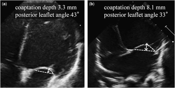
We searched our echocardiographic image databases and evaluated mitral valve geometry at the initial presentation using two-dimensional cineloop recordings of apical four-chamber views stored in the databases. Mitral valve geometry was evaluated by measuring coaptation depth and posterior leaflet angle at mid-systole (Fig 1). Coaptation depth is the distance between the mitral annular plane and the coaptation point of the mitral leaflets.Reference Calafiore, Gallina and Di Mauro 10 The posterior leaflet angle is the angle formed by the mitral annular plane and the line connecting the posterior leaflet hinge point and the coaptation point.Reference Magne, Pibarot and Dagenais 11
Measurements of coaptation depth (double-headed white arrows) and posterior leaflet angle (an angle between two dotted lines) in a healthy 3.3-year-old boy ( a ) and in a 1.1-year-old male dilated cardiomyopathy patient with severe secondary mitral regurgitation (patient 2) ( b ).

As normative values of coaptation depth should vary according to body sizes, we measured coaptation depth in healthy children to establish the reference values of coaptation depth. The healthy controls comprised 58 children (23 boys, 35 girls) who were <15 years of age and had undergone echocardiographic screening for a heart murmur or chest pain. All the control subjects had a structurally normal heart without mitral regurgitation, with sinus rhythm and with left ventricular fractional shortening >0.25 and body weight and height between the 3rd and 97th percentiles. 16
Data are expressed as frequencies, means±standard deviations, or medians and ranges, as appropriate. The correlation between coaptation depth and body surface area in the control group was evaluated by Pearson’s correlation coefficient, and a linear regression equation for the coaptation depth, with log-transformed body surface area as the explanatory variable, was derived. The z-score of the coaptation depth in the patients was obtained using the linear regression equation. The Mann–Whitney U-test was used to compare demographic data and echocardiographic measurements between the patient groups. Moreover, a multivariate linear regression analysis was performed to identify independent variables predicting whether patients had moderate/severe mitral regurgitation or not. A value of p<0.05 was considered statistically significant. All the analyses were performed using R version 2.13.0.
Results
Normative values of coaptation depth in children
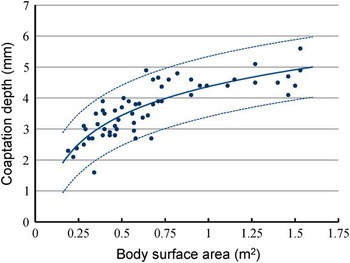
In control subjects, a strong positive correlation was found between coaptation depth and log-transformed body surface area (r=0.83, p<0.0001). The linear regression plot of coaptation depth versus log-transformed body surface area is shown in Figure 2. The regression equation to predict coaptation depth was as follows:
 $$\eqalignno{ {\rm Coaptation}\,{\rm depth}\:{\rm [mm]}= {\rm 4}.{\rm 37}{\plus}{\rm 1}.{\rm 34}\cr {\times}{\rm ln}\,{\rm (body}\,{\rm surface}\,{\rm area}\:{\rm [m}^{{\rm 2}} {\rm ])} \cr & {\rm (residual}\,{\rm standard}\,{\rm error},\,0.{\rm 49},\,{\rm adjusted}\,\cr {\rm R}^{2} ,\,0.{\rm 68};\,{\rm p}\,\lt\,0.000{\rm 1)} $$
Correlation between body surface area and coaptation depth in healthy control subjects. Logarithmic curves represent the univariate regression model of the coaptation depth (solid line) and z-score±2 lines (dotted lines). There is a strong positive correlation between body surface area and coaptation depth (r=0.83, p<0.0001).

Dilated cardiomyopathy patients
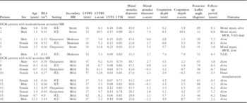
Table 1 shows the clinical characteristics and echocardiographic measurements of the 16 dilated cardiomyopathy patients included in this study. The median age at the initial presentation was 1.2 years (range, 0.4–12.3 years), and the median body surface area was 0.42 m2 (range, 0.29–1.14 m2). The initial echocardiographic assessment was performed in the intensive care setting in 10 patients, whose haemodynamic status had been stabilised with an intravenous infusion of catecholamine (six patients) and milrinone (10 patients). In the remaining six patients, who were haemodynamically stable with oral medications including diuretics, β-blockers, and angiotensin-converting enzyme inhibitors, the initial echocardiographic assessment was performed on an outpatient basis.
Patient characteristics.

BSA=body surface area; DCM=dilated cardiomyopathy; ICU=intensive care unit; LVDD=left ventricular dimension at end diastole; LVFS=left ventricular fractional shortening; LVSI=left ventricular sphericity index; MR=mitral regurgitation; MVR=mitral valve replacement; VAD=ventricular assist device
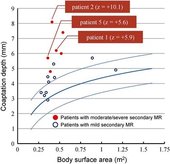
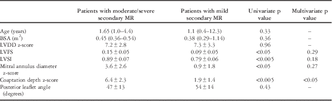
At the initial assessment, six patients had moderate/severe secondary mitral regurgitation, and 10 patients had mild secondary mitral regurgitation. Table 2 shows the differences in demographic data and echocardiographic measurements between the groups. No significant differences were found in age, body surface area, and left ventricular dimension at end-diastole z-scores between the groups. Left ventricular fraction shortening was significantly higher in patients with moderate/severe secondary mitral regurgitation than in patients with mild secondary mitral regurgitation (0.15±0.05 versus 0.09±0.05, p<0.05). Left ventricular sphericity index was significantly increased in patients with moderate/severe secondary mitral regurgitation than in patients with mild secondary mitral regurgitation (0.89±0.07 versus 0.79±0.06, p<0.005). Patients with moderate/severe secondary mitral regurgitation had significantly larger mitral annulus diameter z-scores (3.6±2.6 versus 0.9±1.8, p<0.05) and coaptation depth z-scores (6.4±2.3 versus 1.9±1.4, p<0.005) compared with patients with mild secondary mitral regurgitation. There was no significant difference in posterior leaflet angle between the groups. A multivariate linear regression analysis involving the left ventricular fraction shortening, left ventricular sphericity index, mitral annulus diameter z-score, and coaptation depth z-score revealed that the coaptation depth z-score was the sole independent determinant of secondary mitral regurgitation severity (Table 2). All the patients with moderate/severe secondary mitral regurgitation and only half of the patients with mild secondary mitral regurgitation showed coaptation depth z-scores >2 (Fig 3).
Correlation between body surface area and coaptation depth in dilated cardiomyopathy patients. Logarithmic curves represent the normative values of the coaptation depth (solid line) and z-score±2 lines (dotted lines), which are derived using the univariate regression equation. Patients with moderate/severe secondary mitral regurgitation (closed circles) had higher coaptation depths compared with patients with mild secondary mitral regurgitation (open circles). Note that three patients (patients 1, 2, and 5) underwent mitral repair.

Comparison of demographic data and echocardiographic measurements between the patient groups.

BSA=body surface area; DCM=dilated cardiomyopathy; LVDD=left ventricular dimension at end diastole; LVFS=left ventricular fractional shortening; LVSI=left ventricular sphericity index; MR=mitral regurgitation
Surgical intervention for secondary mitral regurgitation
Mitral repair was performed in three patients with severe secondary mitral regurgitation (patients 1, 2, and 5) at a median of 15 days (range, 13–113 days) after the initial presentation. The surgical procedure included a semicircular suture annuloplasty and an edge-to-edge mitral repair for patients 1 and 2 and a semicircular suture mitral annuloplasty in patient 5. Surgical intervention led to improved mitral regurgitation in two patients (patients 1 and 5) who were discharged on post-operative days 75 and 109, respectively, although patient 5 underwent mitral valve replacement because of worsening mitral regurgitation 2.1 years after mitral repair. In the remaining patient (patient 2), mitral repair failed to alleviate mitral regurgitation, and mitral valve replacement was required before he was discharged. This patient had the largest coaptation depth (8.1 mm; z-score, +10.1) at the initial assessment.
Clinical outcomes
With a median follow-up period of 1.7 years (range, 0.1–5.5 years), the overall survival rate without transplantation or ventricular assist device implantation was 69% in our patients. Of the six patients with moderate/severe secondary mitral regurgitation, two patients were on left ventricular assist device support since 0.6 and 0.1 years after the initial presentation. Of the 10 patients with mild secondary mitral regurgitation, two patients died from heart failure at 0.2 and 0.7 years after the initial presentation; one patient underwent heart transplantation at 1.1 year after the initial presentation. Normalisation of echocardiographic function was observed in two patients with mild secondary mitral regurgitation at 0.3 and 1.1 years after the initial presentation; in these patients, left ventricular fraction shortening increased to >0.25 and left ventricular dimension at end-diastole z-score decreased to <2.0, and all medications were discontinued thereafter.
Discussion
This is the first study to use body surface area-adjusted coaptation depth of the mitral valve as an index to demonstrate that a mitral valve geometric alteration is associated with the development of secondary mitral regurgitation in children with dilated cardiomyopathy. The coaptation depth z-score is an independent determiner of secondary mitral regurgitation severity. Impairment of left ventricular contractility does not contribute to worsening of secondary mitral regurgitation. Our findings might warrant surgical intervention for secondary mitral regurgitation in the paediatric populations.
It is not uncommon for dilated cardiomyopathy patients to develop secondary mitral regurgitation, which results in progressive worsening of left ventricular function and is associated with unfavourable prognosis in childrenReference Fernandes, Manlhiot and McCrindle 2 , Reference Patange, Thomas and Ross 3 as well as adult patients.Reference Grigioni, Enriquez-Sarano, Zehr, Bailey and Tajik 17 – Reference Trichon, Felker, Shaw, Cabell and O’Connor 19 Furthermore, mitral repair has been established as an effective therapy leading to reverse left ventricular re-modelling in adult dilated cardiomyopathy patients with secondary mitral regurgitation refractory to medical therapy.Reference De Bonis, Lapenna and Verzini 20 Certain patients require mitral valve replacement because of significant residual mitral regurgitation after mitral repair.Reference De Bonis, Ferrara and Taramasso 21 Although the number of reports is limited, surgical intervention for severe secondary mitral regurgitation has been attempted in paediatric dilated cardiomyopathy patients.Reference Breinholt, Fraser and Dreyer 4 – Reference Sugiyama, Hoshiai and Naitoh 6 Assessment of the mechanism underlying secondary mitral regurgitation is of great clinical significance for determining the operative indications.
We demonstrated that the geometric alteration of the mitral valve had a pivotal role in the development of secondary mitral regurgitation in children with dilated cardiomyopathy. Paediatric dilated cardiomyopathy patients with moderate or severe secondary mitral regurgitation at presentation had significantly larger coaptation depth, mitral annulus diameter, and left ventricular sphericity index compared with patients with mild secondary mitral regurgitation. Although the geometric alteration of the mitral valve and the left ventricular spherisation were associated with the severity of secondary mitral regurgitation, the impairment in left ventricular fraction shortening and enlarged left ventricular dimension did not contribute to worsening of secondary mitral regurgitation. The evidence for the effectiveness of mitral repair and mitral valve replacement to alleviate secondary mitral regurgitation and control heart failure in children is currently limited.Reference Breinholt, Fraser and Dreyer 4 – Reference Sugiyama, Hoshiai and Naitoh 6 Our findings would provide a theoretical background to surgical intervention for secondary mitral regurgitation in paediatric populations.
Interestingly enough, the posterior leaflet angle did not have any influence on the severity of secondary mitral regurgitation in paediatric dilated cardiomyopathy patients. The increase in both coaptation depth and mitral annulus diameter observed in patients with moderate or severe secondary mitral regurgitation may offset each other, keeping the posterior leaflet angle unchanged.
Patients with moderate or severe secondary mitral regurgitation showed higher left ventricular sphericity index, which highlighted the close relationship between the change in the left ventricular configuration and the geometric alteration of the mitral valve. Secondary mitral regurgitation observed in dilated cardiomyopathy develops through the following mechanism. Patients with dilated cardiomyopathy show marked left ventricular sphericity.Reference Rusconi, Gómez-Marín and Rossique-González 22 In the presence of left ventricular dilatation and spherisation, the papillary muscles shift medio-laterally and posteriorly, and the direction of papillary muscle tension deviates from the long axis of the heart, which hinders the mitral valve from effectively opposing the left ventricular contracting force.Reference Otsuji, Handschumacher and Schwammenthal 7 Geometric alteration of the mitral apparatus leads to mitral annular dilatation, mitral valve leaflet tethering, apical displacement of the coaptation point, and increased posterior leaflet angle.Reference Gogoladze, Dellis and Donnino 23
Multivariate analysis showed that the coaptation depth z-score was the sole independent determinant of secondary mitral regurgitation severity. Although mitral annular dilatation is a worsening factor for secondary mitral regurgitation, it independently can rarely cause significant secondary mitral regurgitation in the absence of papillary muscle displacement, because the normal mitral valve leaflets have a degree of redundancy, which adds a high safety margin against mitral insufficiency.Reference He, Lemmon and Weston 24
Severe alteration of mitral valve geometry precludes effective mitral repair. If there is a marked displacement of the posterior papillary muscle and mitral leaflet tethering, an annuloplasty could further increase the posterior leaflet angle and aggravate the restriction of leaflet motion.Reference Magne, Pibarot and Dagenais 11
Pre-operative assessment of coaptation depth allows prediction of the feasibility of mitral repair in adult dilated cardiomyopathy patients with secondary mitral regurgitation.Reference Calafiore, Gallina and Di Mauro 10 , Reference Magne, Pibarot and Dagenais 11 In adults, the normative value of coaptation depth is 5.8±1.4 mm.Reference Magne, Pibarot and Dagenais 11 Calafiore et al reported that mitral repair resulted in higher residual mitral regurgitation in adult dilated cardiomyopathy patients with a coaptation depth ⩾11 mm than in those with a coaptation depth <11 mm.Reference Calafiore, Gallina and Di Mauro 10 In another study by Magne et al, a coaptation depth of ⩾10 mm had 64% sensitivity and 90% specificity for predicting clinically significant residual mitral regurgitation.Reference Magne, Pibarot and Dagenais 11
Nevertheless, these results cannot be applied to paediatric populations with widely varying body sizes. In this study, we showed that coaptation depth in healthy children aged <15 years had a strong linear positive correlation with log-transformed body surface area, and we derived a regression equation to calculate the body surface area-adjusted coaptation depth z-scores and compare the coaptation depths among children with varying body sizes.
Our experience provides anecdotal evidence that the coaptation depth z-score might be useful for predicting whether a secondary mitral regurgitation is amenable to mitral repair in paediatric dilated cardiomyopathy patients. The only patient in whom mitral repair failed to alleviate mitral regurgitation had the largest coaptation depth (8.1 mm; z-score, +10.1) at the initial assessment; however, because a small sample of patients with short follow-up periods was included in this study, we were unable to determine whether reverse left ventricular re-modelling was observed after mitral repair. Further investigation is required to determine the cut-off value of the coaptation depth z-score predicting the effectiveness of mitral repair in paediatric dilated cardiomyopathy patients with secondary mitral regurgitation.
Study limitations
Our study has certain limitations. First, this is a retrospective study including a small numbers of patients. Second, the determinants of mitral valve geometry assessed in our study were derived solely from two-dimensional echocardiographic images. The mitral valve is a remarkably complex structure; the mitral annulus has a non-planar saddle-like shape, and the geometry of the mitral valve and the left ventricle interact with each other through the basal chords connecting the basal portion of the mitral valve leaflets to the papillary muscles.Reference Silbiger and Bazaz 26 A study using three-dimensional echocardiography and pathological examination of the explanted heart revealed that the insertion sites of the basal chords were associated with mitral valve geometry and the severity of secondary mitral regurgitation.Reference Song, Kim and Ha 27 Third, factors other than mitral valve geometric alteration were not fully assessed. Although mitral valve geometry is the main determinant of secondary mitral regurgitation, some haemodynamic factors such as left atrial pressure and left ventricular dyssynchrony have been reported to contribute to the development of secondary mitral regurgitation.Reference Maréchaux, Pinçon and Poueymidanette 9 , Reference Kanzaki, Bazaz and Schwartzman 25 In addition, the patient condition at the initial echocardiographic evaluation was not standardised. As described in the Results section, this study included patients whose echocardiographic assessments were performed in the intensive care unit as well as those whose echocardiographic assessments were performed in the outpatient clinic. Although the patient backgrounds vary, we consider that our data effectively reflect actual clinical settings.
Acknowledgements
None.
Financial Support
This study was supported by a Grant-in-Aid (KAKENHI, 26860783) from the Japan Society for the Promotion of Science.
Conflicts of Interest
None.
Ethical Standards
The authors assert that all procedures contributing to this work comply with the ethical standards of the relevant Japanese guidelines on human experimentation and with the Helsinki Declaration of 1975, as revised in 2008, and has been approved by the institutional committee.