The extra cardiac conduit has allowed the routine repair of complex anomalies that include pulmonary atresia and complex tetralogy of fallot, truncus arteriosus, transposition of the great arteries with pulmonary stenosis, and other forms of complex congenital heart disease. The homograft conduit has been the gold standard in right ventricle outflow tract reconstruction ever since its inception in 1966.Reference Ross and Somerville 1 However, the long-term result of homografts are far from optimal, due to very limited ability of growth and early calcification leading to progressive obstruction, resulting in unavoidable re-intervention.Reference Boethig, Goeler and Westhoff-Bleck 2 To address this problem, valved bovine jugular vein conduit was developed that incorporates a tri-leaflet valve and affords off-the-shelf availability, good handling characteristics, and excellent haemodynamics. Over the last decade, valved bovine jugular vein conduit is considered a suitable choice for paediatric population with congenital heart defect requiring right ventricle to main pulmonary artery connection because of the availability of an adequate range of sizes (12–22 mm), the relatively low cost, and the low reported incidence of calcification.Reference Breymann, Boethig, Goerg and Thies 3 – Reference Corno, Qanadli and Sekarski 5 An aneurysmal dilatation of valved bovine jugular vein conduit is usually confined to the proximal anastomosis (pseudoaneurysm) and is commonly associated with distal anastomotic stenosis, without distal stenosis is a rare complication.Reference Delmo-Walter, Alexi-Meskishvilli, Abdul-Khaliq, Meyer and Hetzer 6 Here, we describe a severe dilatation of a valved bovine jugular vein conduit in a 7-year-old boy with truncus arteriosus.
Case
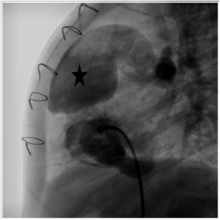
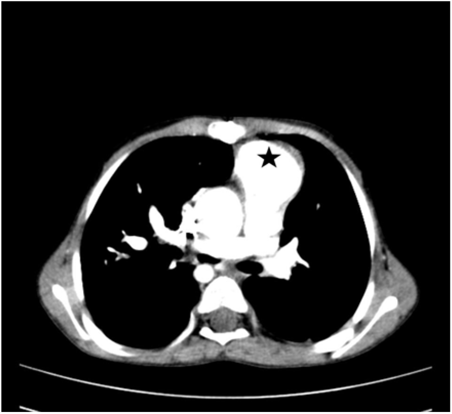
A 7-year-old boy had a history of truncus arteriosus (type 2), who underwent complete repair at the age of 2.5 years. Pre-operative right ventricle systolic pressure was 73 mmHg in the right heart catheterisation. The right ventricular outflow tract reconstruction was performed with a 14-mm valved bovine jugular vein conduit. During the 2 years of follow-up after surgery, he was asymptomatic and had no right ventricular outflow obstruction in echocardiography. Subsequently he was lost to follow-up for 2 years. He was presented again at 7 years of age and echocardiography showed right ventricular dilatation and aneurysmal dilatation of the conduit. Cardiac catheterisation confirmed iso-systemic right ventricular hypertension and aneurysmal dilatation of the conduit extending from the ventricular anastomosis (Fig 1). Computed tomography confirmed the angiographic findings, recording a giant aneurysm of the conduit (Fig 2). The patient was operated for conduit change. The conduit was uniformly dilated from the proximal ventricular anastomosis to the distal anastomosis with no distal stenosis. The conduit was replaced with a 23-mm xenograft. He was discharged on the sixth post-operative day and was asymptomatic at 2 months follow-up. The right ventricle systolic pressure, which was estimated from tricuspid regurgitation jet maximum velocity by continuous wave spectral Doppler, was 40 mmHg with mild conduit stenosis at latest echocardiographic follow-up.
Aneurysmal dilatation of conduit (angiogram lateral view).

Computerised tomography angiography images showing large aneurysmal dilatation of the conduit and wall calcification and unobstructed distal branch pulmonary arteries.

Discussion
Valved conduits are frequently used in surgical correction of a variety of congenital right ventricular outflow tract anomalies to establish continuity between the right ventricle and the pulmonary artery in the paediatric population. In 1999, the bovine jugular vein conduit (Contegra®, Minneapolis, Minnesota, USA, Medtronic) was introduced as a suitable choice, as it is extremely pliable and available in various sizes (12–22 mm) with a relatively low cost. Breymann et al reported a significantly lower conduit-related rate of reoperation after 4 years of follow-up in their bovine jugular vein conduit (Contegra®, Medtronic) recipients versus their homograft patients.Reference Breymann, Boethig, Goerg and Thies 3 Brown et al showed excellent early and midterm outcomes in right ventricular outflow tract reconstruction with bovine jugular vein conduit (Contegra®, Medtronic).Reference Brown, Ruzmetov, Rodefeld, Vijay and Darragh 4 Despite these encouraging results, several other groups have identified proximal aneurysmal dilatation of bovine jugular vein conduit (Contegra®, Medtronic).Reference Corno, Qanadli and Sekarski 5 , Reference Delmo-Walter, Alexi-Meskishvilli, Abdul-Khaliq, Meyer and Hetzer 6 When identified, it is usually confined to the proximal anastomosis (pseudoaneurysm) and is commonly associated with distal anastomotic stenosis. However, true aneurysmal dilatation of the bovine jugular vein conduit has been rarely reported.Reference Bautista-Hernandez, Kaza, Benavidez and Pigula 7 – Reference Qureshi, Abbaker, Sivalingam and Latiff 9 In our patient, true aneurysmal dilatation of the 14-mm bovine jugular vein conduit (Contegra®, Medtronic) device was observed without evidence for distal conduit or branch pulmonary artery stenosis. The mechanism of failure is unknown, but mechanical stress and/or immunologic factors could be involved.Reference Calvaruso, Rubino, Ocello, Cipriani and Marcelletti 8
True aneurysmal dilatation in the absence of distal obstruction is a rare complication of valved bovine jugular vein conduit. Close follow-up of this device is recommended to prevent conduit failure as well as other potentially serious consequences.
Acknowledgements
None.
Financial Support
This research received no specific grant from any funding agency, commercial or not-for-profit sectors.
Conflicts of Interest
None.
Ethical Standards
The authors assert that all procedures contributing to this work comply with the Helsinki declaration of 1975, as revised in 2008, and has been approved by the local Institutional Ethics Committee.