Introduction
Aortic valvar atresia as a congenital cardiovascular malformation in which there is no patency of the aortic valve, and thus no communication between aorta and either cardiac ventricle. A hypoplastic ascending aorta is a constant feature. The Congenital Heart Surgery Nomenclature and Database Project classifies aortic atresia under hypoplastic left heart syndrome, hierarchy level 2. Reference Tchervenkov, Jacobs and Tahta1 But the subgroup of patients in which there is aortic valve atresia with ventricular septal defect with well-developed mitral valve and left ventricle can potentially undergo a biventricular repair, and hence (if we accept that we can define anatomic entities by their most appropriate treatment) should not be classified as hypoplastic left heart syndrome.
The 11th iteration of the International Classification of Disease defines hypoplastic left heart syndrome (ICD-11 code LA89.3) as a spectrum of congenital cardiovascular malformations with normally aligned great arteries without a common atrioventricular junction, characterised by underdevelopment of the left heart with significant hypoplasia of the left ventricle including atresia, stenosis, or hypoplasia of the aortic or mitral valve, or both valves, and hypoplasia of the ascending aorta and aortic arch. Aortic atresia (ICD-11 code LA8A.23) is defined as a congenital cardiovascular malformation in which there is no orifice of the aortic valve, with an additional information that aortic valvar atresia will most often not be coded independently, as it is frequently included within the “Hypoplastic left heart syndrome” code as part of this spectrum of cardiovascular malformations. However, there is a small subset of patients with aortic valve atresia who have a well-developed left ventricle and mitral valve and a large ventricular septal defect (nonrestrictive or restrictive).
The categorisations proposed by the Congenital Heart Surgery Nomenclature and Database Project have been overtaken by those provided by the International Nomenclature Committee. The recommendations made by the International Nomenclature Committee, furthermore, have been adopted by the World Health Organisation for the 11th iteration of the International Classification of Disease. This classification defines aortic valvar atresia as a congenital cardiovascular malformation in which there is no orifice of the aortic valve (IPCCC code 09.15.06). Reference Franklin, Béland and Colan2 Aortic valve atresia will most often be coded under the hypoplastic left heart syndrome / complex diagnostic codes since it most often occurs as part of a spectrum of cardiovascular malformations. However, a small subset of patients with aortic valve atresia (approximately 4%) will have a well-developed left ventricle and mitral valve and a large ventricular septal defect. Reference Roberts, Perry, Chandra, Myers, Shapiro and Scott3
This is the subset we are describing in this manuscript, which is often misdiagnosed as common arterial trunk, based on investigations (echocardiography and / or CT) and on gross examination. Reference Moharkar and Bharati4–Reference Kochi, Sugimoto, Kawamoto, Inoue and Machida7
Common arterial trunk is a congenital malformation in which a single great artery arises from the heart, overrides the interventricular septum, and supplies the systemic, pulmonary, and coronary circulations. Reference Franklin, Béland and Colan2,Reference Jacobs8–Reference Anderson, Penny, Anderson, Baker, Penny, Redington, Rigby and Wernovsky10 Herein, we describe an outflow tract malformation, aortic atresia with a discrete pulmonary trunk with ventricular septal defect and well-developed mitral valve and balanced ventricles, which was misdiagnosed as common arterial trunk, despite some definitive morphological differences. We do not suggest that this is a newly described morphological entity, as many teams have encountered such anatomy, and have performed biventricular repairs. Others have misdiagnosed this entity as common arterial trunk. Reference Austin, Jonas, Mayer and Castaneda11–Reference Moorthy, McGuirk, Jones, Brawn and Barron19 We emphasise the morphological differences, as well as the surgical importance, including technical considerations, conduct of cardiopulmonary bypass, and myocardial protection.
Case series
Over the past three years, three patients, with a referral echocardiographic diagnosis of common arterial trunk, were found to have aortic valve atresia with a discrete pulmonary trunk and a separate hypoplastic ascending aorta with an interventricular communication, with a well-developed mitral valve and balanced ventricles. Echocardiography, cardiac computerised tomography, operative findings, and postoperative course details were reviewed. All were tachypnoeic, with chest retractions, systolic murmurs, cardiomegaly, and pulmonary plethora.
Neither patient had any non-cardiac congenital anomalies. Both had thymus on sternotomy, and there was no 22q11 deletion on karyotyping.
One patient’s family declined treatment, and the remaining two patients were treated surgically. In both the operated cases, the diagnosis of common arterial trunk was made echocardiographically and “confirmed” with CT scan as common arterial trunk with single coronary artery arising from brachiocephalic artery. In our two cases, after consultation with a number of colleagues, the diagnosis was changed to aortic valvar atresia with ventricular septal defect and balanced ventricles. This diagnosis eventually was confirmed with transoesophageal echocardiography and intra-operative findings. Echocardiography, cardiac computerised tomography, operative findings, and postoperative course details were reviewed, and these two cases are the subject of this discussion (Figures 1 and 2).
Characteristics of patient-1. (a) Three-dimensional reconstruction of preoperative CT scan. 1—pulmonary trunk, 2—right pulmonary artery, 3—left pulmonary artery, 4—persistent arterial duct, 5—minor indentation on the greater curvature of aortic arch, 6—aortic arch, 7—descending thoracic aorta, arrow—hypoplastic ascending aorta. (b) Intraoperative findings just before commencing of cardiopulmonary bypass, where pulmonary trunk is retracted inferiorly to expose the hypoplastic ascending aorta (arrow) with coronary artery origin. (c) Preoperative transoesophageal echocardiography demonstrating a trileaflet ventriculo-arterial valve (broken arrow) committed to right ventricle (RV) without any override. There is no continuity between the ventriculo-arterial valve and anterior mitral leaflet as the atretic aortic root portion (circle) is interposed between the two. Perimembranous ventricular defect (flower bracket) is the only outlet for left ventricle. LA = left atrium; MV = mitral valve; LV = left ventricle; RV = right ventricle.

Figure 1 Long description
Panel A: A three-dimensional reconstruction of a preoperative CT scan showing the pulmonary trunk, right pulmonary artery, left pulmonary artery, persistent arterial duct, minor indentation on the greater curvature of the aortic arch, aortic arch, and descending thoracic aorta. The hypoplastic ascending aorta is indicated by an arrow. Panel B: An intraoperative photo taken just before commencing cardiopulmonary bypass. The pulmonary trunk is retracted inferiorly to expose the hypoplastic ascending aorta with the origin of the coronary artery. Panel C: A preoperative transoesophageal echocardiography image demonstrating a trileaflet ventriculo-arterial valve committed to the right ventricle without any override. There is no continuity between the ventriculo-arterial valve and the anterior mitral leaflet as the atretic aortic root portion is interposed between the two. A perimembranous ventricular defect is the only outlet for the left ventricle. Labels include LA for left atrium, MV for mitral valve, LV for left ventricle, and RV for right ventricle.
Characteristics of patient-2. (a) Intraoperative findings just before commencing of cardiopulmonary bypass, showing the hypoplastic ascending aorta (arrow) with coronary artery origin and pulmonary trunkReference Tchervenkov, Jacobs and Tahta1 leading to aortic arch. (b) Three-dimensional reconstruction of preoperative CT scan, with arrow pointing the hypoplastic ascending aorta. (c) Intraoperative findings after separation of cardiopulmonary bypass, with arrow pointing the hypoplastic ascending aorta, reconstructed neo-aorta,Reference Franklin, Béland and Colan2 and valved polytetrafluoroethylene conduit Reference Roberts, Perry, Chandra, Myers, Shapiro and Scott3 between right ventricle and pulmonary artery. (d) Three-dimensional reconstruction of postoperative CT scan, with arrow pointing the hypoplastic ascending aorta, reconstructed neo-aorta,Reference Franklin, Béland and Colan2 and valved polytetrafluoroethylene conduitReference Roberts, Perry, Chandra, Myers, Shapiro and Scott3 between right ventricle and pulmonary artery.

Both infants underwent a single stage surgical repair. Under general anaesthesia and endotracheal intubation, with femoral and arterial line and central line in situ, a median sternotomy was performed. After full heparinization (4 mg/kg), normothermic cardiopulmonary bypass was established employing transverse aortic arch and bi-caval cannulation, and both branch pulmonary arteries were controlled with vascular slings. The left heart was vented through the left atrial body. Then a 5 mm polytetrafluoroethylene vascular tube graft was sutured to right common carotid artery in an end to side fashion using 8-0 polypropylene suture, and an additional arterial inflow cannula was placed. All the arch vessels and aortic arch proximal to the inflow cannula were snared. Aortic root cardioplegia (Del Nido cardioplegia solution at 8o Celsius) was delivered to achieve diastolic arrest of the heart. The pulmonary trunk was transected, and the pulmonary bifurcation was mobilised. A distal anastomosis between the valved polytetrafluoroethylene conduit and the pulmonary artery confluence was performed in an end to end fashion using 6-0 polytetrafluoroethylene continuous sutures. The ductal portion (from branch pulmonary artery take-off up to the junction with the arch) was excised and an anastomosis of the neo-aortic root with the arch was performed with 7-0 polypropylene suture. A right ventriculotomy was performed avoiding injury to coronaries and crossing muscle bundles were divided. Routing of left ventricle to ventriculo-arterial valve across the interventricular communication was performed using a glutaraldehyde treated autologous pericardial patch, using continuous 6-0 polypropylene suture. Then the proximal anastomosis between the conduit and the ventriculotomy was performed in a side to side fashion using 6-0 polytetrafluoroethylene continuous sutures. Reference Kasturi and Prabhu20 Direct closure of the atrial septal defect was performed via a limited right atriotomy. We did not perform a Norwood type of arch reconstruction, and instead left the hypoplastic ascending aorta in its native state, filling retrogradely through the arch and supplying only the coronary arteries. In this way, we avoided a complex procedure and the need for hypothermia and circulatory arrest (Figure 3).
Conduction of cardiopulmonary bypass (CPB) and cardioplegic arrest. Intracardiac component of the surgery is not shown in the figure. (a) Anatomy of great vessels in our cases (one had aberrant right subclavian artery, which is not shown here). (b) Cannulation strategy, establishment of CPB, and cardioplegia delivery: a 5 mm polytetrafluoroethylene graftReference Anderson, Penny, Anderson, Baker, Penny, Redington, Rigby and Wernovsky10 is anastomosed to brachiocephalic arteryReference Li, Li and Chen5 in an end to side fashion using 8-0 polypropylene continuous suture. Arterial inflow cannula (solid arrow) in the graft and in proximal descending thoracic aorta.Reference Jacobs and Anderson9 Bicaval venous drainage cannula with vent in the pulmonary veins (not shown in the figure) to establish the cardiopulmonary bypass after snaring (broken line) the right and left pulmonary arteries. Star mark is the site of cardioplegia needle, which is delivered after snaring (broken line) the brachiocephalic artery, left common carotid artery, left subclavian artery, and descending thoracic aorta proximal to the cannulation site. (c) Cardioplegia site isolated by snaring the delivery site on both sides. Ductal segment completely excised and pulmonary artery end sutured. (d) Anastomosis of pulmonary trunkReference Tchervenkov, Jacobs and Tahta1 to aortic arch in an end to side fashion using 7-0 polypropylene continuous suture. Valved polytetrafluoroethylene conduitReference Austin, Jonas, Mayer and Castaneda11 used to establish right ventricle to pulmonary artery continuity (using 6-0 polytetrafluoroethylene continuous sutures for both the anastomosis, right ventricular end not shown in the figure). 1— pulmonary trunk, 2—right pulmonary artery, 3—left pulmonary artery, 4—persistent arterial duct, 5—brachiocephalic artery, 6—right subclavian artery, 7—right common carotid artery, 8—left common carotid artery, 9—left subclavian artery, 10 —descending thoracic aorta, 11—polytetrafluoroethylene graft (5 mm), 12—valved polytetrafluoroethylene conduit, arch—aortic arch, RCA—right coronary artery, LM—left main coronary artery, solid arrow—arterial inflow cannula, curved arrow—hypoplastic ascending aorta with atretic valve, star—site of cardioplegia delivery needle, broken line—site of snare (using vascular slings).

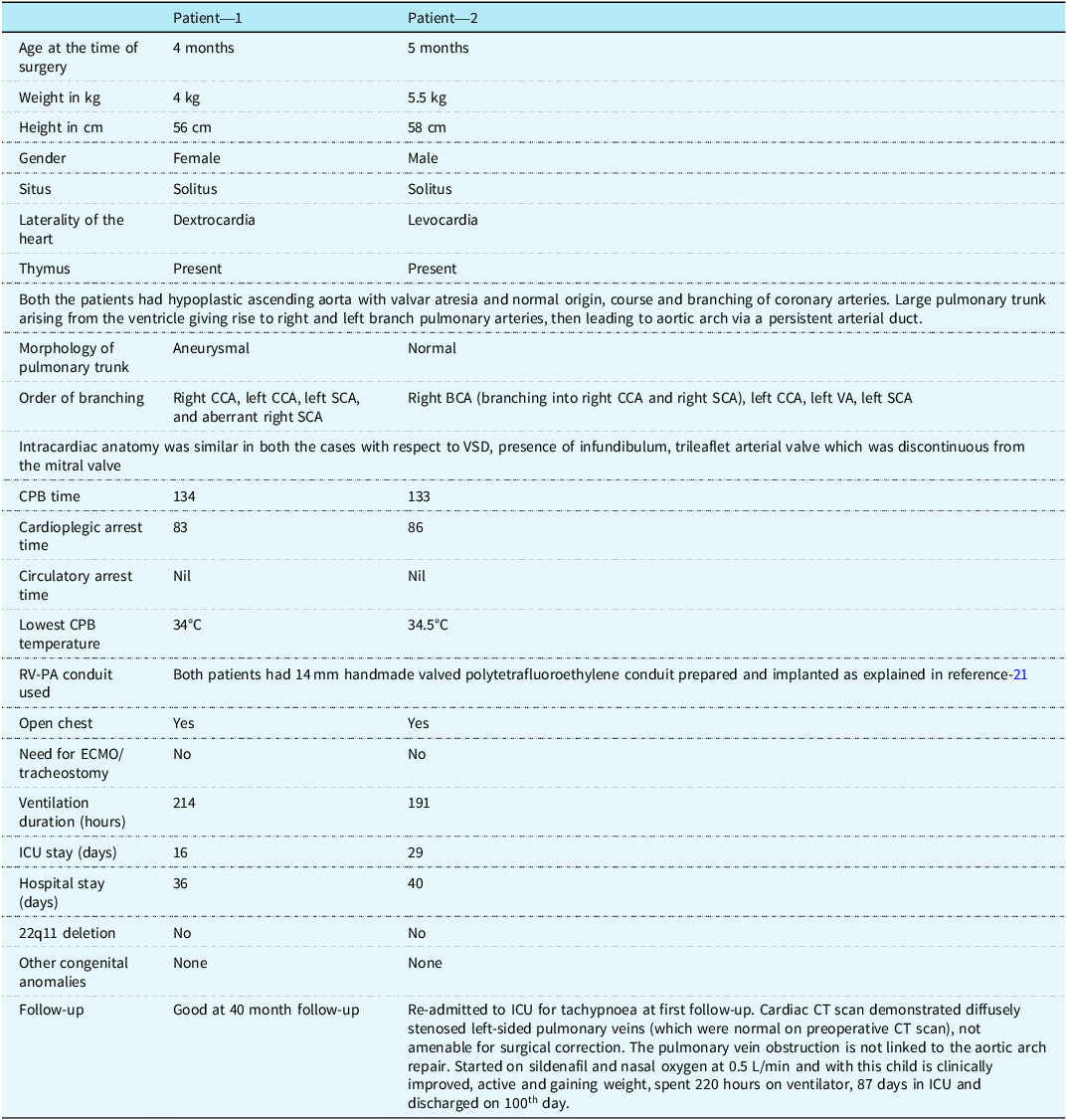
Root deairing done and arch vessels were un-snared, establishing myocardial perfusion. Spontaneous sinus rhythm ensued. After weaning from cardiopulmonary bypass, both patients were shifted to intensive care unit with an open chest, drains, and two atrial and ventricular pacing wires. Elective delayed sternal closure was done the next day. Patient characteristics are as per the Table 1. Both were discharged on aspirin (5 mg/kg), oral diuretics, and sildenafil citrate.
Patient characteristics

Both patients survived surgery and are currently well 40 months and 6 months post-operatively at the time of submission.
Morphology
Both patients had usual arrangement of their organs, with one having a right-sided heart, and the other with the heart in the left chest, with left aortic arch and left-sided descending aorta. One had an aberrant right subclavian artery.
External anatomy: Both the patients had cardiomegaly with biventricular hypertrophy. Diminutive aortic sinuses of Valsalva were present in both cases, giving origin to normally positioned coronary arteries with a normal distribution pattern. The ascending aorta was hypoplastic up to the junction with the brachiocephalic artery, measuring 3 to 4 mm. Intra-operatively we did not directly visualise the atretic aortic valve, as we did not open the hypoplastic ascending aorta to perform a classical Norwood type of repair. The portion of aorta (hypoplastic) between the atretic valve and brachiocephalic artery served only as a conduit for coronary blood flow and did not require intervention. Reference Kouchoukos, Blackstone, Hanley and Kirklin21
A discrete pulmonary trunk was arising from the right ventricle in both cases (connected to right ventricle), giving rise to right and left branch pulmonary arteries and then continuing as aortic arch via a large patent arterial duct. There was no arch hypoplasia or focal coarctation, but both patients had mild infolding of the aortic wall opposite to the ductal insertion site.
Intracardiac anatomy: Both patients had a remnant of a separate aortic valve, but no communication with either ventricle (we could not visualise the atretic valve, but it was clearly demonstrated on transoesophageal echocardiography). Both patients had 3 leaflet ventriculo-arterial (pulmonary) valves, connected to the right ventricle, supported by an infundibulum, and hence there was no override.
The left ventricular cavity was of normal size in both patients, with a normal mitral valve tensor apparatus on direct inspection. Both patients had small atrial septal defects. Systemic veins were draining into right atrium, and all pulmonary veins were draining into left atrium.
Interventricular communication was in the form of perimembranous ventricular septal defect in both patients (superior border related to tricuspid valve), with the postero-inferior margin probably related to the conduction tissue. A sub-pulmonary infundibulum was present in both. The hinges of the leaflets of the pulmonary valve were well separated from mitral valve (arterial valve and mitral valve discontinuity), due to the fact that the pulmonary trunk was connected to right ventricle. There was right ventricular hypertrophy and dilatation.
Discussion
Patients with aortic valvar atresia with ventricular septal defect with well-developed mitral valve and balanced ventricles have a hypoplastic ascending aorta and a discrete pulmonary trunk leading to the aortic arch via a persistent arterial duct. These patients clinically and echocardiographically may resemble those with common arterial trunk, in the absence of arch hypoplasia or coarctation. Presentation can be late, as the patients are not overtly cyanotic and usually present with failure to thrive and features of increased pulmonary blood flow. Surgical management differs from that of common arterial trunk, and early surgical intervention (staged or total correction) is needed to avoid establishment of irreversible pulmonary vascular disease or Eisenmenger syndrome. If the patient is operated on a misdiagnosis of common arterial trunk with aortic dominance and single coronary artery arising from brachiocephalic artery (which is extremely rare), then the ductal tissue will form a part of the aortic arch and in future might cause narrowing, necessitating future arch interventions.
Patients with aortic valvar atresia with ventricular septal defect with well-developed mitral valve and balanced ventricles have a sub-pulmonary infundibulum, a remnant of a separate aortic valve committed to left ventricle (atretic valve without luminal continuity), and a discrete pulmonary trunk arising from the right ventricle supported by an infundibulum and hence no override. Unlike common arterial trunk, there is a true ventricular septal defect. There is no fibrous continuity between the ventriculo-arterial valve (this being the pulmonary valve) and the anterior mitral leaflet. These are five features, which are conspicuous by their absence in common arterial trunk. In patients with common arterial trunk, the infundibular septum is absent from the right ventricular outflow tract (although the truncal valve can be supported by a complete infundibulum when it arises exclusively from the right ventricle). Reference Kouchoukos, Blackstone, Hanley and Kirklin22, Reference Anderson, Spicer, Hlavacek, Cook and Backer23 A phenotypical feature of common arterial trunk is the commonality of the ventriculo-arterial junction, guarded by a common arterial valve, without any evidence of another valve. In common arterial trunk, there is typically fibrous continuity between the leaflets of the truncal and mitral valves in the roof of the left ventricle. The interventricular communication is high (outlet) and anterior, with the truncal valve forming the superior margin. This is consistent with the absence of infundibular septum to form the superior margin of the interventricular communication, leaving the superior margin to be formed by the valve itself.
Another major difference of surgical importance is that no portion of the arch is formed by ductal tissue in common arterial trunk (there are cases in which the duct is present and important in a common arterial trunk such as pulmonary dominance with interrupted aortic arch), unlike in aortic atresia with balanced ventricles, wherein the solitary pulmonary trunk arising from the ventricle leads to the aortic arch via a persistent arterial duct.
The anatomical knowledge of aortic valvar atresia with discrete pulmonary trunk with ventricular septal defect with well-developed mitral valve and balanced ventricles helps us in performing a relatively less extensive surgery, as compared to a Norwood or Damus–Kay–Stansel or double homograft replacement type of surgeries. Reference Austin, Jonas, Mayer and Castaneda11–Reference Moorthy, McGuirk, Jones, Brawn and Barron19 Leaving the hypoplastic ascending aorta as the source for coronary blood flow, with the knowledge that this portion of aorta is not going to become atretic, lets us perform the surgery with normothermic cardiopulmonary bypass. Ductal tissue (between the origin of branch pulmonary arteries and arch junction where the recurrent laryngeal nerve courses around) is the only portion to be excised, before anastomosing the pulmonary (neo-aortic) trunk with the aortic arch.
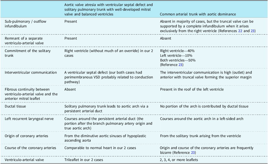
The morphological differences between aortic valvar atresia with ventricular septal defect with well-developed mitral valve and balanced ventricles and common arterial trunk are shown in Table 2.
Anatomical differences

Conclusion
We have described an outflow tract malformation, aortic valvar atresia with discrete pulmonary trunk with ventricular septal defect with well-developed mitral valve and balanced ventricles, which can be misdiagnosed as common arterial trunk, but has definitive morphological differences. This anatomical knowledge helps us in planning the conduction of cardiopulmonary bypass, myocardial protection, and avoiding more extensive procedures while performing the biventricular repair. The long-term outcome of this simpler procedure to treat such anatomy is yet to be discerned.
Acknowledgements
None.
Financial support
None.
Competing interests
None.
Ethical standard
The ethics committee of the Narayana Institute of Cardiac Sciences approved the study (NHH/AEC-CL-2025-1545), and parental individual consents were obtained for publication.