Introduction
Different groups of nematodes have acquired parasitism lifestyles against insects. Molecular phylogenetic analyses suggest that parasitism of invertebrates independently evolved from 10 origins (Bird et al., Reference Bird, Jones, Opperman, Kikuchi and Danchin2014; Blaxter and Koutsovoulos, Reference Blaxter and Koutsovoulos2014). Another study based on the morphology suggests that at least 20 lineages have acquired insect parasitism (Sudhaus, Reference Sudhaus, Makarov and Dimitrijevic2008). Because insect parasitism independently evolved in different groups of nematodes, some basic underlying mechanisms likely enable nematodes to evolve insect parasitism lifestyles but remain to be identified.
The primary requirement for nematodes to parasitize insects is to evade/suppress the immunity of host insects. There are two types of innate immunity in insects: cellular immunity mediated by haemocytes circulating in the haemocoel and humoral immunity mediated by antimicrobial peptides and the phenoloxidase pathway for melanization (Bulet and Stocklin, Reference Bulet and Stocklin2005; Eleftherianos and Revenis, Reference Eleftherianos and Revenis2011). For larger invading multicellular pathogens such as nematodes, cell-mediated immunity is generally effective and the pathogens can be confined through encapsulation by haemocytes (Lavine and Strand, Reference Lavine and Strand2002). Lepidopterans typically have five types of haemocytes: prohaemocytes, spherule cells, oenocytoids, granular cells and plasmatocytes (Lavine and Strand, Reference Lavine and Strand2002; Silveira et al., Reference Silveira, Ribeiro and Báo2003). Granular cells and plasmatocytes are both involved in encapsulation: first, granular cells attach to the foreign target, and then encapsulation is completed through multiple layers of plasmatocytes (Pech and Strand, Reference Pech and Strand1996; Lavine and Strand, Reference Lavine and Strand2001). A morphological change called spreading is the important step in the activation of the cellular immune responses which occurs when plasmatocytes adhere to foreign surfaces (Tsuzuki et al., Reference Tsuzuki, Matsumoto, Furihata, Ryuda, Tanaka, Sung, Bird, Zhou, Shears and Hayakawa2014). Entomopathogenic nematodes (EPNs) of the genera Steinernema and Heterorhabditis use their symbiotic bacteria to suppress host immunity (Dowds and Peters, Reference Dowds, Peters and Gaugler2002; Kim et al., Reference Kim, Ji, Cho and Park2005, Reference Kim, Choi, Jung and Kim2018), but nematodes themselves can suppress the host insect immunity as well. Substances secreted from Steinernema nematodes impair host immune responses through various mechanisms including killing haemocytes and suppressing haemocyte adhesion to invaders (Walter et al., Reference Walter, Dunphy and Mandato2008; Li et al., Reference Li, Cowles, Cowles, Gaugler and Cox-Foster2009a). Moreover, the cuticle of Steinernema species lowers the encapsulation ability of haemocytes (Brivio et al., Reference Brivio, Pagani and Restelli2002; Yi et al., Reference Yi, Wu, Lv and Li2016; Sanda et al., Reference Sanda, Muhammad, Ali and Hou2018). Not only EPNs but also the mermithid nematode Ovomermis sinensis suppress host immunity by inducing apoptosis of haemocytes (Jiao et al., Reference Jiao, Wen, Tao, Wang and Wang2018). Additionally, O. sinensis lowers encapsulation ability also by suppressing haemocyte activity (Li et al., Reference Li, Sun, Wang and Liu2009b; Jiao et al., Reference Jiao, Wen, Tao, Wang and Wang2018). These reports indicate that various kinds of mechanisms are utilized by nematodes to suppress host immunity and some factors secreted from nematodes contribute to trigger such mechanisms.
Changes in total haemocyte population after nematode infection have been previously reported (Ayaad et al., Reference Ayaad, Dorrah, Shaurub and Sadawy2001; Walter et al., Reference Walter, Dunphy and Mandato2008; Manachini et al., Reference Manachini, Schillaci and Arizza2013; Yi et al., Reference Yi, Wu, Lv and Li2016; Lalitha et al. Reference Lalitha, Karthi, Vengateswari, Karthikraja, Perumal and Shivakumar2018). Injection of Heterorhabditis bacteriophora or Steinernema glaseri decreased haemocyte density in the larval haemocoel of the greater wax moth Galleria mellonella (Alvandi et al., Reference Alvandi, Karimi and Dunphy2014). Conversely, haemocyte density was reported to increase in the haemocoel of the cotton bollworm Helicoverpa armigera larvae infected with O. sinensis (Jiao et al., Reference Jiao, Wen, Tao, Wang and Wang2018). However, the mechanisms by which the haemocyte density changes after nematode infection and the link between haemocyte density and encapsulation remains unclear.
Host immune responses against insect parasitic nematodes have been actively studied while non-parasitic nematodes, which are important for understanding the evolution of parasitism, have been paid little attention in this research field. In our previous study, we demonstrated that a non-parasitic nematode Caenorhabditis elegans, when injected into G. mellonella larva, is not encapsulated and instead grows and propagates in the living insect's haemocoel, leading to the death of the host insect (Ono and Yoshiga, Reference Ono and Yoshiga2018). This suggested that non-parasitic nematodes such as C. elegans potentially have the ability to evade and/or suppress the cellular immunity of the insect. Here we aimed to clarify the mechanism by which non-parasitic nematodes evade/suppress cellular immunity by focusing on the nematodes' effect on the haemocytes of host G. mellonella larvae.
Materials and methods
Nematodes and insects
Caenorhabditis elegans N2 and feeding-delay mutants (due to abnormal pharyngeal muscles) DA467 (eat-6), DA493 (phm-3) and DA607 (pha-3) were obtained from the Caenorhabditis Genetics Center (University of Minnesota, St. Paul, MN, USA). Nematodes were maintained on nematode growth medium (NGM) plates (1.7% agar) seeded with Escherichia coli strain OP50 (Stiernagle, Reference Stiernagle and Hope1999) at 20 °C. Caenorhabditis elegans first-stage (L1) larvae were used in experiments because bacteria-free nematodes can be easily prepared by surface-sterilization of eggs. Nematodes that harboured eggs were collected from a Rich NGM plate (Stiernagle, Reference Stiernagle and Hope1999) and treated with sodium hypochlorite and NaOH (Stiernagle, Reference Stiernagle and Hope1999) to induce egg release, and the collected eggs were left overnight in an incubator at 20 °C. Hatched axenic L1 larvae were subjected to the modified Baermann method (Whitehead and Hemming, Reference Whitehead and Hemming1965) to obtain active L1 larvae.
Bacteriophagous nematodes Oscheius myriophila Sg1 was isolated from millipede Riukiaria sp. collected from Hinokuma Mountain, Saga, Japan. Bacteriophagous nematodes Pristionchus pacificus PS312 and Panagrolaimus rigidus AF36 were obtained from the Caenorhabditis Genetics Center. The three species of nematodes were maintained on NGM plates seeded with E. coli strain OP50 at 20 °C. Mixed-stage nematodes recovered from the MGM plate and washed several times with M9 buffer were used for the experiments.
The fungivorous nematode Aphelenchus avenae M1 was cultured on Botrytis cinerea growing on autoclaved barley grain medium at 20 °C. Mixed-stage nematodes extracted using the modified Baermann method were used for experiments.
The EPNs H. bacteriophora TT01 (kindly provided by Dr Ann Burnell, National University of Ireland-Maynooth) and Steinernema carpocapsae All strains are maintained using G. mellonella larvae. Axenic L1 larvae hatched from surface-sterilized eggs were used for experiments.
Galleria mellonella was cultured at 30 °C (Ono and Yoshiga, Reference Ono and Yoshiga2018), and the last instar larvae were used for the experiments because of the ease of handling and injection of nematodes.
Change in haemocyte density after nematode injection
An aliquot of 50 µL of nematode suspension containing ~200 intact L1 larvae of C. elegans N2 or eat-6, pham-3, pha-3 in sterile M9 buffer (3 g of KH2PO4, 6 g of Na2HPO4, 5 g of NaCl, 1 mL of 1 M MgSO4, H2O to 1 L) was injected into a G. mellonella larva using a 1-mL tuberculin syringe with a 26 G × 1/2″ needle (SS-01T2613S: Terumo Corporation, Tokyo, Japan). For comparison, 200 heat-killed L1 or 200 L1 treated with 2 ppm abamectin (Abamectin standard, Wako Pure Chemical Industries, Ltd., Osaka, Japan), a nematicide that inhibits a glutamate-gated ion channel in the invertebrate neural system resulting in nematode inactivation, were injected also. Heat-killed nematodes were prepared by soaking the nematode suspension in a water bath at 60 °C for 10 min. Abamectin treated nematodes were prepared by soaking nematodes in a final concentration of 2 ppm abamectin followed by several washes with M9 buffer. One drop of haemolymph was collected by pinching the prolegs of insects with forceps 24 and 72 h after nematode injection. The 10 µL of haemolymph was mixed with 10 µL of saline buffer (0.25 g of NH4H2PO4, 0.25 g of K2HPO4, 0.1 g of MgSO47H2O, 2.5 g of NaCl, H2O to 1 L) and the number of haemocytes in 10 µL of the mixture was counted with a Thoma cell counting chamber under a phase contrast microscope (×400–1000 magnification; Olympus BH2, Tokyo, Japan). Five insects were used at each time point, and the experiment was repeated three times.
Spreading assay of plasmatocytes in the insect after nematode injection
An aliquot of 50 µL of nematode suspension containing ~200 L1 larvae in sterile M9 buffer was injected into a G. mellonella larva. Twenty-four hours after nematode injection, the haemolymph of four insects was added to 500 µL of anticoagulant buffer (7.877 g of citric acid, 10.87 g of NaCl, 0.497 g of EDTA, 2.8 g of NaOH, H2O to 1 L) on a piece of parafilm placed on ice and the mixture was pipetted. The mixture was transferred into a 1.5-mL tube. The tube was centrifuged (1379 × g, 2 min) to remove the supernatant and 500 µL of anticoagulant buffer was added to the tube. The centrifugation and supernatant removal were repeated twice. The tube was placed on ice for 15 min, centrifuged (1379 × g, 2 min) to remove the supernatant and 500 µL of medium (EX-CELL 420 with L-glutamine: Nichirei Biosciences Inc, Tokyo, Japan) was added to the tube. Fifty microlitre of haemocyte suspension was added into a well of a 96-well plate (Sanplatec, Osaka, Japan). After 90 min, spreading of plasmatocytes was observed under a phase contrast microscope. All the plasmatocytes in randomly chosen fields of view were counted and the percentage of spreading plasmatocytes was calculated as the number of spreading plasmatocytes observed/total number of plasmatocytes observed × 100. As a comparison, spreading of plasmatocytes in intact or 50 µL of M9 buffer-injected insects was quantified also. Four insects were used for each treatment, and the experiment was repeated three times.
Mortality of haemocytes in the insect after nematode injection
An aliquot of 50 µL of nematode suspension containing ~200 L1 larvae in sterile M9 buffer was injected into a G. mellonella larva. At 24 and 72 h after nematode injection, one drop of haemolymph was collected from one insect on a piece of parafilm. An aliquot of 10 µL haemolymph was mixed with 10 µL of 10 mm Trypan Blue (Katayama Chemical, Osaka, Japan) on a Thoma cell counting chamber and incubated for 15 min at room temperature. Stained dead haemocytes and non-stained live haemocytes in the haemolymph sample were counted under a phase contrast microscope. As a comparison, live and dead haemocytes of insects injected with 50 µL of M9 buffer were counted. Four insects were used for each treatment, and the experiment was repeated three times.
Effect on the insect haematopoietic organs after nematode injection
An aliquot of 50 µL of nematode suspension containing ~200 L1 larvae in sterile M9 buffer was injected into a G. mellonella larva. The haematopoietic organs were collected from insects 24 h after nematode injection and the morphology of the organs were observed. Two insects were used for each treatment, and the experiment was repeated three times. Additionally, the production volume of stem cells released from haematopoietic organs was quantified. The haematopoietic organs collected from insects 24 h after injecting 300 L1 larvae/50 µL were suspended in 50 µL of sterile M9 buffer on a sterile plastic 9-cm petri dish (Sansei medical, Kyoto, Japan). After 1.5 h, 10 µL of neutral red (0.1 mg mL−1) (Waldeck, Munster, Germany) was added and the preparation was incubated for 30 min. The haematopoietic organs were removed and the stem cells released from haematopoietic organs in M9 buffer were transferred into a 1.5 mL tube. The tube was centrifuged (6606 × g, 5 min) to remove the supernatant and this process was repeated twice. The pellet was sonicated with one 130 W bursts, for 10 s in an ultrasonic processor (VC 130PB: Sonics & Materials Inc, Newtown, CT, USA). The absorbance of the supernatant recovered after centrifugation (6606 × g, 2 min) was measured (OD540) using a spectrophotometer (MPS-2000: Shimadzu, Kyoto, Japan). As a comparison, the stem cell production from intact insects was quantified. Three insects were used for each treatment, and the experiment was repeated three times.
Change of haemocyte density in the insect haemocoel after injecting the exudate from nematodes
Approximately 10 000 L1 larvae in 1 mL of sterile M9 buffer were incubated in a 15-mL tube (Greiner Japan, Tokyo, Japan) overnight at 20 °C (Walter et al., Reference Walter, Dunphy and Mandato2008). The nematode suspension was centrifuged (1379 × g, 1 min) and the supernatant recovered. Fifty microlitre of supernatant passed through a filter (Steradisc 25 0.2 µm γ-ray sterile: Kurabou Industries Ltd, Osaka, Japan) was injected into an insect. One drop of haemolymph was collected from the insect prolegs 24 h after supernatant injection on a piece of parafilm and the total number of haemocytes in haemolymph was counted using a Thoma cell counting chamber under a phase contrast microscope. As a comparison, insects injected with 50 µL of M9 buffer were used. Five insects were used for each treatment, and the experiment was repeated three times.
In vitro change in the haemocyte number after co-incubation of nematodes and haemocytes
The haemolymph of five insects was added to 500 µL of anticoagulant buffer on a piece of parafilm on ice. The mixture was pipetted and transferred into a 1.5-mL tube. The tube was centrifuged (1379 × g, 2 min) to remove the supernatant and 500 µL of anticoagulant buffer was added to the tube. The centrifugation and supernatant removal were repeated twice. The tube was placed on ice for 15 min, centrifuged (1379 × g, 2 min) to remove the supernatant and 500 µL of M9 buffer was added to the tube. Fifty microlitre of haemocyte suspension and 50 µL of nematode suspension containing ~200 L1 larvae of C. elegans N2, eat-6, H. bacteriophora, S. carpocapsae or 100 mixed-stage of O. myliophila, P. pacificus, P. rigidus and A. avenae in sterile M9 buffer were co-incubated in a well of a 96-well plate. For comparison 50 µL of nematode suspension containing ~200 abamectin treated L1 larvae in sterile M9 buffer were incubated in a well. A change in the number of haemocytes was observed at 24 h under a phase contrast microscope. Four wells were used for each treatment, and the experiment was repeated three times. For the video recording, 50 µL of haemocyte suspension and two or three adults of C. elegans N2 were co-incubated in a well of a 96-well plate. Nematodes and haemocytes were recorded 15 min after incubation under an inverted microscope (Model CK2, Olympus, Tokyo, Japan) connected to a microscope digital camera (DP21, Olympus, Tokyo, Japan). Microscope imaging software (cellSens® Standard, Olympus, Tokyo, Japan) was used for the video recording.
Encapsulation assay
An aliquot of 50 µL of nematode suspension containing ~100 L1 larvae of C. elegans N2 or eat-6, suspended in sterile M9 buffer, was injected into a G. mellonella larva. Insects were dissected under a microscope 24 and 72 h after nematode injection, the insect carcass was carefully washed with M9 buffer to recover injected nematodes. The nematodes were counted under a stereoscopic microscope as un-encapsulated or encapsulated. For comparison, N2 larvae treated with 2 ppm abamectin were injected. Five insects were used for each treatment, and experiments were repeated three times.
Survival of insects after nematode injection
The survival of insects after the injection of 50 µL of M9 buffer containing no nematodes, 200 live L1 larvae of C. elegans N2 or eat-6 was measured 1, 3 and 8 days after injection. Eight insects were used for each treatment, and the experiment was repeated three times.
Statistical analysis
The percentage data were transformed to arcsin and were compared using one-way analysis of variance (ANOVA). For the haemocyte number, mean values were compared using one-way ANOVA. The statistical analysis was performed by Tukey's test. For the measure of stem cells released from haematopoietic organs, Student's t-test was used. All tests were considered significant at P < 0.05.
Results
The effect of C. elegans injection on haemocytes in G. mellonella larvae
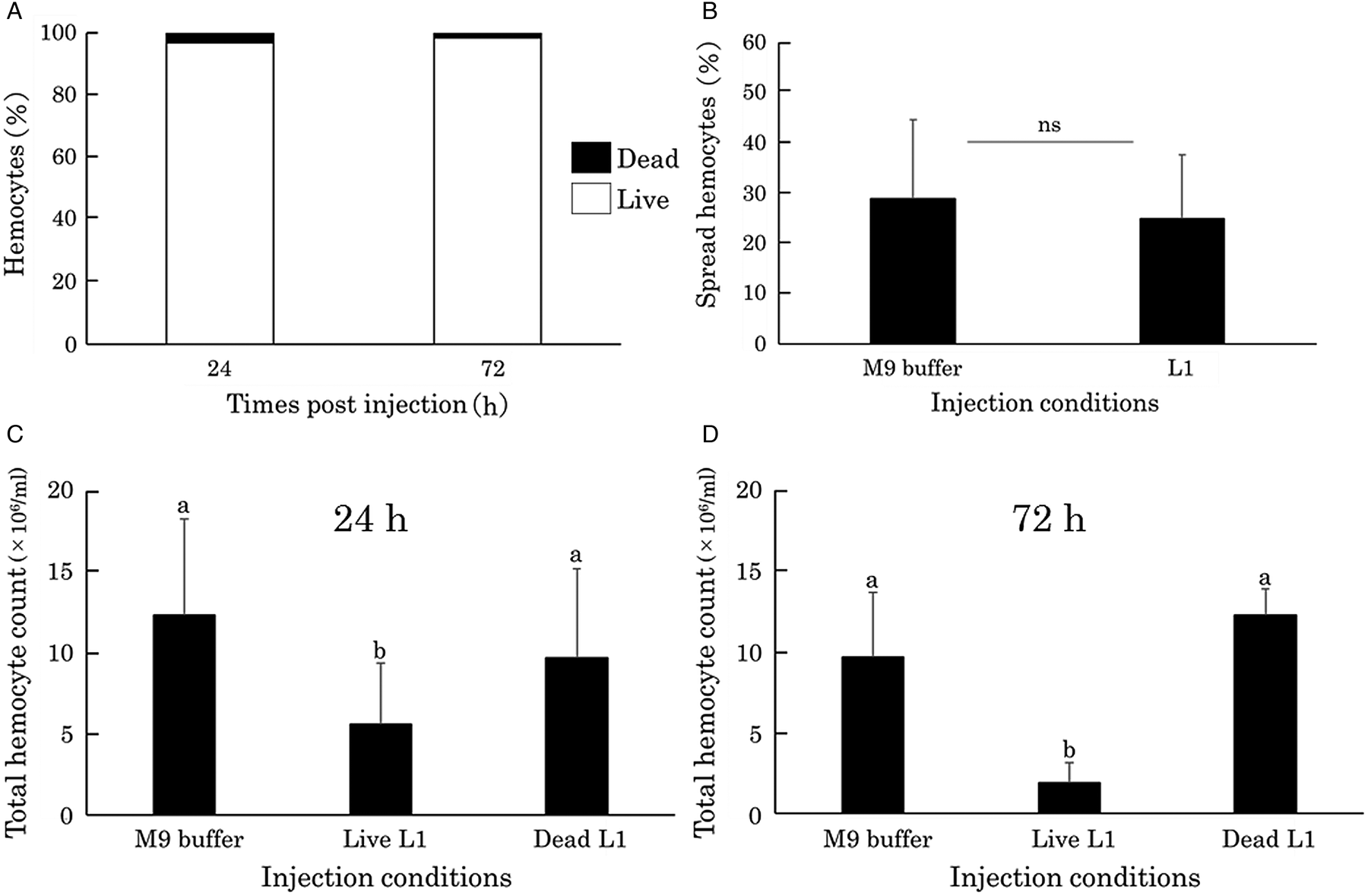
Dead haemocytes were hardly observed 24 and 72 h after injection of live C. elegans N2 L1 larvae into G. mellonella larvae (Fig. 1A) and the ability of plasmatocytes to spread did not change (Fig. 1B, Supplementary 1). However, haemocyte density was significantly lower in host larvae that had been injected with live nematodes than that in those injected with only buffer and dead nematodes (Fig. 1C). Injection of nematode exudate did not decrease haemocyte density in hosts (Supplementary 2B).
Effect on the haemocytes of G. mellonella larvae after injection of C. elegans wild type N2. (A) Haemocyte mortality in a G. mellonella larva 24 and 72 h after the injection of C. elegans first-stage (L1) larvae. (B) Spreading ability of plasmatocytes 24 h after the injection of L1 larvae into a G. mellonella larva. ns, no statistical difference (Student's t-test, P < 0.05). (C) Total number of haemocytes in a G. mellonella larva 24 and 72 h after the injection of L1 larvae. The same letter indicates no statistical difference (Tukey's test, P < 0.05).

We examined the effect of C. elegans injection on haematopoietic organs of host larvae to reveal the mechanism of the lowered haemocyte density. Neither morphological change on the haematopoietic organ, nor stem cell production rate following the nematode challenge was observed (Supplementary 2A). Additionally, we did not observe apparent haemocyte attachment to insect tissues such as fat body and epidermis (data not shown), indicating that it is not the cause of the decrease of haemocyte density.
The decrease in haemocyte density in the G. mellonella haemolymph after C. elegans injection
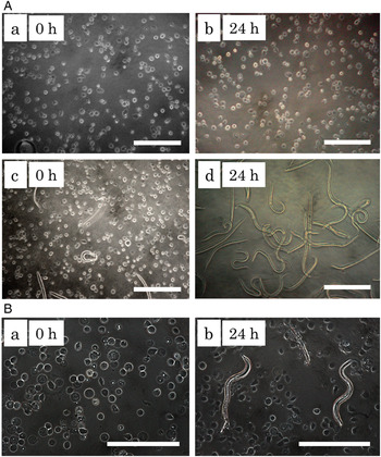
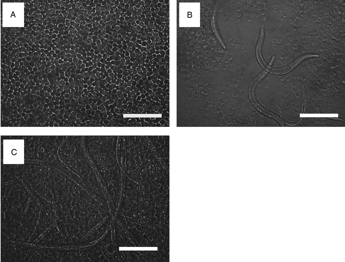
When nematodes and haemocytes were co-incubated in vitro, C. elegans were observed to suck up various types of haemocytes (Movie. 1), which led to a reduction in haemocyte number (Fig. 2A). The EPN S. carpocapsae was also observed to ingest haemocytes and the haemocyte number was decreased (Fig. 3B) as compared with control (Fig. 3A) while H. bacteriophora was not (data not shown). The reduction in haemocyte number was also observed when haemocytes were co-incubated with other non-parasitic bacteriophagous nematodes, O. myliophila, P. pacificus and P. rigidus (data not shown) but not with the fungivorous nematode A. avenae (Fig. 3C).
In vitro observation of the effect of C. elegans on haemocytes of G. mellonella larvae. (A) Haemocytes in the absence (a, b) and presence (c, d) of C. elegans N2 first-stage (L1) larvae in a 96-well plate. Scale bar indicates 100 µm. (B) Haemocytes in the absence (a) and presence (b) of C. elegans feeding-delay mutant eat-6 L1 larvae. Scale bar indicates 150 µm.

In vitro observation of the effect of an EPN and a non-parasitic fungivorous nematode on haemocytes in G. mellonella larvae. (A) Haemocytes in the absence of nematodes. (B) Haemocytes co-incubated with S. carpocapsae first-stage (L1) larvae. (C) Haemocytes co-incubated with mixed stages of fungivorous nematode A. avenae. Haemocytes and nematodes were co-incubated in a well of a 96-well plate for 24 h. Scale bar indicates 100 µm.

The decrease in haemocyte density of G. mellonella larvae by C. elegans feeding
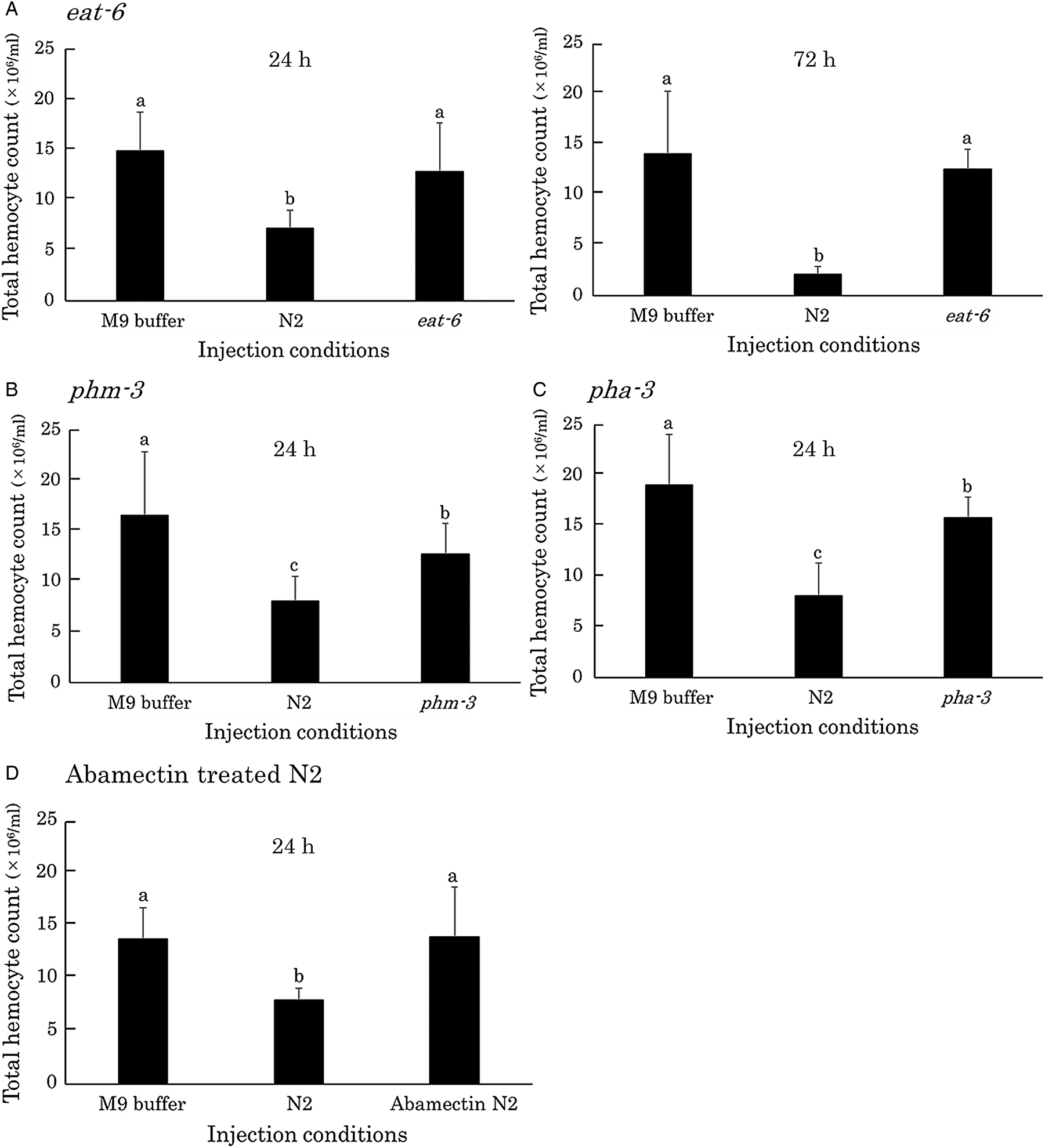
To verify the possibility that the reduction in haemocyte density could be due to ingestion by nematode, a C. elegans feeding-delay mutant eat-6 was co-incubated with haemocytes but no decrease of haemocytes was observed (Fig. 2B). Injection of eat-6 into G. mellonella larvae hardly decreased haemocyte density 24 and 72 h after injection (Fig. 4A), indicating that feeding abilities are important to the reduction in host haemocyte density. Injection of other feeding-delay mutants of C. elegans (phm-3, pha-3) slightly decreased total haemocyte density but the haemocyte densities were significantly higher than that of wild type N2 (Fig. 4B and C). Additionally, injection of immobilized C. elegans N2 treated with abamectin did not change the haemocyte density (Fig. 4D).
Change of haemocyte density in G. mellonella larvae after the injection of C. elegans feeding-delay mutants and immobilized C. elegans N2 by abamectin treatment. (A) Haemocyte densities in a G. mellonella larva 24 and 72 h after the injection of C. elegans eat-6 first-stage (L1) larvae. The haemocyte densities 24 h after the injection of L1 larvae of C. elegans phm-3 (B), pha-3 (C) and immobilized C. elegans N2 by abamectin treatment (D). The same letter in a figure indicates no statistical difference (Tukey's test, P < 0.05).

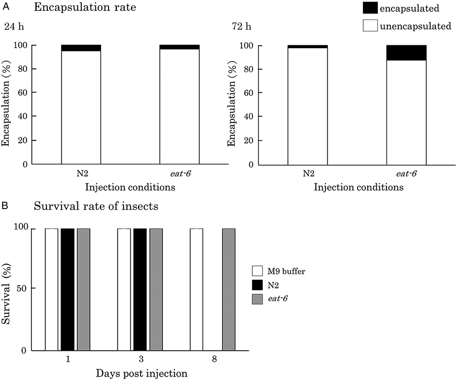
Caenorhabditis elegans feeding-delay mutants and wild type N2 were hardly encapsulated in the haemocoel of insects (Fig. 5A). Interestingly, the mortalities of hosts were different between the mutant and wild type nematode injection: all of test host larvae died by 8 days after wild type injection but did not at all after mutant injection (Fig. 5B).
Comparison of nematode encapsulation and host insect survival after injection of C. elegans N2 and a feeding-delay mutant eat-6 into the haemocoel of G. mellonella larvae. (A) Percentages of eat-6 and N2 first-stage (L1) larvae that were encapsulated in the haemocoel of G. mellonella larvae 24 and 72 h after the injection of C. elegans. ns, no statistical difference. (B) Survival of G. mellonella larvae 1, 3 and 8 days after the injection of C. elegans.

Discussion
The decrease of haemocyte density by nematode's feeding
In our previous study, we demonstrated that C. elegans injected into G. mellonella larvae are not encapsulated by host haemocytes and the nematodes propagate in the insect, resulting in the host death (Ono and Yoshiga, Reference Ono and Yoshiga2018). In the present study, we focused on the influence of nematodes on the insect haemocytes that are known to play important roles for the inactivation of foreign organisms such as nematodes by encapsulation. We found that the injection of C. elegans resulted in a decrease of haemocytes in the host haemolymph due to feeding of haemocytes by C. elegans in vitro. Injection of feeding-delay C. elegans mutants and paralysed nematodes did not cause any apparent decrease in haemocytes compared to that by wild type, suggesting that feeding of haemocytes by the nematode is the main cause of the reduction in the number of haemocytes. Caenorhabditis elegans is a bacterial feeding nematode and feeds on substances that can be sucked up through its mouth. The nematodes injected into insect haemocoel seem to suck up haemocytes together with haemolymph, resulting in a decrease of haemocytes. To the best of our knowledge, this is the first report of feeding of haemocytes by nematodes.
The effects of feeding of haemocytes by the nematode on insect
Granular cells and plasmatocytes play important roles for encapsulation (Jiang et al., Reference Jiang, Vilcinskas and Kanost2010). In lepidopteran insects, granular cells are the type of haemocytes that trigger the encapsulation process (Pech and Strand, Reference Pech and Strand1996). Elimination of these cells could protect nematodes from encapsulation. In the present study, both granular cells and plasmatocytes were observed to be almost equally fed by nematodes, indicative of no preference for a haemocyte type for feeding. It is reasonable for us to expect that the reduction of haemocyte number by nematode's feeding helps them from evading encapsulation by host haemocytes. However, because feeding-delay mutants as well as paralysed nematodes which do not lower haemocyte density were not encapsulated, the decrease of haemocyte density by nematode's feeding appears not to contribute to encapsulation evasion. The effects of reduced haemocyte number may affect physiological aspects such as metabolic and endocrine activities. We observed that all host insects died by 8 days after the injection of wild type C. elegans while did not at all after the injection of the mutant. Given that haemocytes serve as a pivotal role in maintaining homeostasis in insect bodies, they could not survive after the decline of total haemocyte number to lower than a certain threshold level. Additionally, the stresses caused by the propagation of nematodes in the haemocoel (Ono and Yoshiga, Reference Ono and Yoshiga2018) may also be the cause of insect death.
The ability to feed on haemocytes by the bacterial feeding nematodes
EPNs in the genera Heterorhabditis and Steinernema are bacterial feeders, forming an intimate mutualistic association with their symbiotic bacteria in the genera Photorhabdus and Xenorhabdus, respectively. Interestingly, Heterorhabditis and Caenorhabditis share a common ancestor (Blaxter et al., Reference Blaxter, De Ley, Garey, Liu, Scheldeman, Vierstraete, Vanfleteren, Mackey, Dorris, Frisse, Vida and Thomas1998) and it is thought that these EPNs have evolved from free-living bacterial feeding nematodes through phoretic, necromenic (feeding on decaying insects) and facultative parasitic associations with insects (Sudhaus, Reference Sudhaus, Makarov and Dimitrijevic2008). However, the evolution of their insect parasitism, especially in early stages of parasitism establishment, has not been still well elucidated. EPNs usually grow by feeding on the insect haemolymph and tissues digested by their symbiotic bacteria (Shapiro-Ilan and Gaugler, Reference Shapiro-Ilan and Gaugler2002). Additionally, S. carpocapsae is reported to eat bacteria-free insect cultured cell (Sf9) and propagate in vitro (Kikuta et al., Reference Kikuta, Kiuchi, Aoki and Nagata2008), suggesting that S. carpocapsae is able to suck up not only bacteria but also insect cells. In the present study, we demonstrated that not only S. carpocapsae, but also non-parasitic bacterial feeding nematodes, C. elegans, O. myliophila, P. pacificus and P. rigidus from different phylogenetic clades (Blaxter et al., Reference Blaxter, De Ley, Garey, Liu, Scheldeman, Vierstraete, Vanfleteren, Mackey, Dorris, Frisse, Vida and Thomas1998; Kiontke et al., Reference Kiontke, Gavin, Raynes, Roehrig, Piano and Fitch2004) feed on G. mellonella haemocytes in in vitro experiments. Bacteria and small eukaryotes serve as a food source for C. elegans but its pharyngeal pumping does not appear to be very selective (Félix and Braendle, Reference Félix and Braendle2010). Pristionchus pacificus has been described as an omnivorous feeder, which can live on a diet of bacteria, protozoans, fungi or yeast (Sanghvi et al., Reference Sanghvi, Baskaran, Röseler, Sieriebriennikov, Rödelsperger and Sommer2016). These reports suggest that so-called bacterial feeding nematodes appear to have an ability to live on different food sources. The feeding behaviour and ability to feed on haemocytes conserved in these nematodes may have been an important character as a pre-adaptation for the evolution of the early stages of parasitism. On the other hand, an EPN H. bacteriophora did not ingest haemocytes, despite being a closely related species to C. elegans. Unlike Steinernema species, there is no report on the cultivation of Heterorhabditis nematodes in the absence of their symbiotic bacteria and the cultivation is not successful without their symbiotic bacteria (Yoshiga, unpublished data), indicating their more intimate and strict mutualistic association with their symbiotic bacteria. During the process of intimate coevolution, the ability to ingest insect cells including haemocytes might have been lost or ingestion is not stimulated in the absence of their symbiotic bacteria in the Heterorhabditis nematodes.
The effect of nematode exudate on haemocytes and evasion from encapsulation
Factors secreted from Steinernema possess the ability to kill haemocytes (Walter et al., Reference Walter, Dunphy and Mandato2008; Li et al., Reference Li, Cowles, Cowles, Gaugler and Cox-Foster2009a) and plasmatocytes prepared from the host insect infected with O. sinensis show abnormal spreading (Li et al., Reference Li, Sun, Wang and Liu2009b). In the present study, cellular immune abilities of haemocytes were not affected in host insects after injection of C. elegans: normal spreading of haemocytes prepared from C. elegans injected-host larvae was observed both in vivo and in vitro. Although it has been reported that parasitization by a parasitic wasp decreased haemocyte density in a host lepidopteran insect larva due to the damaged haematopoietic organ (Teramoto and Tanaka, Reference Teramoto and Tanaka2004; Huang et al., Reference Huang, Shi, Yang, Li and Chen2009), we observed no damage of haematopoietic organs in G. mellonella injected with C. elegans. Damaging haemocyte as well as haematopoietic organs results in the failure of recognition of foreignness by haemocytes and the following encapsulation. Our results suggest that C. elegans escapes host immunity by not damaging them but different mechanisms. Caenorhabditis elegans may escape it by not being recognized by the physical structure and/or chemical components of nematode's body surface cuticle.
Supplementary material
The supplementary material for this article can be found at https://doi.org/10.1017/S0031182019001550.
Acknowledgements
The authors would like to thank the Caenorhabditis Genetics Center for providing nematode strains and Enago (https://www.enago.jp) for the English language review of the manuscript.
Financial support
This work was supported by Grant-in-Aid for Challenging Exploratory Research (T.Y.) under Grant number 23658051 and Grant-in-Aid for Scientific Research (C) (T.Y.) under Grant number 18K05676.
Conflict of interest
The authors report no conflict of interest.
Ethical standards
Not applicable.