Introduction
Malignant (necrotising) otitis externa is a progressive infection of the external auditory canal and adjacent structures. It was first described as a clinical entity in 1968 by Chandler, who described the disease as a pseudomonal infection of the external ear causing cranial base osteomyelitis in the elderly and diabetics.Reference Chandler1
As early as 1992,Reference el-Silimy and Sharnuby2 it was stated that there was no proven, agreed regime for treating malignant otitis externa. Many different therapeutic options have been used to treat this condition, including hyperbaric oxygen therapy,Reference Phillips and Jones3 but there has been very little progress in establishing a unified treatment protocol.
Prior to the introduction of effective antibiotic regimes, malignant otitis externa was fatal in nearly 50 per cent of cases, hence the renaming to include the word ‘malignant’.Reference Chandler1 In early reviews, radical surgical debridement was the mainstay of treatment. This has since been replaced by antibiotic treatment, with local debridement in some cases.
This review aims to assess recent case reports and to summarise current knowledge regarding the management of patients with malignant otitis externa.
Epidemiology
Malignant otitis externa is predominantly seen in elderly and/or diabetic patients. It is most commonly associated with diabetes mellitus; recent reviews have quoted the prevalence of diabetes in malignant otitis externa cases as 90–100 per cent.Reference Berenholz, Katsenell and Harell4, Reference Mani, Sudhoff, Rajagopal, Moffat and Axon5 Other immunocompromised states also represent risk factors for malignant otitis externa development (with or without diabetesReference Shpitzer, Stern, Cohen, Levy, Segal and Feinmesser6), including human immunodeficiency virus (HIV) infection, acquired immunodeficiency syndrome (AIDS), neoplasia, leukaemia, lymphoma, splenectomy and post-transplantation immunosuppression.
Malignant otitis externa is rarely seen in paediatric practice. However, when encountered it is commonly associated with immunocompromise (either malignancy or malnutrition). Diabetes is a less common comorbid factor in paediatric cases.Reference Sobie, Brodsky and Stanievich7
Pathophysiology
Malignant otitis externa is an invasive, granulomatous infection of the external auditory canal and skull base. It usually originates at the junction of the cartilaginous and bony portions of the external auditory canal. Pseudomonas aeruginosa is nearly always the causative organism,Reference Rubin and Yu8 although the use of topical antibiotics before collection of microbiology culture specimens often prevents isolation of the pathogen. Fungal malignant otitis externa is also seen, although usually in patients with HIV infection.
Malignant otitis externa has been associated with irrigation of the external auditory canal with water as treatment for cerumen impaction.Reference Ford and Courteney-Harris9
Both diabetes and age are associated with microvascular abnormalities. For this reason, diabetic patients and the elderly are both predisposed to malignant otitis externa,Reference Chandler1 caused by pseudomonas (not a commensal organism of the external auditory canal) invading blood vessels to cause a vasculitis with thrombosis and coagulation necrosis of surrounding tissue.
There has been speculation that the higher pH of cerumen found in the external auditory canal of diabetic patients,Reference Driscoll, Ramachandrula, Drezner, Hicks and Schaffer10 together with the increase in microbial growth seen following application of water to cerumen,Reference Campos, Betancor, Arias, Rodríguez, Hernández and López Aguado11 contributes to the development of invasive pseudomonal infection. In addition, diabetic patients' polymorphonuclear cells have impaired immune function, which could add to the relative ease with which infection occurs.Reference Oya-Ito, Naitou, Masuda, Kinae and Ohashi12
Diagnosis
Malignant otitis externa is diagnosed based on three parameters: clinical findings (e.g. polypoidal granulation tissue arising from the external auditory canal); raised serum inflammatory markers (notably the erythrocyte sedimentation rate,Reference Rubin and Yu8 although many laboratories now only routinely perform C-reactive protein measurement); and radiographic evidence of soft tissue (with or without bone erosion) in the external auditory canal and infratemporal fossa. If granulation tissue is not evident in the external auditory canal, exposed bone on the canal floor at the bone–cartilage junction may lead to suspicion of malignant otitis externa.
Patients have often received multiple courses of treatment within the primary care setting, which have failed to make any improvement to their severe otalgia. Children with malignant otitis externa often have an acute illness and tend to develop facial nerve palsies earlier in the course of the infection,Reference Rubin, Yu and Stool13 due to anatomical differences (i.e. the more medial location of the fissures of Santorini, and the underdeveloped mastoid process).
The patient will typically complain of otorrhoea, unremitting otalgia, aural fullness and hearing loss (which will be conductive in nature). Headache, temporomandibular joint pain and related trismus may also be experienced, with a subsequent reduction in oral intake, which in the diabetic patient may hamper glycaemic control.
Fungal malignant otitis externa is seen to originate more often in the middle ear or mastoid, in contrast with the late middle-ear involvement observed in pseudomonal malignant otitis externa. In patients with malignant otitis externa and HIV infection, there is often a lack of typical granulation tissue. When granulation tissue from such patients is examined, the histopathologist will typically report that the external auditory canal shows epidermal inflammation, with acute and chronic components of inflammation in the dermis.
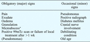
In 1987, Cohen and Friedman published diagnostic criteria in an attempt to stratify the outcome of malignant otitis externa, and to differentiate between severe otitis externa and malignant otitis externa;Reference Cohen and Friedman14 these are given in Table I.

All of the obligatory signs must be present in order to diagnose true type one malignant otitis externa. The less severe type two form (or severe otitis externa) is diagnosed if one of the obligatory criteria is absent and the patient improves within a week of hospitalisation.Reference Joshua, Sulkes, Raveh, Bishara and Nageris15
The extent of disease influences the prognosis. For example, if nasopharyngeal involvement is seen at clinical examination or as a radiological finding, then the patient is deemed to have true, type one malignant otitis externa.Reference Joshua, Sulkes, Raveh, Bishara and Nageris15 In Joshua and colleagues' series,Reference Joshua, Sulkes, Raveh, Bishara and Nageris15 this form had a fatal outcome in all patients within 40 months of hospitalisation. However, facial nerve involvement at presentation is not deemed to be a poor prognostic factor.Reference Soudry, Joshua, Sulkes and Nageris16
Radiological investigation
In Friedman and Cohen's classification guidance, the positive radiograph requirement alludes to the presence of soft tissue in the external auditory canal. This is seen on computed tomography (CT) scans of the temporal bones and skull base, which are often undertaken early in the patient's admission. However, if the patient has been treated over a prolonged period of time in the community, then the disease process may have been active for months by the time CT scanning is performed. Bone erosion is only demonstrated on CT scans once bone demineralisation has occurred, which can take several months and rarely reverses. Hence, CT scanning is a useful initial investigation for assessing the anatomical extent of disease in patients with malignant otitis externa.Reference Sudhoff, Rajagopal, Mani, Moumoulidis, Axon and Moffat17
Unfortunately, there is no one single radiological investigation that provides sufficient anatomical and physiological detail to diagnose malignant otitis externa and subsequently to monitor treatment. Hence, more complex imaging modalities are often utilised.
These include technetium scanning (using Tc 99m methylene diphosphonate), which can be useful in malignant otitis externa cases as the radiotracer accumulates at the sites of osteoblastic activity. However, such abnormal features are non-specific and can also occur in patients with simple otitis externa.Reference Levin, Shary, Nichols and Lucente18 Gallium scans (using gallium citrate Ga67) viewed in conjunction with single photon emission CT scans have also been used in patients with malignant otitis externa, as this gives more detailed information about specific areas of focally increased uptake (and active infection). However, such modalities are unavailable in most acute settings.
The need to confirm and subsequently monitor bony involvement (i.e. osteomyelitis) requires radiological and radionuclide investigations. In practice, CT remains a useful initial imaging tool, with progression or resolution monitored using magnetic resonance imaging (MRI). In 2005, Okpala et al. Reference Okpala, Siraj, Nilssen and Pringle19 suggested that CT scanning should be used to outline the anatomical extent of disease, while MRI scanning was felt by Grandis et al.,Reference Grandis, Curtin and Yu20 to be useful in cases of skull base involvement to outline soft tissue changes (particularly medullary bone space involvement and dural enhancement). Supported by case reports, these authors also proposed the use of technetium bone scanning as a valuable additional investigation if the initial CT scan is negative despite a high clinical index of suspicion of malignant otitis externa, and also during patient follow up.
Microbiology
The commonest causative pathogen for malignant otitis externa, and indeed any otitis externa, is Pseudomonas aeruginosa.Reference Ninkovic, Dullo and Saunders21 Positive culture results have been reported for as many as 75 per cent of patients,Reference Joshua, Sulkes, Raveh, Bishara and Nageris15 although pseudomonas is often not isolated in patients receiving community-prescribed antibiotics prior to culture specimen collection. In some cases, more virulent pseudomonal species have been isolated which contain a mucoid surface layer protecting the bacterium from phagocytosis.Reference Carfrae and Kesser22 Pseudomonal strains two, six and 11 are associated with more aggressive otological infections in general,Reference Englender, Harrell, Guttman and Segal23 including malignant otitis externa.
It is important to consider malignant otitis externa as a diagnosis, even if pseudomonas cannot be isolated from the culture, especially if the patient has received multiple antibiotics in the primary care setting. Other bacteria which have been isolated from (and hence implicated in) cases of malignant otitis externa include klebsiella,Reference Garcia Rodriguez, Montes Martinez, Gómez González, Ramos Macías and López Alburquerque24Staphylococcus aureus Reference Bayardelle, Jolivet-Granger and Larochelle25 and S epidermidis,Reference Barrow and Levenson26 although it is unclear if these bacterial species are true pathogens.
In the immunosuppressed, non-diabetic patient with malignant otitis externa, fungi are relatively frequently isolated, particularly in patients with HIV–AIDS. However, fungal malignant otitis externa is extremely rare overall. The most commonly implicated fungi are the aspergillus subtypes Aspergillus fumigatus,Reference Menachof and Jackler27A niger Reference Bellini, Antonini, Ermanni, Dolina, Passega and Bernasconi28 and A flavus.Reference Kountakis, Kemper, Chang, DiMaio and Stiernberg29 In a minority of cases, candidal species have also been implicated.Reference Bae, Lee, Park, Bae, Ma and Kim30 The wide variety of implicated organisms has lead some clinicians to speculate that malignant otitis externa is polymicrobial; however, when more than one organism is isolated on culture, the additional species are likely to represent normal skin flora.
Histopathology
Pseudomonas is implicated in an overwhelmingly large proportion of malignant otitis externa cases. It has therefore been suggested by Rubin Grandis et al. Reference Rubin Grandis, Branstetter and Yu31 that if P aeruginosa is not isolated from a patient suspected of having malignant otitis externa, then a bone biopsy should be taken for culture. Biopsies are also required to differentiate malignant otitis externa from temporal bone cancer and squamous cell carcinoma.
Treatment
Patients with malignant otitis externa may have been treated for a considerable period of time in the community before being seen in a hospital setting. Once admitted to hospital, many patients may have no positive results from ear swab cultures. If there is a high level of clinical suspicion, empirical intravenous antibiotics are given. Once culture specimens have been taken, a fluoroquinolone (usually ciprofloxacin) or an antipseudomonal cephalosporin (e.g. ceftazidime) are prescribed.
There are many regional and international variations to this regime. The key management strategies are: close monitoring with regular aural toilet, systemic antipseudomonal therapy, and tight glycaemic control in diabetic patients.
In those who are immunocompromised and unresponsive to treatment, a microbiological diagnosis is even more important, and a careful search for an unusual pathogen (such as candida) should be commenced.Reference Bae, Lee, Park, Bae, Ma and Kim30 Oral fluoroquinolones remain the first line of treatment, together with careful monitoring of disease progression.
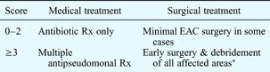
Oral quinolones (such as ciprofloxacin) have been shown to have excellent penetration into bone, together with a low toxicity profile.Reference Barza32 Peleg et al. Reference Peleg, Perez, Raveh, Berelowitz and Cohen33 have described a malignant otitis externa severity scoring system: points ranging from one to four are allocated in order to guide treatment, with more than two points indicating a categorisation of ‘severe’. Points are allocated as follows: one point for diagnosed type one diabetes mellitus, and one point each for CT evidence of bony destruction of the temporal bone or the skull base or the temporomandibular junction (see Table II).
Malignant otitis externa severity scoring system

Adapted with permission.Reference Peleg, Perez, Raveh, Berelowitz and Cohen33 *Removal of bony sequestrum and necrosis. Rx = therapy; EAC = external auditory canal
A large, systematic review of topical antimicrobial treatment of acute otitis extermaReference Rosenfeld, Singer, Wasserman and Stinnett34 reported a clinical cure rate of 65–80 per cent within 10 days of commencing therapy. Malignant otitis externa was an exclusion criterion in many of the papers assessed by this review, by virtue of its chronicity. However, the review did show that quinolone drops increased bacteriological cure rates by 8 per cent, compared with non-quinolone drops.
Fluoroquinolones target bacterial topoisomerases, rendering the bacterial DNA physiochemically unstable. Specifically, quinolone antibiotics are strong inhibitors of type II enzymes (DNA gyrase and topoisomerase IV).Reference O'Donnell and Gelone35 Older fluoroquinolones such as ciprofloxacin have predominant activity against aerobic Gram-negative bacilli, with ciprofloxacin having the most activity of this group of drugs against P aeruginosa.Reference Schentag, Scully, Yu, Merigan and Barriere36
Osteomyelitis due to Gram-negative bacilli has a cure rate of 70–80 per cent when treated with either ofloxacin or ciprofloxacin.Reference Lew and Waldvogel37 Where P aeruginosa is implicated in osteomyelitis, an oral fluoroquinolone is the preferred treatment.Reference O'Donnell and Gelone35 Care must be taken in patients with renal impairment, and an adjusted dose used where the creatinine clearance is less than 30 ml/min.Reference Flor, Guay, Opsahl, Tack and Matzke38
Rubin et al. Reference Rubin, Stoehr, Yu, Muder, Matador and Kamerer39 have suggested that the addition of rifampicin complements ciprofloxacin treatment. Despite the fact that all these authors' patients' infections involved organisms sensitive to ciprofloxacin (given as an oral dose of 750 mg twice daily), there was still only a minimal increase in inhibition and bactericidal action when this drug was added to the treatment regime. Rifampicin (600 mg twice daily) was added to the therapy regime as it did not alter the pharmacokinetics of ciprofloxacin when used concurrently, and could also be given as an oral preparation.
Other groups have favoured urgent treatment conversion to intravenous ceftazidime as an alternative to ciprofloxacin,Reference Johnson and Ramphal40 with the addition of an aminoglycoside if no improvement is seen or if resistance is encountered.Reference Giamarellou41 In such cases, there are the associated issues of administration route and prolonged hospitalisation.
The role of surgery, and of debridement in particular, is controversial in the management of malignant otitis externa. Surgical management is instigated in order to gain biopsy material and to remove areas of bony necrosis; however, only a few isolated reports have observed an improvement in symptoms following surgical debridement alone.Reference Joshua, Sulkes, Raveh, Bishara and Nageris15
Discussion
At first presentation, malignant otitis externa is essentially a clinical diagnosis made in the presence of severe pain plus granulation tissue in the external auditory canal. In all cases, priority must be given to obtaining early microbiological culture specimens, in order to guide treatment. In addition, regular culture specimens should be obtained throughout the course of treatment to ensure that antibiotic resistance has not developed.
Studies involving gallium citrate scanning have proposed that antipseudomonal treatment should continue for up to three months,Reference el-Silimy and Sharnuby2 as guided by the presence of osteomyelitis. The role of surgery is generally limited to local debridement and biopsy collection, the latter both to exclude malignancy and to guide antimicrobial therapy; this is a significant change from the management described in early case reports.
There is no dispute that the incidence of diabetes mellitus is increasing in developed countries worldwide, and that more children are now presenting with type two diabetes mellitus. Hence, there will in future be a larger percentage of the population with the most significant risk factor for development of malignant otitis externa. The increasing longevity of patients with HIV infection, due to antiretroviral therapy, will also potentially increase the incidence of malignant otitis externa.
When combined with the increasing life expectancy and increasing cumulative antibiotic usage worldwide, malignant otitis externa could become a real problem when multi-resistant pathogen strains are encountered as the causative organism.
Recent studies have shown that ciprofloxacin usage does not have a significant impact on the speed with which P aeruginosa acquires resistance to fluoroquinolones.Reference Lee, Liu, Lin, Sun, Chun and Hsueh42 However, this is in contrast to other fluoroquinolones (e.g. levofloxacins), which if used may predispose the patient to infection by resistant organisms (especially in the setting of multiple community-based antibiotic treatments prior to malignant otitis externa diagnosis).
One reviewReference Bernstein, Holland, Porter and Maw43 has also implicated purely topical administration of fluoroquinolones as adding to the problem of resistant organisms. Consequently, it has been suggested that such treatment be limited to those patients who have had a full 10-day course of topical aminoglycoside drops in the first instance.Reference Joshua, Sulkes, Raveh, Bishara and Nageris15 There are of course issues relating to the theoretical risk of ototoxicity in a potentially open middle ear, although a 2007 review suggested a maximum treatment duration of two weeks' topical aminoglycoside usage in such cases.Reference Phillips, Yung, Burton and Swan44
Fluoroquinolone resistance has been related to inadequate oral doses, short (and incomplete) courses, use in upper respiratory tract infections, and topical preparation use in simple otitis externa. Carfrae and Kesser's reviewReference Carfrae and Kesser22 reported that, in the out-patient setting, the incidence of P aeruginosa ciprofloxacin resistance was 20 per cent in simple otitis externa cases and 33 per cent in malignant otitis externa cases. It is of concern that fluoroquinolone resistance has been documented for more than a decade in malignant otitis externa cases.Reference Cooper, Andrews and Wise45
Should treatment with ciprofloxacin fail, the emergence of drug-resistant strains may cause significant problems.Reference Berenholz, Katsenell and Harell4, Reference Bernstein, Holland, Porter and Maw43 When ciprofloxacin is given, it is important that a full course of more than six weeks be commenced as early as possible, at full dose (i.e. 750 mg orally twice daily). If resistance is encountered, debridement and subsequent use of intravenous ceftazidime is advocated.Reference Berenholz, Katsenell and Harell4
Antimicrobial resistance has also been documented in the fungal species seen in malignant otitis externa.Reference Hitchcock, Pye, Troke, Johnson and Warnock46 However, it should be remembered that the small sub-set of patients with fungal malignant otitis externa will often have a multi-factorial cause for their condition, which may render their clinical course much more variable by comparison.
Fluoroquinolones are used cautiously in children because of the problems (identified in animals) related to joint development;Reference Tucker47 however, reviews highlight ciprofloxacin as being safe in selected cases.Reference Forsythe and Ernst48 Successful treatment of children with ciprofloxacin has been reported following failure of penicillin and aminoglycoside therapy.Reference Paul, Justus, Balraj, Job and Kirubakaran49 Paediatric cases are usually treated with intravenous cephalosporins.Reference Smart, Lemay and Kellner50
Conclusion
Malignant otitis externa is a rare condition, and may cause concern in patients who have had prolonged courses of treatment prior to diagnosis. The key management steps are to obtain a culture specimen, to undertake appropriate imaging, and to instigate suitable initial medical (and if necessary surgical) management followed by long-term antibiotics. Follow-up imaging has also been deemed necessary, from the literature reviewed. It is important that causative organisms (and their respective resistance to antibiotics) are recorded and published, in order to identify any new trends.