Introduction
The complete life cycles of the marine parasites of Chile are mostly uncharacterized. This great paucity of information is because most studies have focused on the parasites of marine vertebrates: for many years, invertebrates have not been included in parasitological analyses (Muñoz & Olmos, Reference Muñoz and Olmos2008), even though they are intermediate hosts for many parasites. Therefore, to date, intermediate hosts have been identified for only a few of the known parasite species.
Some studies on the life cycles of parasites have been based on morphological comparisons between developmental stages, which have been complemented with experimental infections between potential intermediate and definitive hosts (Carvajal, Reference Carvajal1977; George-Nascimento et al., Reference George-Nascimento, Carmona and Riffo1994, Reference George-Nascimento, Balboa, Aldana and Olmos1998; González, Reference González1998; Torres et al., Reference Torres, Villalobos, Woelfl and Puga2004; Aldana et al., Reference Aldana, González, Loot, Pulgar and Marquet2009). However, many parasites have complex life cycles that require one or more intermediate hosts. Furthermore, the parasites in each host may be at a developmental stage that differs from the adult stage, thus morphological comparisons between larvae and adult parasites may only be possible for certain species. Experimental infection in the laboratory has been a complementary and useful tool to elucidate parasite life cycles; however, such attempts are not always successful because infections may depend on environmental variables or on the suitability of the host species used for the experimental parasite transmission. Because the characterization of the life cycle of a given parasite becomes more complex as the number of intermediate hosts increases, molecular analyses have been applied (e.g. Nolan & Cribb, Reference Nolan and Cribb2005; Pina et al., Reference Pina, Barandela, Santos, Russell-Pinto and Rodrigues2009, Peribáñez et al., Reference Peribáñez, Ordovás, Benito, Benejam, Gracia and Rodellar2011) and have become a convenient tool to study parasites, as these methods are rapid and now relatively inexpensive. In Chile, molecular analyses have recently been used to determine whether marine parasites found in different hosts belong to the same species (Oliva et al., Reference Oliva, Valdivia, Cárdenas, George-Nascimento, González, Guiñez and Cuello2010; Valdivia et al., Reference Valdivia, Cárdenas, González, Jofré, George-Nascimento, Guiñez and Oliva2010; Muñoz & Bott, Reference Muñoz and Bott2011).
This study focused on Perumytilus purpuratus (Mytilidae), a common bivalve that is widely distributed throughout the rocky intertidal zone from northern to southern Chile. Trematodes exist at the sporocyst stage in P. purpuratus, harbouring cercariae, and three different morphologies have indicated the presence of three different species. One of these trematodes is an undetermined bucephalid (Lasiak, Reference Lasiak1991), which is distinguished by the morphology of the sporocysts and cercariae. Another trematode has been identified as Proctoeces lintoni (Fellodistomidae) based on morphology and experimental infection of mytilids with trematode eggs (Aldana et al., Reference Aldana, González, Loot, Pulgar and Marquet2009). The third species was determined by molecular analysis to be close to the Fellodistomidae family, although the genus has not been determined (Oliva et al., Reference Oliva, Valdivia, Cárdenas, George-Nascimento, González, Guiñez and Cuello2010). None of the parasitological studies performed in P. purpuratus to date have focused on all of the trematode species, thus it may be possible to find other trematode species due to the high abundance of this bivalve in the intertidal zone.
The trematodes in P. purpuratus may complete their life cycles in vertebrates that are in the same habitat, thus intertidal fish are good candidates to be definitive hosts for the larval trematodes of this bivalve. There are approximately 15 fish species (Muñoz & Ojeda, Reference Muñoz and Ojeda1998; Muñoz & Cortés, Reference Muñoz and Cortés2009) that reside seasonally or permanently in the intertidal zone. Each intertidal fish species normally harbours 1–4 adult trematodes (Muñoz & Cortés, Reference Muñoz and Cortés2009) of nine possible species, including one bucephalid, one haploporid, one opecoelid, three fellosdistomid, two lecithasterid and one derogenid species. Accordingly, this study sought to compare the DNA sequences among the sporocyst species found in P. purpuratus and the adult trematodes present in the most common fish species residing in the intertidal rocky zone of central Chile.
Methods
Collection and examination of bivalves
Individual specimens of P. purpuratus (n= 1000) were collected from the rocky intertidal zone of central Chile (~33°S, 71°W), El Tabo (33°27′S), Las Cruces (33°13′S) and Montemar (32°58′S) during 2010. The bivalves were dissected when fresh, and parasitized bivalves contained sporocysts in their gonads and mantle. The sporocysts were separated from the host tissues and fixed in 100% ethanol for molecular analyses or in 10% formalin for morphological analyses.
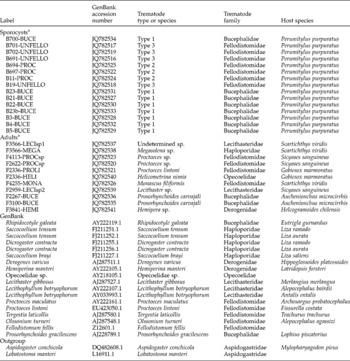
We referred to the morphospecies of the sporocysts according to the available literature: type 1 (Bucephalidae), type 2 (Proctoeces) and type 3 (undetermined fellodistomid). The parasite specimens used in the analyses and their IDs are indicated in table 1.
Molecular analyses of trematodes and hosts together with GenBank accession numbers. Sporocysts correspond to the first stage of trematodes obtained from bivalves (B); adults correspond to trematode individuals obtained from fish (F), and GenBank refers to the samples obtained from the public database.

a Each label is composed of host identification (B, bivalve; F, fish) followed by specimen number, and then by the abbreviation of the trematode species.
Morphology of sporocysts and cercariae
The morphological characteristics of the sporocysts and cercariae among the morphotypes were observed using optical microscopy. In addition, several cercariae were observed by scanning electron microscopy (SEM). For this purpose, the cercaria larvae were dehydrated through an ethanol series (70–100%), followed by critical point drying in CO2 using a Samdri-780A machine (Tousimis, Rockville, Maryland, USA). The cercariae were then sputter-coated with gold using a JFC-1100 ion sputter machine (JEOL Ltd, Tokyo, Japan) and examined using a JEOL T-300 SEM. Some specimens were stained in haematoxylin for collecting measurements using an eye-piece micrometer and drawings using a camera lucida, both attached to a Leica DMLS2 light microscope (Leica Microsystems, Wetzlar, Germany). Length and width measurements, given in micrometres (μm), were obtained for the sporocysts and cercariae.
Molecular analysis
The parasites were isolated, and each morphotype was transferred to a 1.5-ml microcentrifuge tube. DNA extraction was performed by adding 500 μl of 5% Chelex® and 2.5 μl of 20 mg/ml proteinase K to each tube. The samples were incubated at 60°C for 4 h and then boiled for 8 min (Leung et al., Reference Leung, Donald, Keeney, Koehler, Peoples and Poulin2009).
The V4 region of the 18S rRNA gene was amplified by polymerase chain reaction (PCR) using the primers described by Hall et al. (Reference Hall, Cribb and Barker1999). Each PCR was performed using 0.025 U of Taq polymerase, 1 × buffer, 0.2 mm deoxynucleotide triphosphate (dNTP), 4 mm MgCl2, 0.4 pmol/μl of each primer, 2.5–5 μl of concentrated DNA, 1.5 μl of 10 mg/ml bovine serum albumin (BSA) (New England Biolabs, Ipswich, Massachusetts, USA) and water to a final volume of 25 μl. The DNA was amplified using a Perkin Elmer thermal cycler (Perkin Elmer, Santiago, Chile) and optimal cycling parameters for the V4 region, including an initial denaturation step at 94°C (5 min) followed by 35 cycles of 94°C (30 s), 45°C (30 s) and 72°C (3 min) and a final extension step at 72°C (10 min). The PCR products were visualized on a 1.5% agarose gel and purified using the EZNA kit (BioTec, Winooski, Vermont, USA). The PCR products for each specimen were sequenced using an automated capillary electrophoresis sequencer (ABI 3730XL, Macrogen Inc., Seoul, Korea) (http://www.macrogen.com). The sequences were edited using ProSeq v3.0 beta (Filatov, Reference Filatov2002) and aligned with Clustal 2 (Larkin et al., Reference Larkin, Blackshields, Brown, Chenna, McGettigan, McWilliam, Valentin, Wallace, Wilm, Lopez, Thompson, Gibson and Higgins2007).
A total of nine adult trematode species belonging to six families (Bucephalidae, Derogenidae, Fellodistomidae, Haploporidae, Lecithasteridae and Opecoelidae) found in five intertidal fish species were considered for our molecular analysis (table 1); the species and sample size of the fish dissected are based on the study by Muñoz & Cortés (Reference Muñoz and Cortés2009). In addition, sequences of other trematode members of the six families considered were obtained from the GenBank database (http://www.ncbi.nlm.nih.gov/genbank/) to compare with the samples of the present study (table 1).
A phylogenetic rooted tree was used to establish molecular sequence differences among the sporocysts from P. purpuratus and the adult trematodes from intertidal fish; the tree was generated using Mega v5.05 software (Tamura et al., Reference Tamura, Peterson, Peterson, Stecher, Nei and Kumar2011) with the neighbour-joining (NJ) algorithm (Nei & Kumar, Reference Nei and Kumar2000) and the maximum composite likelihood (ML) evolution model (Tamura & Nei, Reference Tamura and Nei1993). As outgroup species, members of Aspidogastrea (Aspidogaster conchicola and Lobatostoma manteri) were used as sister taxa, according to Olson et al. (Reference Olson, Cribb, Tkach, Bray and Littlewood2003); their sequences were obtained from GenBank (table 1). The nodes were statistically evaluated by 1000 bootstrap resamplings, according to Efron (Reference Efron1982). The genetic distance matrices were computed with the total number of mutations, and the divergences were calculated among individuals by applying the ML model.
Results
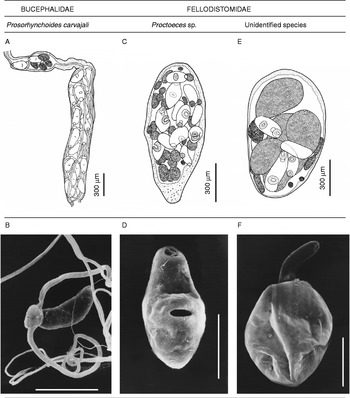
The sporocysts in P. purpuratus were easily distinguished as one of three known morphospecies. The type 1 sporocysts were long (n= 32, several millimetres in length, 105–250 μm wide) and ramified, with several cercariae at similar developmental stages (fig. 1A); these cercariae (n= 16, 81–163 length × 0.050–0.70 width) have two long posterior filaments and a tegument with spines, a typical morphology among bucephalids (fig. 1B). The type 2 sporocysts were long and oval (n= 49, 700–1925 length × 250–465 width), with several cercariae at different development stages (fig. 1C); these cercariae were short (n= 55, 175–256 length × 75–125 width), with a wide acetabulum in an equatorial position, a smooth tegument (fig. 1D), and no tail was observed. The type 3 sporocysts contained oval, nearly round sacs (n= 40, 825–2000 length × 550–950 width), with 2–4 large cercariae and several small and undeveloped cercariae (fig. 1E). These cercariae were oval and small (n= 36, 225–525 length × 65–130 width), with a large reddish/brownish and apparently empty sac (523–881 length × 285–413 width) at the posterior end and a smooth tegument (fig. 1F). No other trematode morphospecies was found in P. purpuratus.
Sporocysts (A, C, E) and SEM micrographs of cercariae (B, D, F) of the three species found in the bivalve P. purpuratus. Scale bars = 100 μm.

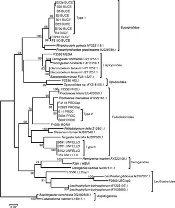
In total, 26 sequences were obtained for the specimens analysed using the V4 region of the 18S rRNA (adult trematodes and sporocysts). The PCR products of the V4 region were between 402 and 448 base pairs in length. The branching pattern of the tree reconstruction grouped into six major clades, according to each trematode family considered (fig. 2). The type 1 sporocysts were placed within the Bucephalidae clade, and types 2 and 3 were placed in Fellodistomidae. In the Bucephalidae clade, Prosorhynchoides gracilescens and Rhipidocotyle galeata comprised one group, whereas Prosorhynchoides carvajali (F2267BUCE and F3100BUCE) and the type 1 sporocysts comprised another (96% bootstrap support). The mean genetic distance between these two trematodes was 0.78%, with a mean number of mutations of 6.3 bp (table 2A, fig. 2).
Phylogenetic tree of the relationship between sporocysts of P. purpuratus and adult trematodes of fish based on the V4 region of 18S rRNA.

Pairwise sequence divergences for the V4 region of the rRNA gene among species of the (A) Bucephalidae and (B) Fellodistomidae clades. The divergence distance was calculated using the maximum composite likelihood model, and it is shown as a percentage (below the diagonal). The mean number of mutations between pairwise comparisons is also shown in each clade (above the diagonal).

The Fellodistomidae clade was divided into two subclades, as follows: one was composed of Monascus filiformis (F4255 MONA), Proctoeces lintoni (F2336PROLI, EU423050.1), Proctoeces maculatus (AY222161.1), Proctoeces sp. (F4113PROCsp and F2622PROCsp) and the type 2 sporocysts (B11PROC, B694PROC, B697PROC). The latter split node had 99% bootstrap support (fig. 2), and there was no genetic divergence among them (table 2B). The other subclade was composed of Fellodistomum fellis, Olssonium turneri, Tergestia laticollis and the type 3 sporocysts. The latter two species formed one subclade, with 100% bootstrapping support but with 8.16% genetic divergence (table 2B, fig. 2). Thus, the genetic sequence of the undetermined sporocysts (type 3) did not match any of the known adult trematode species from intertidal fish in Chile (table 2B, fig. 2).
Discussion
Two sporocyst morphospecies found in the bivalve P. purpuratus genetically matched known trematode genera of intertidal fish from central Chile. The type 1 sporocyst was genetically similar to the genus Prosorhynchoides (Bucephalidae) (fig. 2), the type 2 sporocyst was classified as Proctoeces sp. (Fellodistomidae), and the type 3 sporocyst did not match any of the adult trematode species used in this study, although it was placed within Fellodistomidae (fig. 2).
The type 1 sporocysts were close to P. carvajali (fig. 2) and showed a mean divergence of 0.78% (table 2A). Nolan & Cribb (Reference Nolan and Cribb2005) suggested that a conserved gene, such as the V4 region of the 18S gene, normally shows no more than 1% genetic divergence between specimens of the same species. Similarly, Sorensen et al. (Reference Sorensen, Curtis and Minchella1998) reported that the maximum divergence was 0.9% for Echinostoma revolutum, whereas other authors have indicated up to 2.5% genetic divergence among specimens within a species as a consequence of intraspecific genetic variation (see Luton et al., Reference Luton, Walker and Blair1992). Therefore, because of the little divergence variation between sporocyst type 1 and the adults of P. carvajali, we conclude that they are the same trematode species, using P. purpuratus as the first intermediate host and maturing in intertidal Auchenionchus spp. and occasionally in the clingfish Sicyases sanguineus (see Muñoz & Bott, Reference Muñoz and Bott2011).
The type 2 sporocysts were placed in the Proctoeces clade, and the sequences perfectly matched (0% genetic divergence) Proctoeces sp. from the clingfish S. sanguineus (fig. 2, table 2B). Moreover, the type 2 sporocysts exhibited 5.57% genetic divergence from P. lintoni specimens. This finding indicates that the type 2 sporocysts belong to the Proctoeces genus but are definitely not P. lintoni, as was suggested by Aldana et al. (Reference Aldana, González, Loot, Pulgar and Marquet2009), in accordance with the results of other studies (e.g. Luton et al., Reference Luton, Walker and Blair1992; Sorensen et al., Reference Sorensen, Curtis and Minchella1998; Nolan & Cribb, Reference Nolan and Cribb2005). This is an interesting result because, for many years, P. lintoni was the only trematode species documented in S. sanguineus, and an undescribed Proctoeces was commonly found in young clingfish only recently (Muñoz & Zamora, Reference Muñoz and Zamora2011). Aldana et al. (Reference Aldana, González, Loot, Pulgar and Marquet2009) performed experimental infections of the bivalve P. purpuratus with trematode eggs from a species that was presumed to be P. lintoni; the resulting sporocyst and cercaria morphotypes were similar to those observed in this study (fig. 1C and D). There are two possible explanations for the apparent discrepancy. First, whereas Aldana et al. (Reference Aldana, González, Loot, Pulgar and Marquet2009) likely utilized sporocysts from Proctoeces sp., they may not have used those of P. lintoni. Alternatively, it is possible that these authors may have successfully infected P. purpuratus with P. lintoni eggs in the laboratory; however, P. lintoni sporocysts are not a parasite of P. purpuratus in nature. Consequently, based on the results of the present study, P. purpuratus is clearly not the first intermediate host for P. lintoni. Although Oliva et al. (Reference Oliva, Valdivia, Cárdenas, George-Nascimento, González, Guiñez and Cuello2010) reached a similar conclusion, they only compared the genetic sequences of P. lintoni and the type 3 sporocyst, but it was necessary to analyse other fellodistomid species from Chile to confirm the identity of this trematode. Therefore, in this study, we found that the type 2 sporocysts belong to Proctoeces sp. and that the clingfish is the definitive host. This is an undescribed species (see Muñoz & Zamora, Reference Muñoz and Zamora2011), which is currently being identified by Chilean parasitologists.
Although we have considered all of the trematode species found in intertidal fishes, which are the most abundant vertebrates in the rocky coastal zone where P. purpuratus resides, the identity of the type 3 sporocysts remains a mystery. The morphology of the type 3 sporocyst cercariae is quite distinct because of the large, floppy posterior sac (fig. 1F); to our knowledge, this has not been documented for other Fellodistomidae species (e.g. Stunkard & Uzmann, Reference Stunkard and Uzmann1959; Aldana et al., Reference Aldana, González, Loot, Pulgar and Marquet2009; this study). Our study obtained a similar result as that reported by Oliva et al. (Reference Oliva, Valdivia, Cárdenas, George-Nascimento, González, Guiñez and Cuello2010) in that this type of sporocyst may be a fellodistomid species, close to Tergestia laticollis. Although the type 3 sporocysts were in a clade within ‘Fellodistomidae’, the sequences were not close to any genera considered in the group. Furthermore, the type 3 sporocysts showed genetic divergences between 18.3% and 20.6% from the three other fellodistomid species found in Chile (M. filiformis, P. lintoni and Proctoeces sp.) and 8.16% from T. laticollis. Thus, we still do not know to which genus the type 3 sporocysts might belong. In future investigations, it would be helpful to incorporate other species within the superfamily Gymnophalloidea (of which Fellodistomidae is a member) for the molecular analyses. Moreover, it is also necessary to include different perspectives to advance this subject, as follows: (1) the type 3 sporocyst may not belong to Fellodistomidae, which necessitates considering more trematode species from other families in future studies; (2) the definitive host for the type 3 sporocyst may be a subtidal fish, thus trematodes of this fish group should be included; and (3) the definitive host may be a bird, which would require the analysis of trematodes from coastal bird species.
This study has contributed to the knowledge of the genetic sequences for trematode species (in sporocyst stages) parasitizing P. purpuratus and intertidal fish. Two sporocyst types were identified at least to the genus level (Prosorhynchoides carvajali and Proctoeces sp.), which were sequenced for the first time, whereas one morphospecies was determined to the family level (possibly Fellodistomidae). These results demonstrate that adult trematode species have not been well documented for the Chilean coast, indicating that additional surveys and taxonomic classification of trematodes in the marine organisms of this country need to be performed.
Acknowledgements
This study was supported by the grant DIPUV 12-2008 from Universidad de Valparaiso, Chile, awarded to G.M.