

The vertebrate eye, a delicate soft tissue structure, in gnathostomes is protected by a number of different skeletal tissues (Ritchie Reference Ritchie1968; Burrow et al. Reference Burrow, Jones and Young2005; Franz-Odendaal & Vickaryous Reference Franz-Odendaal and Vickaryous2006). These skeletal tissues within the eye consist of two types of tissues, cartilage (e.g. scleral cartilage) and bone (e.g. scleral ossicles, os opticus) (Franz-Odendaal & Vickaryous Reference Franz-Odendaal and Vickaryous2006). Furthermore, skeletal tissues are found directly associated with the eye (e.g. within the eyelids). These tissues are usually composed of bony tissues that include, for example, palpebrals in reptiles, sesamoid-like elements found in some avians (e.g. owls), and neomorphic bones in reptiles (e.g. parafrontal bones in geckos, supraorbitals in falconiform birds) (Franz-Odendaal & Vickaryous Reference Franz-Odendaal and Vickaryous2006). This collection of skeletal tissues within or among the eye has been a focus of studies examining the development of the eye in extant gnathostomes (e.g. Coulombre Reference Coulombre, De Haan and Ursprung1965) and using the extant phylogenetic bracket (sensu Witmer Reference Witmer and Thomason1995) to elucidate the evolution of skeletal features in the vertebrate eye (Burrow et al. Reference Burrow, Jones and Young2005; Franz-Odendaal & Vickaryous Reference Franz-Odendaal and Vickaryous2006; Vickaryous & Sire Reference Vickaryous and Sire2009). To examine the origin of these features, we must turn to the fossil record. Unfortunately, the fossil record ordinarily only preserves bony elements of the orbit, as cartilage is only preserved in extraordinary cases (e.g. Dal Sasso & Maganuco Reference Dal Sasso and Maganuco2011). Therefore, we are limited to hard parts in our understanding of the origin and evolution of skeletal elements associated with the eye.
With few exceptions, these bony elements are limited to palpebral bone(s), supraorbital(s) and scleral ossicles in a variety of extinct reptiles. Additionally, the morphology and presence of these elements have been used as behaviour indicators (e.g. the size of the sclerotic ring suggests diving depth in Ichthyosauria, Motani et al. Reference Motani, Rothschild and Wahl1999; Hall et al. Reference Hall, Kirk, Kamilar and Carrano2011; Schmitz & Motani Reference Schmitz and Motani2011). Scleral ossicles are found across the family tree of Reptilia and more distantly related clades within Vertebrata, suggesting that the presence of scleral ossicles is plesiomorphic for all of Reptilia (Fig. 1). Palpebrals or supraorbitals are found in a variety of reptiles including microsaurs (Daly Reference Daly1973), lizards (lacertids, scincoids, and anguimorphan lizards; Estes et al. Reference Estes, De Queiroz, Gauthier, Estes and Pregrill1988), pterosaurs (Coombs Reference Coombs1972; Wang et al. Reference Wang, Kellner, Zhou and Campos2007), ornithischian dinosaurs (Coombs Reference Coombs1972; Maidment & Porro Reference Maidment and Porro2010) and crocodylomorphs (including Crocodylia) and other pseudosuchians (Walker Reference Walker1961; Desojo & Baez Reference Desojo and Baez2007; Schoch Reference Schoch2007; Weinbaum Reference Weinbaum2011). Although all of these elements in the dorsal portion of the orbit have been referred to as either palpebrals or supraorbitals, the homology of these structures among extinct groups with the structures in extant reptile clades remains untested. Furthermore, the terms palpebral and supraoccipital have been used interchangeably (Romer Reference Romer1956). For example, the orbital structure(s) in ornithischian dinosaurs have been called ‘palpebrals,’ (Romer Reference Romer1956), but the homology of these structures with those of crocodylians is contentious; the structures in ornithischians could be neomorphic elements that are not homologous with palpebrals of crocodylians.
The distribution of bony skeletal elements in the orbits of extant amniotes. The possible relationships of Chelonia among amniotes includes a phylogenetic position as a non-saurian sauropsid, a lepidosauromorph and an archosauromorph. The presence of a sclerotic ring is plesiomorphic for Amniota and secondarily lost among the crocodilian-line archosaurs, as demonstrated by the crocodylian Paleosuchus located at the Shedd Aquarium in Chicago, IL. The white arrow highlights the palpebral above the eye.

Recent developmental studies of crocodylian palpebrals (Vickaryous & Hall Reference Vickaryous and Hall2008) have made it possible to determine the origin of tissues that lead to the formation of palpebrals. Palpebral elements are part of the dermal skeletal system (i.e., metaplastic ossification, not preformed in cartilage) that develops within the upper eyelid, and this type of development is not the same as dermal elements of the skull (Vickaryous & Hall Reference Vickaryous and Hall2008). In crocodylians, the mode and pattern of skeletogenesis of the palpebral matches identically with that of postcranial osteoderms (Vickaryous & Hall Reference Vickaryous and Hall2008). Essentially, crocodylians have an osteoderm in the orbit. The development of the palpebrals of crocodylians may be understood, but the evolutionary history of the crocodylian palpebral has not been examined in detail.
Extant crocodylians, one of the two living groups of archosaurs, bear a distinct palpebral at the dorsal margin of their orbit and lack sclerotic ossicles. In contrast, avians, the other group of living archosaurs, lack palpebral elements and nearly all possess sclerotic ossicles, a plesiomorphic condition within Reptilia. Given this distribution of ossified elements in the orbits of crocodylians, the origin of the palpebral and the loss of the sclerotic ring must have occurred among taxa more closely related to crocodylians than to avians, or within the clade Pseudosuchia (=Crurotarsi of Sereno et al. Reference Sereno, Mcallister and Brusatte2005; =crocodylian-line archosaurs). In the present contribution, we survey the distribution and forms of skeletal elements in the orbit of crocodylians and among their closest relatives and discuss the origin of the palpebral and the loss of the sclerotic ossicles within Pseudosuchia (Fig. 1). After recent revisions of the anatomy of non-crocodylomorph pseudosuchians such as Postosuchus kirkpatricki (Weinbaum Reference Weinbaum2011) and Aetosaurus ferratus (Schoch Reference Schoch2007), it is clear that skeletal elements were present in or around the eye in close relatives of crocodylomorphs. These key pseudosuchian taxa have triggered the revaluation of other closely related non-crocodylomorph pseudosuchians and illuminated the origins and early evolution of the skeletal elements in the orbit of pseudosuchians.
This study does observe the following limitations when examining skeletal elements in the orbit of extinct taxa. First, skeletal elements of the eye are prone to loss during the fossilisation process, given that most are not articulated to other hard parts. Given that the eye decomposes soon after death (Gordon & Shapiro Reference Gordon and Shapiro1975), the usually fragile skeletal elements become disassociated from the rest of the skeleton at a much faster rate than other hard tissues. Furthermore, once skeletal elements of the orbit are displaced from the orbital area, they become difficult to orient, articulate with other skeletal elements near or within the orbit, or even identify the bone in question as a skeletal element of the eye. Skeletal elements formed in the orbit of vertebrates also are thin, sometimes poorly ossified structures prone to collection and preparation biases in vertebrate palaeontology. For example, delicate skeletal features of the orbit are easily removed if special care is not taken within the orbit of a fossil vertebrate.
Secondly, the identification of certain elements may be impossible without developmental sequences (e.g. Hall Reference Hall2005; Vickaryous & Hall Reference Vickaryous and Hall2008). For example, the identification of a palpebral (osteoderm) versus a neomorphic circumorbital dermal bone may not be determinable from morphology, composition or position, without an understanding of the underlying development process in the taxon being examined (see Hall Reference Hall2005).
Thirdly, the timing of ossification of the skeletal elements in the orbit during ontogeny may be variable across taxa, and determining the ontogenetic age of archosaurs outside of Aves and Crocodylia is problematic with our current interpretations of skeletal chronological correlates (Brochu Reference Brochu1996; Irmis Reference Irmis2007). For example, crocodylians only ossify osteoderms and the palpebral after the first year of life (Vickaryous & Hall Reference Vickaryous and Hall2008). So, the presence or absence of certain skeletal elements of the orbit may be related to ontogenetic stage of an individual. This is further exemplified by the apparent absence of a palpebral in the long snouted crocodylian Gavialis in adults, even though a palpebral is present in hatchlings (see below).
Clade names utilised in this contribution derive from Sereno et al. (Reference Sereno, Mcallister and Brusatte2005) and recent revisions and additions from Nesbitt (Reference Nesbitt2011).
Institutional abbreviations. AMNH, American Museum of Natural History, New York NY, USA; BSPG, Bayerische Staatssammlung für Paläontologie und Geologie, Munich, Germany; CM, Carnegie Museum of Natural History, Pittsburgh PA, USA; DGM, Departmento de Produção Mineral, Rio de Janeiro, Brazil; FMNH, The Field Museum of Natural History, Chicago IL, USA; IGM, Mongolian Institute of Geology, Ulaanbaatar, Mongolia; IVPP, Institute of Vertebrate Paleontology and Paleoanthropology, Beijing, China; MCZ, Museum of Comparative Zoology, Harvard University, Cambridge MA, USA; MG, Museo Geológico, Lisbon, Portugal; MOZ, Museo Profesor J. Olsacher, Zapala, Argentina; MPMA, Monte Alto Museum of Paleontology, Monte Alto SP, Brazil; MTM, Hungary Natural History Museum, Budapest, Hungary; NHMUK, Natural History Museum, London, UK; PIN, Paleontological Institute of the Russian Academy of Sciences, Moscow, Russia; PVL, Instituto Miguel Lillo, Tucuman, Argentina; PVSJ, Division of Paleontology of the Museo de Ciencias Naturales de la Universidad Nacional de San Juan, Argentina; SAM, South African Museum, Cape Town, South Africa; SMNH, Royal Saskatchewan Museum (formerly the Saskatchewan Museum of Natural History), Regina, Canada; SMNS, Staatliches Museum für Naturkunde, Stuttgart, Germany; TMM, Vertebrate Paleontology Laboratory, Texas Natural Science Center, Austin TX, USA; TTU-P, Texas Tech University Museum, Lubbock TX, USA; UA, University of Antananarivo, Madagascar; UCMP, University of California Museum of Paleontology, Berkeley CA, USA; UH, Urweltmuseum Hauff, Holzmaden, Germany; UNC, University of North Carolina, Chapel Hill NC, USA; YPM, Yale Peabody Museum, New Haven CT, USA; ZPAL, Institute of Paleobiology of the Polish Academy of Sciences in Warsaw, Poland.
1. Osseous skeletal element(s) in the orbit of pseudosuchians
1.1. Extant crocodylians
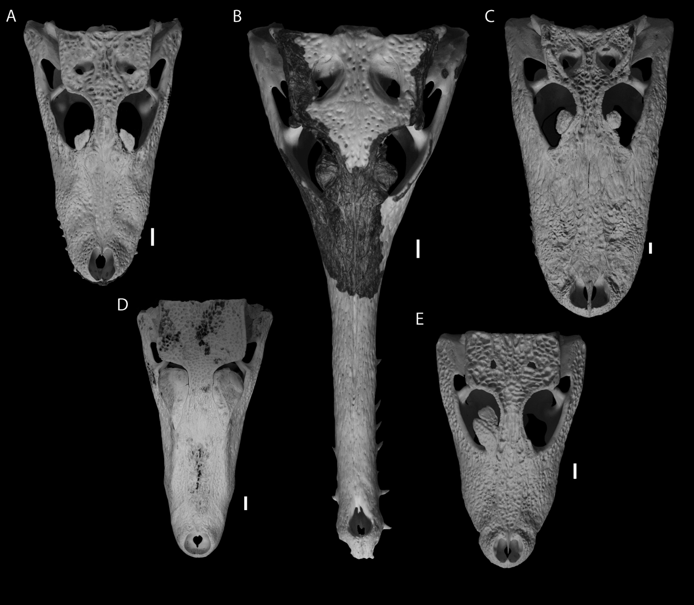
Crocodylians are the only extant group of reptiles to possess a palpebral and lack any scleral ossicles. With the exception of Paleosuchus and Osteolaemus, all extant crocodylians possess a palpebral that is typically small and situated in the anteromedial corner of the orbit. The shape and thickness of the palpebral differs across Crocodylia, but generally consists of a sub-rounded element that is mediolaterally compressed. The dorsal surface is rugose, similar to that of the surrounding skull roof elements and osteoderms, and the ventral surface is almost smooth. The palpebral rests upon a facet on the orbital edge of the prefrontal and may or may not contact the frontal along its medial margin. The prefrontal and the palpebral articulate by connective tissue. The shape of the palpebral is variable among crocodylians, but is usually amygdaloidal with the long axis aligned anteroposteriorly (Fig. 2). Furthermore, the size and shape of palpebrals can vary within a species (e.g. Alligator mississippiensis), or even an individual (Fig. 2C). The palpebral in most crocodylians does not reach the posterior or lateral margins of the orbit.
Extant crocodylians with articulated palpebral(s) in dorsal view: (A) Caiman crocodylus yacare (TMM M-7365) with a palpebral composed of a single ossification; (B) Tomistoma schlegelii (TMM M-6432) with a palpebral still embedded in dried skin (black); (C) Alligator mississippiensis (TMM M-7487) with a palpebral element composed of a single ossification; (D) Paleosuchus palpebrosus (YPM R11407) with palpebrals composed of three ossification centers forming a single element; (E) Osteolaemus tetraspis (TMM M-6774) with palpebrals composed of two ossifications (the left element has been removed). Scale bars=1 cm.
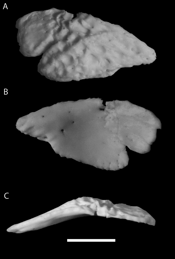
The palpebral in extant crocodylians forms from a single ossification centre, except in Paleosuchus and Osteolaemus. In these taxa, the single palpebral is formed from either two or three ossifications (Osteolaemus tetraspis; Figs 2E, 3), or three ossifications (Paleosuchus; Fig. 2D). Nearly all specimens of Osteolaemus tetraspis possess a single palpebral consisting of two ossifications (Christopher Brochu, pers. comm. 2012), but the specimen presented in Figure 3 possesses a single palpebral composed of three ossifications. In Osteolaemus tetraspis and Paleosuchus, the palpebral is a single structure, in contrast with the two distinct palpebrals in some extinct pseudosuchians (see below).
The right palpebral of Osteolaemus tetraspis (TMM M-6774) in dorsal (A), ventral (B), and lateral (C) views. Scale bar=1 cm.
Paleosuchus and Osteolaemus also show the most deviation in palpebral morphology from the typical crocodylian palpebral, as exemplified by Alligator mississippiensis (Fig. 2C). The two species of Paleosuchus have large palpebrals that traverse most of the dorsal aspect of the orbit (Fig. 2D, B). The palpebral reaches or nearly reaches the postorbital (but does not have a facet on the bone) and overhangs the dorsolateral portion of the orbit. The medial margin of the palpebral tightly follows the lateral edge of the frontal, but does not share a cranial suture with the bone. In Paleosuchus trigonatus, the palpebral is often elevated above the dorsal surface of the skull table. Osteolaemus tetraspis also has an enlarged palpebral relative to other crocodylians, but not to the extent present in the two Paleosuchus species. The palpebral of Osteolaemus does not reach the postorbital and the medial margin does not parallel the frontal (Fig. 2E). In general form and location, the palpebral of Osteolaemus is very similar to the anterior palpebral in most fossil crocodyliforms.
1.2. Postosuchus and other rauisuchids (sensu Nesbitt Reference Nesbitt2011)
In 1985, Chatterjee described one of the most complete “rauisuchian” (rauisuchid of Nesbit Reference Nesbitt2011) archosaurs to date, Postosuchus kirkpatricki from the Late Triassic of the Dockum Group of Texas. The well-preserved, nearly complete skull (TTU-P 9000) possesses an unusual triangular bone lying dorsal to the orbit that Chatterjee (Reference Chatterjee1985) interpreted as a prefrontal. This interpretation was followed by Long & Murry (Reference Long and Murry1995) and Peyer et al. (Reference Peyer, Carter, Sues, Novak and Olsen2008) for Postosuchus alisonae. However, in a revised description of the skull of TTU-P 9000, Weinbaum (Reference Weinbaum2011) identified a small, but complete prefrontal incorporated into the dermatocranium, suggesting that the triangular “prefrontal” observed by Chatterjee (Reference Chatterjee1985) had been misinterpreted. Given the revised interpretation of the prefrontal (sensu Weinbaum, Reference Weinbaum2011) and the unorthodox topological relationship of the putative “prefrontal” sensu Chatterjee (Reference Chatterjee1985), a reassessment of the homology of this latter element is given here. We, following Weinbaum (Reference Weinbaum2011), hypothesise that this element represents a palpebral that was firmly attached to the surrounding skull roof elements in P. kirkpatricki. Our interpretation is consistent with the morphology of palpebrals of crocodylomorphs, the spatial associations (connectivity) of the surrounding bones, and the development of the palpebral in Alligator (see Vickaryous & Hall Reference Vickaryous and Hall2008). Furthermore, the reinterpretation of the bone in P. kirkpatricki has also led to a reinterpretation of similar elements in other rauisuchids and closely related taxa (e.g. Batrachotomus and Saurosuchus).
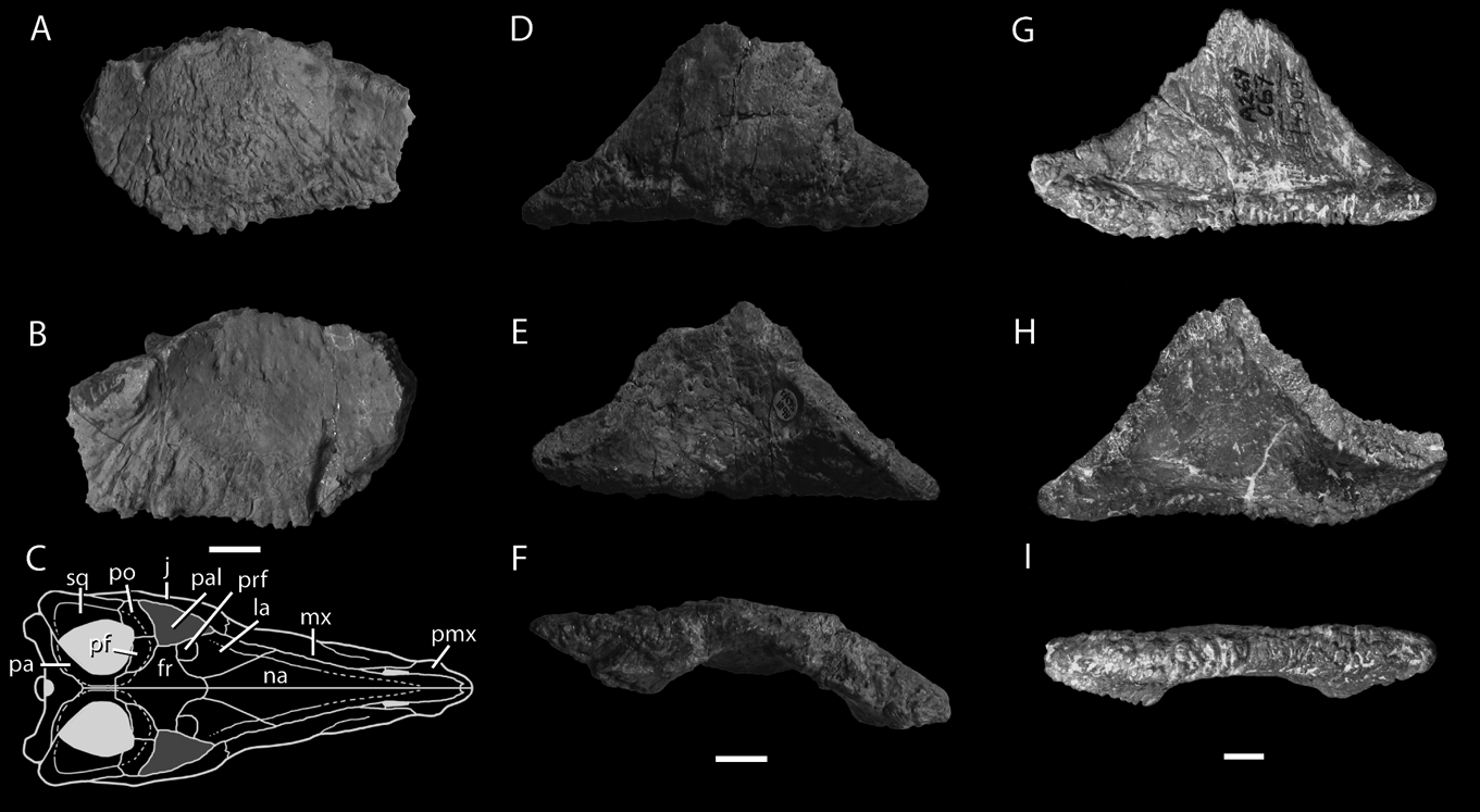
The external surface of the palpebral of Postosuchus kirkpatricki (TTU-P 9000, 9002, Fig. 4G, H, I) is ornamented with an irregular pattern of striations and interwoven bone fibres penetrated by numerous small foramina – a pattern typical of osteoderms of pseudosuchians and other archosaurs (Hill Reference Hill2005). The irregular ornamentation of the palpebral of P. kirkpatricki contrasts with the relatively smooth unornamented bone texture of the adjacent frontal, postorbital and lacrimal of the same specimen (TTU-P 9000). The dorsal surface is slightly convex, whereas the ventral surface is slightly concave and smooth.
Palpebrals of members of Rauisuchidae (sensu Nesbitt Reference Nesbitt2011): (A–B) the right palpebral element of Postosuchus alisonae (UNC 15575) in dorsal (A) and ventral (B) views; (C) a reconstruction of the dorsal view of the skull of Postosuchus to illustrate the position and connectivity of the palpebral of rauisuchids; (D–F) the left palpebral of Polonosuchus silesiacus (ZPAL Ab III/563) in dorsal (D), ventral (E) and lateral (F) views; (G–I) the left palpebral of Postosuchus (UCMP 140035) in dorsal (G), ventral (H) and lateral (I) views. Scale bars=1 cm. Arrow indicates the anterior direction. Abbreviations: fr=frontal; j=jugal; la=lacrimal; mx=maxilla; na=nasal; pa=parietal; pal=palpebral; pf=postfrontal; pmx=premaxilla; po=postorbital; prf=prefrontal; sq=squamosal.
The morphology and position of the palpebral of P. kirkpatricki is generally similar to the palpebral of the closely related (from Nesbitt Reference Nesbitt2011) early crocodylomorph Hesperosuchus agilis (CM 29894). In Hesperosuchus, one large, circular palpebral dominates the space at the dorsal margin of the orbit (Clark et al. Reference Clark, Sues and Berman2000; Fig. 9a). Even though the palpebral of P. kirkpatricki is triangular in overall shape, a seemingly homologous circular structure delimited by a roughened boundary occupies the middle portion of the element in ventral view (Fig. 4H; see Nesbitt Reference Nesbitt2011, fig. 25d) and this smooth portion of the element is an extension of the orbital fossa on the ventral surface of the frontal. The distinct circular structure in the centre of the osteoderm, visible on both the dorsal and ventral surfaces, is concave ventrally, convex dorsally, and the ventral surface has less sculpturing than the dorsal surface (see Nesbitt Reference Nesbitt2011, fig. 25). Compared to the size of the orbit, the relative size of the circular structure within the palpebral is similar to the size of the palpebral of the early crocodylomorph Hesperosuchus (CM 29894; see below). Thus, we hypothesise it may be a homologous structure. In Hesperosuchus, the palpebral is composed of a single ossification that spans the dorsal margin of the orbit, but it remains unclear how many ossifications compose the palpebral of P. kirkpatricki, as there are no clear sutures within the triangular element.
Unlike those of extant crocodylians and Hesperosuchus agilis (CM 29894), the palpebral of P. kirkpatricki is fully integrated into the supraorbital region in the largest specimens (e.g. TTU-P 9000). In P. kirkpatricki the palpebral bridges this region and attaches to the frontal medially, the postorbital and postfrontal posteromedially, and the prefrontal and lacrimal anteromedially, obscuring the dorsal portion of the orbit from dorsal view. Interdigitating sutures similar to those among skull elements join the palpebral and the circumorbital skull elements. These sutures illustrate that the palpebral and surrounding bones were tightly integrated. The disarticulated palpebral of a specimen referred to P. kirkpatricki (UCMP 140035) by Long & Murry (Reference Long and Murry1995) shows that the sutural surface is speckled with pits and spires of bone, thus indicating that the interdigitating suture is present throughout the dorsoventral length of the element. In this regard, the shape and connectivity of the palpebral of P. kirkpatricki is most similar to the extant crocodylian species Paleosuchus trigonatus (FMNH 81980) and Paleosuchus palpebrosus (FMNH 69871), in comparison with that of Hesperosuchus agilis (CM 29894). In Paleosuchus (Fig. 2D), the palpebral traverses the dorsal portion of the orbit and contacts the circumorbital elements. The greatest difference between the palpebrals of P. kirkpatricki and Paleosuchus is that in the latter taxon, the body of the palpebrals retain some of the dorsoventral arching common to many crocodyliform palpebrals (Fig. 2).
The degree of integration of the palpebral with the surrounding skull elements may be controlled ontogenetically. The largest specimen of P. kirkpatricki (TTU-P 9000) has a palpebral that has a contact surface for the pre- and postfrontals, frontal, lacrimal and postorbital. In addition, the similarly sized, isolated palpebral mentioned above (UCMP 140035), bears sutural surfaces on all sides of the element save the lateral edge, thus demonstrating that the holotype and UCMP 140035 had a fully integrated palpebral at death. However, in smaller individuals of P. kirkpatricki, the palpebral is not fully integrated into all the dorsal circumorbital elements. For example, the paratype of P. kirkpatricki (TTU-P9002) is about 75% the size of the holotype and the palpebral is firmly attached to the frontal and the postfrontal, but has little contact with the postorbital. Furthermore, even though the lacrimal and the prefrontal are not completely preserved, the anteromedial edge of the palpebral is largely rounded and does not exhibit an obvious contact surface with either element. Additionally, a similarly sized frontal–postfrontal (UCMP 27480) of a referred specimen of P. kirkpatricki shows a sutural contact for the attachment of the palpebral on the lateral side of the frontal, but not on the lateral side of the postfrontal. This pattern of the development of more sutural contacts between the palpebral and surrounding bones in larger individuals is consistent with an ontogenetic trajectory but, at this point, little data is supporting this notion and, additionally, little is known about individual variation in P. kirkpatricki.
Among other rauisuchids, similar elements to what we interpret as a palpebral were found disassociated from the skulls in the holotypes of Postosuchus alisonae (UNC 15575) and Polonosuchus silesiacus (ZPAL Ab III 563). Originally, Peyer et al. (Reference Peyer, Carter, Sues, Novak and Olsen2008) interpreted the element in Postosuchus alisonae as a frontal, whereas Sulej (Reference Sulej2005) interpreted the element as a prefrontal, following Chatterjee (Reference Chatterjee1985) for Postosuchus kirkpatricki. However, the elements in both Postosuchus alisonae and Polonosuchus silesiacus share a nearly identical morphology to that of the palpebral of Postosuchus kirkpatricki (TTU-P 9000); all are concave ventrally and convex dorsally, have a triangular shape in dorsal view, a rugose external surface, and articulation surfaces for skull elements on the anteromedial and posteromedial sides (Fig. 4). Therefore, we reinterpret these elements as homologous with the palpebral of Postosuchus kirkpatricki. The palpebrals of Postosuchus alisonae and Polonosuchus silesiacus do show variation from each other and with Postosuchus kirkpatricki. For example the palpebral of Polonosuchus silesiacus is dorsally arched at its centre relative to the other taxa, and the palpebral of Postosuchus alisonae has relatively deeper sculpturing on the lateral edge of the ventral surface compared to the other taxa (Fig. 4). Furthermore, in both Postosuchus alisonae and Polonosuchus silesiacus, the circular element in the middle of the palpebral described above for Postosuchus kirkpatricki is more prominent and distinct relative to the surrounding portions of the element.
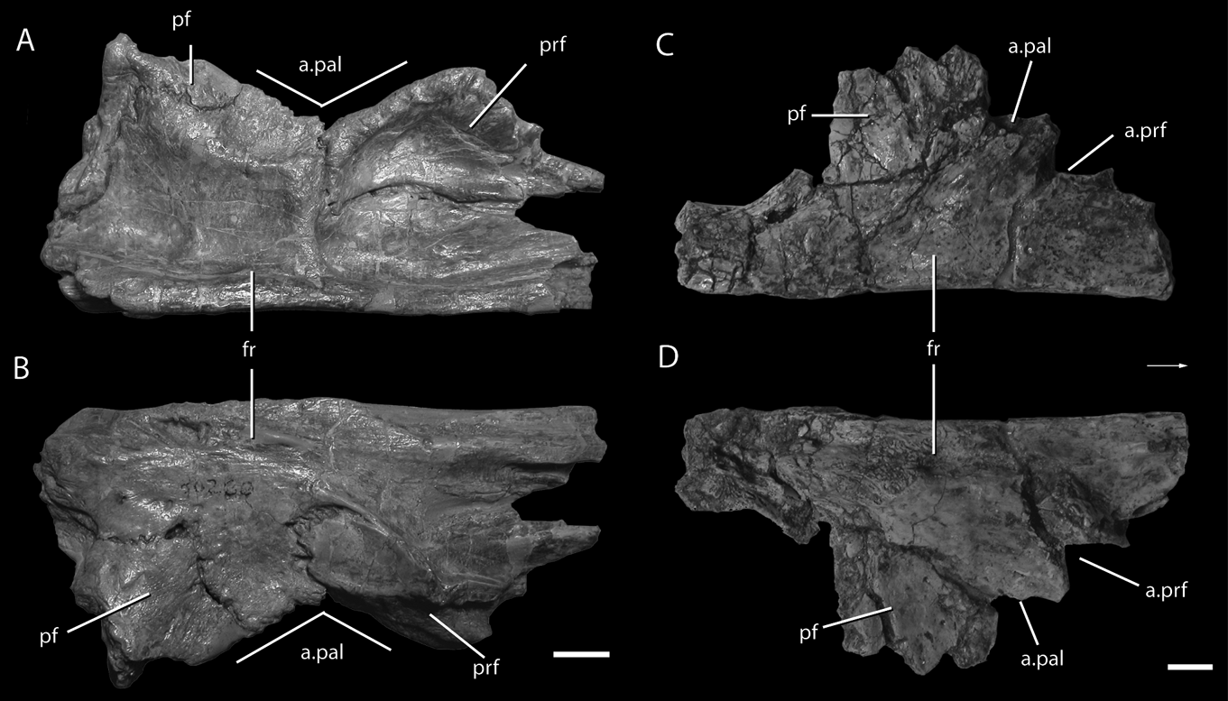
Batrachotomus kupferzellensis, a loricatan pseudosuchian just outside Rauisuchidae and Crocodylomorpha, is represented by at least three partial, well preserved skeletons including mostly disarticulated skull material (Gower Reference Gower1999, Reference Gower2002; Gower & Schoch Reference Gower and Schoch2009). A separate palpebral has not been identified among the remains of B. kupferzellensis; however, a prefrontal–postfrontal–frontal complex of a paratype of B. kupferzellensis (SMNS 80260) preserves evidence of the attachment of the palpebral similar to that on the holotype of Postosuchus kirkpatricki (TTU-P 9000), suggesting that B. kupferzellensis likely had a palpebral attached to the dorsal circumorbital bones. The complete frontal with articulated pre- and postfrontals (Fig. 5) bears a dorsoventrally thick lateral margin that consists of small spires of bone and pits, like that of the medial articulation surface of the isolated palpebral of P. kirkpatricki (UCMP 140035) and the exposed lateral margin of the frontal, postfrontal and prefrontal of P. kirkpatricki (TTU-P 9000). The frontal of B. kupferzellensis, like that of P. kirkpatricki, has little contribution to the orbit, unlike that of paracrocodylomorphs such as Arizonasaurus babbitti (Nesbitt Reference Nesbitt2005). Additionally, the lateral margin of the frontal and postfrontal is straight anterolaterally and the lateral margin of the prefrontal is straight posterolaterally (Fig. 5), a pattern present in P. kirkpatricki (TTU-P 9002). These straight lateral margins of B. kupferzellensis and the morphology of the lateral surfaces are consistent with an articulation surface for a palpebral, as in rauisuchids.
The frontals of the non-crocodylomorph loricatans Batrachotomus kupferzellensis and Postosuchus kirkpatricki: (A–B) the prefrontal, postfrontal and frontal of Batrachotomus kupferzellensis (SMNS 90260) in dorsal (A) and ventral (B) views; (C–D) the frontal and postfrontal of the holotype of Postosuchus kirkpatricki (TTU-P 9000) in dorsal (C) and ventral (D) views. The similarities between the lateral margins of the frontals and the arrangement of the pre- and postfrontals of Postosuchus and Batrachotomus suggest that Batrachotomus may have also had a palpebral integrated into the skull roof. Scale bars=1 cm. Arrow indicates the anterior direction. Abbreviations: a.=articulates with; fr=frontal; pal=palpebral; pf=postfrontal; prf=prefrontal.
1.3. Saurosuchus galilei
Saurosuchus galilei is a large (∼6 m) early loricatan from the late Carnian Ischigualasto Formation of Argentina (Reig Reference Reig1959; Sill Reference Sill1974; Alcober Reference Alcober2000; Trotteyn et al. Reference Trotteyn, Desojo and Alcober2011). The taxon is known from a variety of specimens, including a partial skull (PVL 2062 holotype) and a nearly complete, well-preserved skull (PVSJ 32). Both skulls bear a unique dorsal margin of the orbit and seemingly unique arrangement of circumorbital elements that was discussed by both Sill (Reference Sill1974) and Alcober (Reference Alcober2000) in their diagnoses of the species. Sill (Reference Sill1974) termed the unusual dorsal margin of the orbit as the “orbital arch” and considered that the frontal composed the entire “orbital arch”. Furthermore, Sill (Reference Sill1974) concluded that the pre- and postfrontals were small relative to the frontal. In the description of PVSJ 32, Alcober (Reference Alcober2000) paid particular attention to skull roof elements, but largely followed the interpretation of Sill (Reference Sill1974) in identifying the frontal as the thick element overhanging the dorsal margin of the orbit. Three (numbers 7–9) of the seven autapomorphies of Saurosuchus galilei identified by Alcober (Reference Alcober2000) pertain to the features of the frontal and the arrangement of the frontal to other skull roof elements. In Alcober's (Reference Alcober2000) interpretation, the frontal of S. galilei forms the thick lateral margin of the orbit, excludes the postfrontal from the orbital margin, contacts the postorbital, and has a unique posterolateral projection.
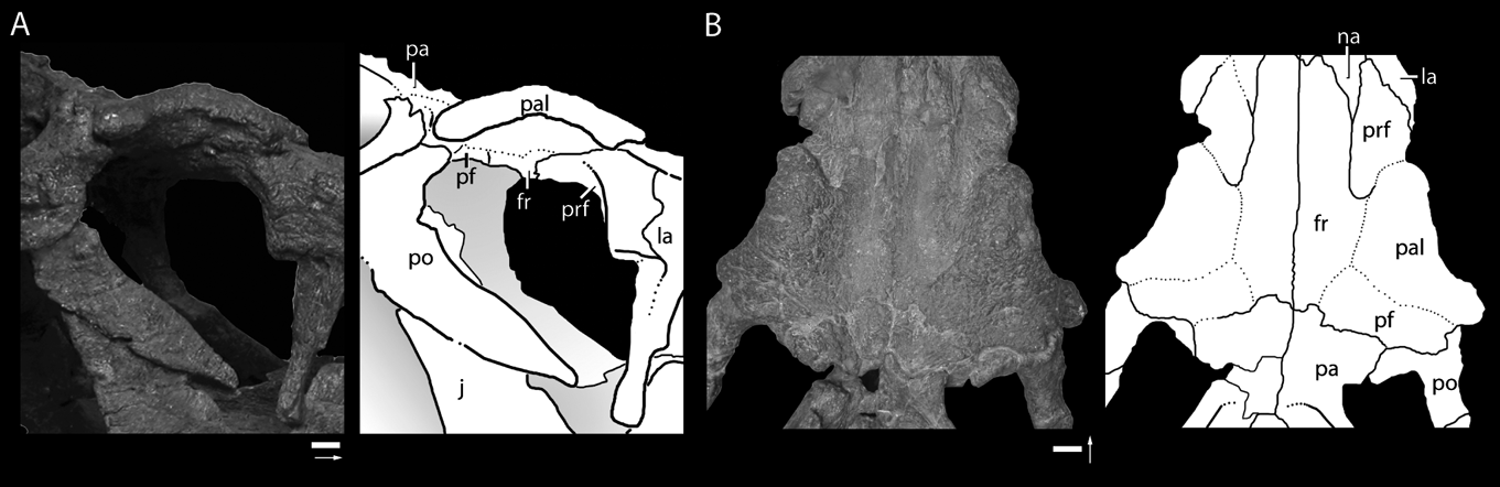
Here, we reinterpret the element at the dorsal portion of the orbit as a palpebral, largely following the re-interpretation of and arguments given for Postosuchus kirkpatricki and closely related taxa (Fig. 6). The separate ossification is almost fully integrated into the skull roof, even more so than in P. kirkpatricki. Like that of P. kirkpatricki (TTU-P 9000) and crocodyliforms, a rugose surface with small foramina characterises the dorsal surface of the palpebral element of S. galilei. This ornamentation, similar to osteoderms in the trunk region (Trottyen et al. 2011), is distinct relative to the rest of the skull table and is utilised to determine the extent of the palpebral, given that sutures on the dorsal surface of the skull roof cannot be readily traced. We outline the extent of the palpebral where the ornamentation of the palpebral meets the less rugose, nearly foramina-free smooth ornamentation of the frontal, prefrontal and postfrontal (Fig. 6). The partially visible sutures present on the ventral surface of skull roof elements (Fig. 6) further support our interpretation of the extent of the palpebral in dorsal view.
The palpebrals of Saurosuchus galilei following our reinterpretation of the identification of the skull roof elements: the right orbit of Saurosuchus galilei (PVSJ 32) in lateral view (A) and a dorsal view (B) of the skull roof elements. Scale bars=1 cm. Arrow indicates the anterior direction. Abbreviations: fr=frontal; j=jugal; la=lacrimal; na=nasal; pa=parietal; pal=palpebral; pf=postfrontal; po=postorbital; prf=prefrontal.
The palpebral of S. galilei is triangular, like that of P. kirkpatricki (TTU-P 9000), but is concave laterally instead of anteroposteriorly straight. The thickened lateral margin of the palpebral arcs dorsally, as does the middle portion, thus resulting in an expansion dorsal to all other skull roof elements. The concave lateral margin is also thickened relative to the other portions of the palpebral. These features of S. galilei differ from P. kirkpatricki (TTU-P 9000), in which the lateral margin of the palpebral is nearly straight and similar in thickness to the middle portion. Only one ossification appears to compose the palpebral of S. galilei, although sutures are difficult to observe on both the dorsal and ventral sides.
The palpebral of S. galilei articulates with the frontal medially, the prefrontal anteromedially, the postfrontal posteromedially, and likely the postorbital posterolaterally. The anterior end of the palpebral terminates in a rounded boss, displaced dorsal to the articulation with the prefrontal. A tongue of the prefrontal lies on the ventromedial surface of the palpebral and, dorsally, the lateral side of the frontal and the anteromedial side of the palpebral constrict the posterior process of the prefrontal. The frontal–palpebral articulation of S. galilei is similar to that of P. kirkpatricki (TTU-P 9000) in which the lateral edge of the frontal is constricted by the prefrontal anteriorly and the postfrontal posteriorly. The lateral edge of the frontal appears to be curving dorsally where the two elements meet. Posteriorly, the palpebral and the postfrontal meet in a mediolaterally oriented contact. Posterolaterally, the palpebral terminates in a rounded, ventrally directed point, just lateral and anterior to its contact with the postorbital. The exact sutural surface between the palpebral and surrounding elements cannot be determined.
The palpebral of S. galilei is unique among loricatans in that the element is completely integrated into the skull roof, with the obliteration of most sutures, and in the dorsal arcing of the element, the thickness of the element, and the presence of an anterolateral boss that lies dorsal to the prefrontal. Without the integrated palpebral, the arrangement of the prefrontal, frontal and the postfrontal would be similar to that of other pseudosuchians, where all three elements form a portion of the orbit. Therefore, of the original autapomorphies cited by Alcober (Reference Alcober2000) that concerned the lateral margin of the orbit, autapomorphies eight and nine (Alcober Reference Alcober2000, p. 304) pertain to the palpebral and not the frontal.
1.4. Poposauroidea
Recent contributions on the relationships of the traditionally termed group “rauisuchians,” (including rauisuchids, and/or poposaurs) have identified a distinct and morphologically diverse group of pseudosuchians now referred to as poposauroids (Gower Reference Gower2000; Nesbitt Reference Nesbitt2003, Reference Nesbitt2005, Reference Nesbitt2007, Reference Nesbitt2011; Weinbaum & Hungerbühler Reference Weinbaum and Hungerbühler2007; Brusatte et al. Reference Brusatte, Benton, Desojo and Langer2010; Butler et al. Reference Butler, Brusatte, Reich, Nesbitt, Schoch and Hornung2011). This varied group of largely osteoderm-free pseudosuchians includes the sail-backed ctenosauriscids (Nesbitt Reference Nesbitt2003; Butler et al. Reference Butler, Brusatte, Reich, Nesbitt, Schoch and Hornung2011), the poorly understood taxon Poposaurus gracilis (Mehl Reference Mehl1915; Colbert Reference Colbert1961; Weinbaum & Hungerbühler Reference Weinbaum and Hungerbühler2007; Gauthier et al. Reference Gauthier, Nesbitt, Schachner, Bever and Joyce2011; Schachner et al. Reference Schachner, Manning and Dodson2011), the sail-backed and edentulous Lotosaurus edentus (Zhang Reference Zhang1975) and the bipedal and edentulous shuvosaurids (Nesbitt & Norell Reference Nesbitt and Norell2006; Nesbitt Reference Nesbitt2007). Only recently has there been unequivocal cranial material assigned to any poposauroid taxa (Nesbitt Reference Nesbitt2003, Reference Nesbitt2007; Li et al. Reference Li, Wu, Cheng, Sato and Wang2006; Gauthier et al. Reference Gauthier, Nesbitt, Schachner, Bever and Joyce2011) and of these specimens, the skulls of the shuvosaurid Effigia okeeffeae (Nesbitt & Norell Reference Nesbitt and Norell2006) and the early poposauroid Qianosuchus mixtus (Li et al. Reference Li, Wu, Cheng, Sato and Wang2006) bear skeletal elements in the orbit.
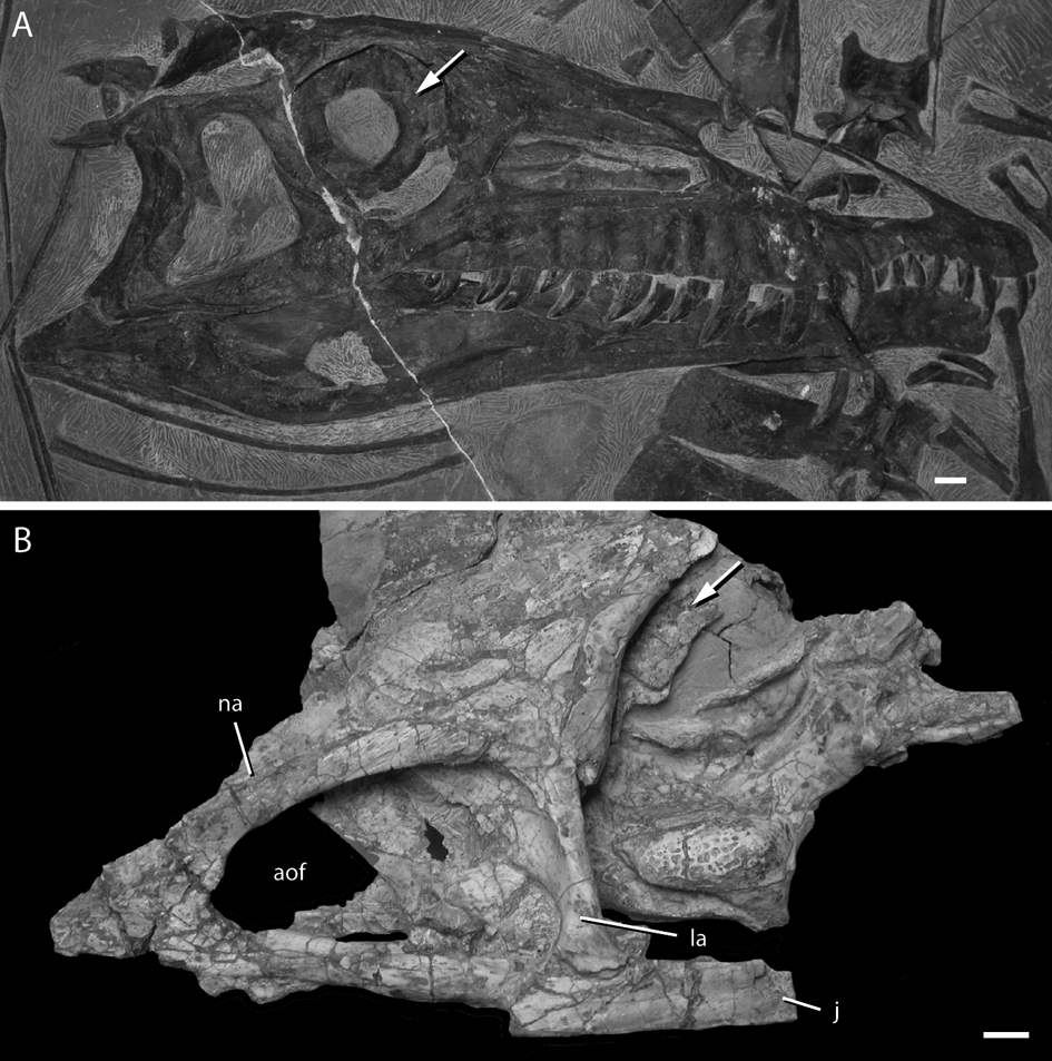
The large orbits of both specimens of E. okeeffeae (Fig. 7B; AMNH FR 30587 and 30589) have a partially preserved sclerotic ring composed of thin osseous plates (Nesbitt Reference Nesbitt2007). Each scleral ossicle has a low keel and the shape of the ossicles varies from oval in AMNH FR 30587 to rectangular in AMNH FR 30589. The portion of the sclerotic ring of AMNH FR 30589 that remains in life position, and the width of each scleral ossicle, indicates that the ring has only a slightly smaller radius than that of the orbit and the ring itself occupied much of the orbit. Likewise, the paratype skull of Q. mixtus (Fig. 7A; IVPP V14300) preserves a sclerotic ring in the right orbit (Li et al. Reference Li, Wu, Cheng, Sato and Wang2006). The sclerotic ring is fully preserved and remains in life position. The sclerotic ring consists of an unknown number of scleral ossicles, but has a similar width and ratio of the radius of the sclerotic ring relative to that of the radius of the orbit as E. okeeffeae.
Sclerotic rings in the orbits of poposauroids: a complete sclerotic ring in the paratype of Qianosuchus mixtus (IVPP V 14300) (A) compared with the partial sclerotic ring in the left orbit of the paratype of Effigia okeeffeae (AMNH FR 30589) (B). Scale bars=1 cm. White arrows highlight the skeletal elements in the orbit. Abbreviations: aof=antorbital fenestra; j=jugal; la=lacrimal; na=nasal.
E. okeeffeae and Q. mixtus represent the only poposauroids with well-preserved skulls and each preserve a sclerotic ring. Furthermore, no palpebral-like elements are present in any poposauroid. Although E. okeeffeae and nearly all other poposauroids lack osteoderms (Nesbitt Reference Nesbitt2003, Reference Nesbitt2005, Reference Nesbitt2007, Reference Nesbitt2011), small osteoderms are present along the vertebral column of Q. mixtus (Li et al. Reference Li, Wu, Cheng, Sato and Wang2006).
1.5. Aetosauria
Aetosaurs were a group of herbivorous or omnivorous pseudosuchians, covered in a dense carapace composed of osteoderms, that had a global distribution during the Late Triassic (e.g. Long & Ballew Reference Long and Ballew1985; Heckert & Lucas Reference Heckert and Lucas2000; Small Reference Small1998). Aetosaurs are one of a few groups of pseudosuchian archosaurs that are currently known to possess skeletal elements within the orbit. The element(s) have been referred to supraorbitals (Walker Reference Walker1961) and palpebrals (Schoch Reference Schoch2007) for Aetosaurus ferratus and a palpebral/supraorbital in Neoaetosauroides engaeus (Desojo & Baez Reference Desojo and Baez2007). Furthermore, a new, undescribed “Aetosaurus-like” taxon from the Chinle Formation of Colorado also possesses structures like those of Aetosaurus ferratus (B. Small, pers. comm. to JCW 2011). Thus far, aetosaurs have not been found to possess any scleral ossicles.
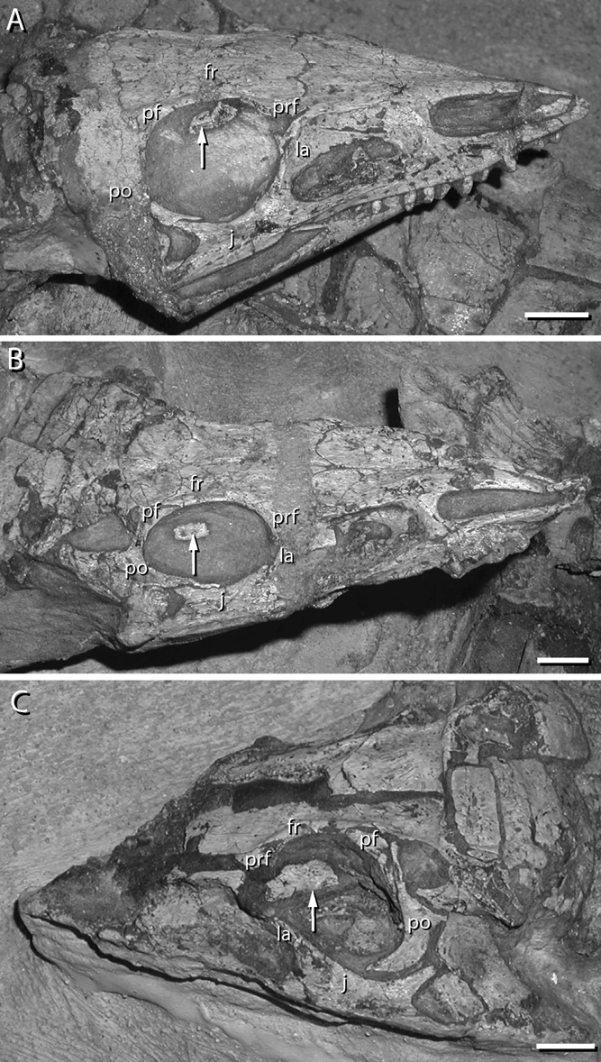
Aetosaurus ferratus is represented by at least 22 nearly complete specimens, including intact skulls and orbital regions (Fraas Reference Fraas1877; Walker Reference Walker1961; Schoch Reference Schoch2007). The skeletal elements in the orbit were first identified by Walker (Reference Walker1961) and then further described by Schoch (Reference Schoch2007). Here, we supplement their descriptions. In A. ferratus (Fig. 8), three small, bony elements lie at the dorsal portion of the orbit and stretch from just posterior to the prefrontal to just anterior of the postfrontal and postorbital in an anteroposteriorly oriented row (Walker Reference Walker1961; Schoch Reference Schoch2007). In most cases (see below), the three elements articulate with each other at interdigitating sutures, but do not have a contact with the dermal bones of the skull. The elements are mediolaterally thin and bear a rugose pattern on the lateral side, like that of the sculpturing of the external surface of the skull table. Additionally, the centre of each element has a weakly developed anteroposterior trending ridge. These elements likely represent palpebrals homologous with those of crocodylians, but the identification of these elements remains uncertain (see below).
Skulls of Aetosaurus ferratus (SMNS 5770) with skeletal elements in the orbit: Aetosaurus ferratus number XVI (A) in right lateral view, VIII (B) in right lateral view, and I (C) in left dorsolateral view. The skulls in (A) and (C) preserve two elements inn the dorsal portion of the orbit whereas (B) preserves only one element. Schoch (Reference Schoch2007) reported that Aetosaurus ferratus had up to three elements in the dorsal portion of the orbit. Scale bars=1 cm. White arrows highlight the skeletal elements in the orbit. Abbreviations: fr=frontal; j = jugal; la = lacrimal; pf = postfrontal; po = postorbital; prf = prefrontal.
The skeletal elements in the orbit of A. ferratus show variation in the following ways: (1) connectivity with each other; (2) relative sizes of the elements; and (3) possibly the number of elements. In some specimens (e.g. SMNS 5770 S-18), the elements meet each other at interdigitating contacts, whereas in other specimens (e.g. SMNS 5770 S-7, S-16) the elements fail to meet. The three elements of Aetosaurus are relatively close in size; however, the anteriormost element is 10–20% longer than the middle and posterior element in several specimens (e.g. SMNS 5770 S-16, S-18). The size of the anteriormost element of Aetosaurus is variable; the anterior element is never more than approximately one and a half times the length of the other palpebral elements combined. The number of skeletal elements in the orbit of A. ferratus varies from one (SMNS 5770 S-8) to two (SMNS 5770 S-16) or three (SMNS 5770 S-18, S-7). The number of elements can differ in each orbit in the same individual. For example, in SMNS 5770 S-18, there are two elements in the right orbit, whereas there are three elements in the left orbit (see Schoch Reference Schoch2007, fig. 7). Nevertheless, it is not clear if the number of elements in the orbit represents variation within A. ferratus, or if it is a product of slight disarticulation and loss, the absence of complete preparation, or the removal during preparation. Given that all are about the same size and thus ontogenetic stage, this variation in the aforementioned features may represent variation with a single population.
A single skeletal orbital element was recently described for Neoaetosauroides engaeus (Desojo & Baez Reference Desojo and Baez2007) on both sides of PVL 5698. The element consists of a rounded, osseous element in the dorsal portion of the orbit near the prefrontal. The skeletal element within the orbit does not contact the prefrontal or any other skull roof element (Desojo & Baez Reference Desojo and Baez2007). The bone may be homologous to the anteriormost palpebral within the orbit of Aetosaurus, but the homology within Aetosauria is unclear. Details of the thickness and surface sculpturing cannot be determined as the result of poor preservation.
As hypothesised by Schoch (Reference Schoch2007) in his thorough review of A. ferratus, the elements within the orbit are unlikely to be components of the sclerotic ring because of the presence of sutures between the elements instead of a simple overlap and the morphology of the anterior element, which is elongate and tapers to a point anteriorly. Furthermore, in all specimens the elements fail to form a ring, as they do in all well preserved taxa with scleral ossicles. Therefore, it appears that no member of Aetosauria possess sclerotic ossicles. The assignment of the elements in the orbit of aetosaurs to a palpebral (homologous with those of crocodylians) or supraorbital bones (neomorphic bones with a similar developmental history of dermal bones) remains unclear. On one hand, the elements appear to have formed in the upper eyelid, as evidenced by the dorsal position within the orbit, which is similar to extant crocodylians. Moreover, the upper eyelid appears to have been completely mobile, given that the ossicles are not articulating with other skull bones. The external texture is similar to those of osteoderms and may have shared a similar developmental history, and the interconnection of the elements is similar to that of Osteolaemus (Fig. 2). On the other hand, the number of elements, shape of those elements, and lack of contact with the prefrontal is different from those of early loricatans and early crocodylomorphs. With that said, we currently interpret these elements as palpebrals, but with the differences between crocodylomorphs and aetosaurs noted (see discussion).
1.6. Extinct crocodylomorphs
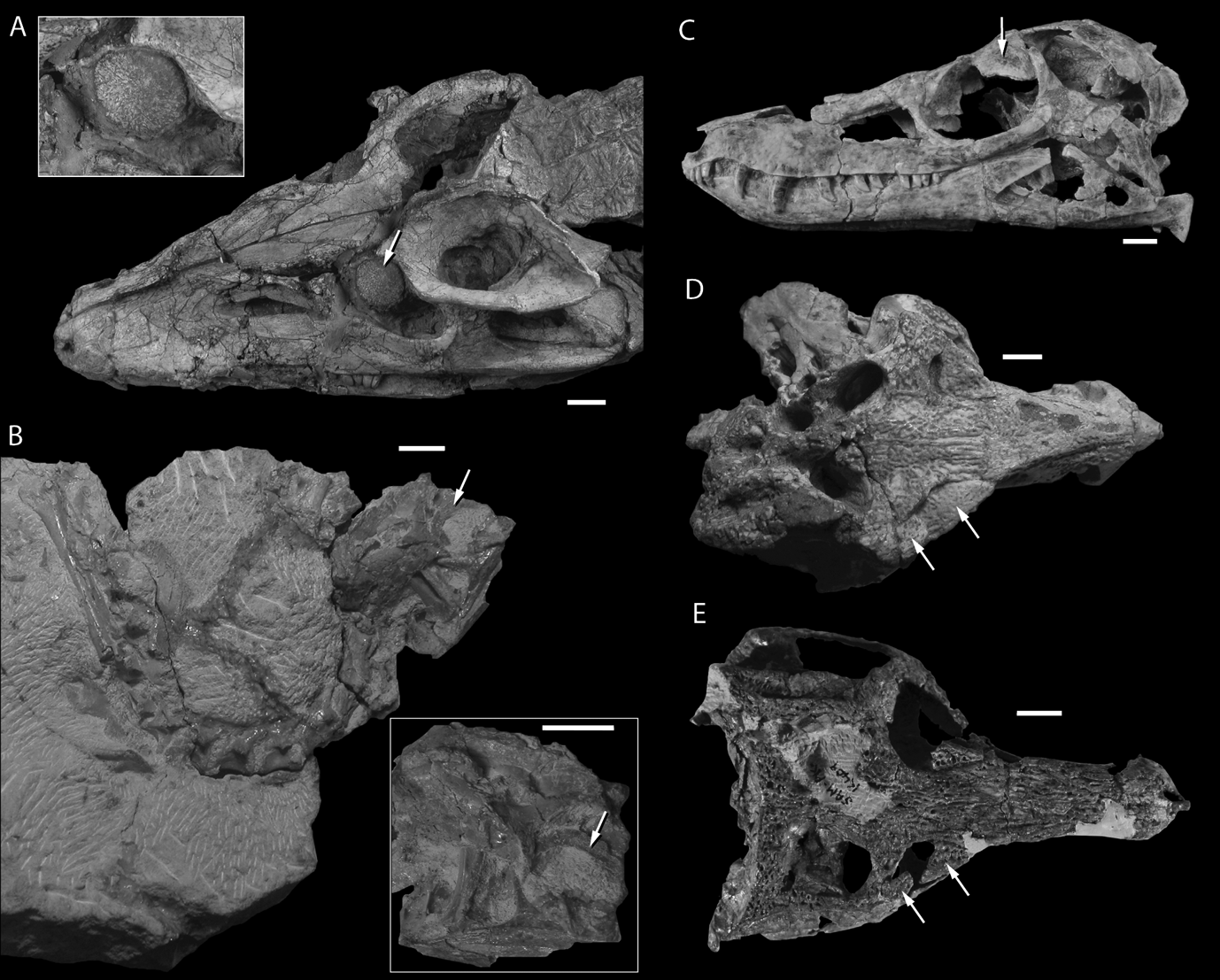
Early crocodylomorphs from either the Triassic or the Jurassic remain rare components of faunal assemblages. Skulls of crocodylomorphs have been particularly difficult to study, given the fragmentary condition (e.g. Hesperosuchus agilis, AMNH FR 6758) or poor preservation (Terrestrisuchus gracilis, Crush Reference Crush1984). Fortunately, a series of recent discoveries of well-preserved skulls of early crocodylomorphs have revolutionised our understanding of crocodylomorph systematics and our understanding of their palaeobiology (Clark et al. Reference Clark, Sues and Berman2000, Reference Clark, Xing, Forster and Wang2004; Clark & Sues Reference Clark and Sues2002; Sues et al. Reference Sues, Olsen, Carter and Scott2003). A specimen referred to Hesperosuchus agilis (CM 29894) preserves the earliest record of an unequivocal palpebral among crocodylomorphs (Clark et al. Reference Clark, Sues and Berman2000). The single, sub-circular element lies in the dorsal portion of both orbits (Clark et al. Reference Clark, Sues and Berman2000). The edges of the palpebral (Fig. 9A) are slightly serrated, consisting of small bone spicules radiating from the centre. The surface of the palpebral consists of small grooves and ridges similar to the ornamentation of osteoderms on the neck and back of the same specimen. The dorsal surface of the palpebral is convex dorsally. Within the orbit, the palpebral extends laterally beyond the orbital margin and lies just ventral to lateral margin of the frontal, without contacting the bone. Additionally, the palpebral covers much of the dorsal margin of the orbit. Of the known Triassic crocodylomorphs, CM 29894 is the only specimen with a palpebral preserved in the orbit. A closely related taxon, Dromicosuchus grallator (Sues et al. Reference Sues, Olsen, Carter and Scott2003) is known from a complete skull and partial skeleton, but there does not appear to be any palpebral.
Skeletal elements in the orbits of early crocodylomorphs: (A) a skull referred to Hesperosuchus agilis by Clark et al. (Reference Clark, Sues and Berman2000) in dorsolateral view, with a close up (inset) of the palpebral in articulation; (B) partial skull and articulated skeleton of Terrestrisuchus gracilis (NHMUK R7591 b) in right lateral view with a close up (inset) of the partially preserved sclerotic ring in articulation; (C) the skull of Junggarsuchus sloani (IVPP V 14010) in left lateral view with a single palpebral; (D) a nearly complete skull of Protosuchus richardsoni (MCZ 6727) in dorsal view exhibiting two large palpebrals; (E) the holotype skull of Orthosuchus strombergi (SAM-K-409) in dorsal view, bearing two palpebrals. Scale bars=1 cm. White arrows highlight the skeletal elements in the orbit.
Terrestrisuchus gracilis is an uncharacteristically gracile crocodylomorph from the Triassic–Jurassic fissure fills of the UK, known from partially articulated and disarticulated remains (Crush Reference Crush1984). Of the skeletal material preserved, only the posterior half of one skull remains in articulation (Fig. 9B; NHMUK R7591b). The skull is crushed ventrolaterally, but still preserves an intact posterior, ventral and partial dorsal orbital margin. A partial sclerotic ring lies within the dorsal portion of the orbit of the T. gracilis. A minimum of five rectangular sclerotic ossicles composes the partial ring. The thin ossicles are slightly concave along the long axis of the elements, but it is not clear if this concavity is the result of crushing. Unfortunately, part of the dorsal and the entire anterior portions of the orbit are incompletely preserved, so it is not clear if a palpebral was also present.
Junggarsuchus sloani (Fig. 9C), a close relative of Crocodyliformes, (Clark et al. Reference Clark, Xing, Forster and Wang2004) was found also to have a palpebral composed of one ossification in near life position. Generally, the palpebral is similar to that of CM 29894 in size, surface features and location in the orbit. The palpebral is distinctly concave ventrally and has a smooth lateral margin. The contact with elements of the skull roof is not clear. The anterior portion of the palpebral appears not to touch the prefrontal and the medial edge of the palpebral appears to be partially attached to the lateral side of the frontal. A sutural contact between the elements is not present. During prepartation of the orbit, a series of scleral ossicles preserved in partial sclerotic ring were discovered in the right orbit (J. Clark pers. comm. Reference Clark2012). The scleral ossicles are longer than tall and the proportions are similar to those of Terrestrisuchus gracilis. Junggarsuchus sloani represents the only confirmed pseudosuchian with both a sclerotic ring and a palpebral.
Palpebrals are likely plesiomorphic for Crocodyliformes, given their presence in a number of crocodylomorph outgroups (as discussed above) and in the most plesiomorphic members of the clade (e.g. protosuchids, gobiosuchids, shartegosuchids), according to our understanding of crocodylomorph relationships (e.g. Clark Reference Clark1986; Brochu Reference Brochu2001; Pol et al. Reference Pol, Turner and Norell2009). A major shift in number and morphology of the palpebrals occurs at Crocodyliformes relative to their proximal outgroups. Instead of a single rounded palpebral, two large similarly sized palpebrals occupy much of the area dorsal to the orbital cavity.
Protosuchus richardsoni (Fig. 9C), one of the earliest diverging taxa in Crocodyliformes, is known from a number of partial to complete skeletons from the Early Jurassic Moenave Formation of Arizona (Brown Reference Brown1933; Colbert & Mook Reference Colbert and Mook1951; Crompton & Smith Reference Crompton, Smith and Jacobs1980). Although not described by Colbert and Mook (Reference Colbert and Mook1951), two palpebrals are present at the dorsal portion of orbit in the holotype (AMNH FARB 3024). An anterior palpebral articulates with the dorsal surface of the prefrontal, whereas a more posterior palpebral articulates with the dorsal surface of the postorbital. A gap is present between the two palpebrals and between the palpebrals and the lateral margin of the orbit (Colbert & Mook Reference Colbert and Mook1951, plate 12). A better-preserved specimen of Protosuchus richardsoni (MCZ 5727) exhibits a similar pattern (Fig. 9C) as the holotype. However, in MCZ 6727, the palpebrals meet and nearly hide the orbit in dorsal view. In MCZ 6727, the palpebrals contact the prefrontal, frontal and the postorbital, but do not suture to those elements. The anterior palpebral is triangular in dorsal view where it tapers to acute angles anteriorly and laterally. The posterior palpebral meets the anterior palpebral in a slightly interdigitating suture, like that of Osteolaemus (Fig. 2) and Simosuchus clarki (UA 8679). This contact is more well developed on the right side compared to the left, where a medial gap still persists between the two elements. The much smaller posterior palpebral is sub-rounded and occupies the posterior third of the dorsal margin of the orbit. Both palpebrals are slightly convex in dorsal view, appear to be concave in ventral view, and are uniformly thick throughout the length of the elements. The dorsal ornamentation of the palpebrals consists of small grooves and ridges, an identical ornamentation to the rest of the skull roof elements.
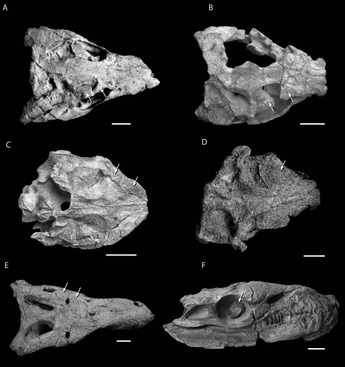
The palpebrals of Protosuchus richardsoni are typical for other protosuchids (e.g. Fig. 9D, Orthosuchus strombergi, SAM-K-409) and many other crocodyliforms (e.g. Fig. 10A, Sichuanosuchus shushanensis, IVPP V10594; Fig. 10BZosuchus davidsoni, IGM 100/1305; Stegomosuchus longipes, Walker Reference Walker1968). However, a number of other early crocodyliforms deviate from this pattern. The Late Cretaceous gobiosuchid Zaraasuchus shepardi (Fig. 10C; Pol & Norell Reference Pol and Norell2004; IGM 100/1321) has anterior and posterior palpebrals that tightly suture to each other and to the margins of the prefrontal, frontal and postorbital. Along the lateral margin of the orbit, the posterior palpebral sends a small anterior projection to lap against the anterior palpebral, as well as a posterior projection that tracks along much of the anterolateral margin of the postorbital. This degree of palpebral development is most similar to what is seen on the right side of the MCZ 6727 specimen of Protosuchus richardsoni, suggesting that multiple levels of variation (i.e., individual, ontogenetic and/or phylogenetic) may exist in the palpebrals of early crocodyliforms.
(A–E) Extinct crocodyliforms with articulated palpebral(s) in dorsal view: (A) Sichuanosuchus shushanensis (IVPP V10594); (B) Zosuchus davidsoni (IGM 100/1305); (C) Zaraasuchus shepardi (IGM 100/1321); (D) Nominosuchus matutinus (PIN 4147-3); (E) Baurusuchus salgadoensis (MPMA 62-0001-02). (F) The thalattosuchian Dakosaurus andiniensis (MOZ 6146P) in right lateral view, illustrating the presence of a complete sclerotic ring and the absence of a palpebral. Scale bars=1 cm (A–D); 5 cm (E–F). White arrows highlight the skeletal elements in the orbit.
Most mesoeucrocodylians share a similar palpebral morphology to early crocodyliforms, in that there are two palpebrals in the orbit and the anterior palpebral is dominant. Palpebral variation among mesoeucrocodylians typically takes three forms: presence or absence of palpebrals; whether or not the anterior palpebral contacts the posterior one; and whether the anterior palpebral tracks along the orbital margin, or if the medial margin is posteriorly concave, thereby creating a fenestra dorsal to the orbit (see Sertich Reference Sertich2011).
The shartegosuchid Fruitachampsa callisoni from the Upper Jurassic Morrison Formation (Clark Reference Clark2012) has a proportionally large anterior palpebral with a greatly concave medial margin forming the stereotypic “comma” shape common to many mesoeucrocodylian crocodyliforms. This anterior palpebral shape results in a fenestra dorsal to the orbit formed by the gap between the frontal and the anterior palpebral. The posterior palpebral is very small and does not contact the anterior palpebral in Fruitachampsa. The early mesoeucrocodylian (and probably notosuchian) taxa Araripesuchus tsangatsangana, A. gomesii, A. patagonicus, Malawisuchus mwakasyungutiensis and Pakasuchus kapilimai have a similar “comma”-shaped anterior palpebral. In these, as in Fruitachampsa, the posterior palpebral is small and does not contact the anterior palpebral.
Asian shartegosuchids, an enigmatic clade of crocodyliforms near the origin of Mesoeucrocodylia (Fiorelli & Calvo Reference Fiorelli and Calvo2007), may have only a single large anterior palpebral. The holotype of Shartegosuchus asperopalatum (PIN 4171/2) has two preserved in left orbit, but it is not clear if they are both from left side. The Nominosuchus matutinus holotype (PIN 4147-3) is one of the better preserved specimens, and no posterior palpebral is evident in this individual. The anterior palpebral is proportionally large, being wider than the interorbital width of the frontal. The palpebral is semicircular in dorsal view, with a straight margin facing laterally and a curved medial margin abutting the lateral surface of the frontal for most of its length. However, the entire medial palpebral margin is not sutured to the frontal; this leaves a gap between the frontal and palpebral, approximately halfway along the interorbital region, and a large posterior gap between the palpebral and postorbital. The orbit in the holotype of Adzhosuchus fuscus (PIN 4174-5) is less well preserved compared to Nominosuchus (PIN 4147-3), but it still preserves a single large palpebral that has been displaced ventrally into the orbit. No indication of a posterior palpebral is present. It is possible that the posterior palpebrals in these taxa were very small (as in Fruitachampsa) and/or poorly attached to the postorbital and therefore simply failed to be preserved with the specimens.
In a number of advanced mesoeucrocodylians and notosuchians, the palpebral rests adjacent to the frontal margin for the entire (or nearly the entire) medial margin of the palpebral. This results in little or no fenestra dorsal to the orbit. However, not all taxa that have a close correspondence between the medial palpebral margin and the lateral frontal margin have palpebrals that are tightly integrated or sutured to the orbital margin. For example, the notosuchians Simosuchus clarki (UA 8679) and Armadillosuchus arrudai (Marinho & Carvalho Reference Marinho and Carvalho2009) have a large anterior palpebral that closely follows the orbital margin, but these palpebrals are not tightly sutured to the margin and are easily dissociated during preparation. Likewise, Sebecus icaeorhinus (AMNH FR 3160) has an anterior element that closely corresponds to the dorsolateral portion of the orbit (Simpson Reference Simpson1937; Brown & Schlaikjer Reference Brown and Schlaikjer1940; Colbert Reference Colbert1946). However, these anterior palpebrals are not tightly integrated to the margin and were discovered separated from the orbital margin.
Other advanced mesoeucrocodylians have highly integrated anterior palpebrals that shared tight sutures to the prefrontal and frontal. These taxa include the baurusuchids Baurusuchus salgadoensis (Carvalho et al. Reference Carvalho, Campos and Nobre2005) and Stratiotosuchus maxhechti (DGM 1477-R) as well as the peirosaurids Uberabasuchus terrificus (Carvalho et al. Reference Carvalho, Ribeiro and Avilla2004), Montealtosuchus arrudacamposi (Carvalho et al. Reference Carvalho, Vascocellos and Tavares2007), and Lomasuchus palpebrosus (Gasparini et al. Reference Gasparini, Chiappe and Fernandez1991). Additionally, the peirosaurids have close contact between the anterior and posterior palpebrals and no indication of a dorsal fenestra. In Baurusuchus and Stratiotosuchus, the anterior and posterior palpebral also contact each other, but in Baurusuchus there remains a small dorsal fenestra above the orbit.
Absence of palpebrals is difficult to assess with fossilised specimens (see above) and this is especially problematic given that, for many mesoeucrocodylians, the palpebral would have been a loose element within the eyelid, and therefore easy to lose once the skin and connective tissue rotted. A number of early mesoeucrocodylians and eusuchians do not preserve palpebrals but, based on the presence of palpebral facets on the prefrontal and postorbital, it seems likely that the elements were present during life. These taxa include Araripesuchus wegeneri (Sereno & Larsson Reference Sereno and Larsson2009), Kaprosuchus saharicus (Sereno & Larsson Reference Sereno and Larsson2009), Mahajangasuchus insignis (Turner & Buckley Reference Turner and Buckley2008), Anatosuchus minor (Sereno & Larsson Reference Sereno and Larsson2009), Hamadasuchus rebouli (Larsson & Sues Reference Larsson and Sues2007), Mariliasuchus amarali (Zaher et al. Reference Zaher, Pol, Carvalho, Riccomini, Campos and Nava2006; Andrade & Bertini Reference Andrade and Bertini2008), Shamosuchus djadochtaensis (Pol et al. Reference Pol, Turner and Norell2009), Yacarerani boliviensis (Novas et al. Reference Novas, Pais, Pol, Carvalho, Mones, Scanferla and Riglos2009) and Adamantinasuchus navae (Nobre & Carvalho Reference Nobre and Carvalho2006).
Thalattosuchians and pholidosaurids are two groups of crocodyliforms that do appear to genuinely lack palpebrals. This is supported by the absence of the element in numerous complete and exceptionally well-preserved individuals (e.g. Geosaurus giganteus, NHMUK 37020; Pelagosaurus, BSPG 1973 VII 592; Cricosaurus suevicus, SMNS 90513). Interestingly, thalattosuchians and some pholidosaurids are the only groups of crocodyliforms that preserve sclerotic rings in a number of its constituent species (e.g. Dakosaurus andiniensis, Pol & Gasparini Reference Pol and Gasparini2009). Within Thalattosuchia, a number of specimens of Pelagosaurus (UH 1, BSPG 1973 VII 592; BSPG 1925 I 34), as well as Geosaurus giganteus (NHMUK 37020), Cricosaurus suevicus (SMNS 90513) and Dakosaurus andiniensis (Pol & Gasparini Reference Pol and Gasparini2009), have sclerotic rings that are indistinguishable from other clades with the same structure (e.g. Ichthyosauria, Aves). Furthermore, Wu et al. (Reference Wu, Russell and Cumbaa2001) reported scleral ossicles in the orbit of the pholidosaurid Terminonaris robustus (SMNH P2411.1), whereas there have been no other reports of sclerotic rings in any other pholidosaurid. All of the taxa within Thalattosuchia and Pholidosauridae with sclerotic rings have been found in marine sediments, ranging from the Early Jurassic through the Late Cretaceous (Buffetaut Reference Buffetaut1982). Additionally, these forms have been considered partially or fully marine, with adaptations for a full aquatic lifestyle (e.g. salt glands, Fernandez & Gasparini Reference Fernandez and Gasparini2008; flipper-like forelimbs and hypocercal caudal region (Gasparini et al. Reference Gasparini, Pol and Spalletti2006) and the sclerotic rings may have helped to stabilise the eye under pressure (Curtis & Miller Reference Curtis and Miller1938).
Among most other neosuchians, there is an apparent trend in palpebral reduction. Alligatorum meyeri, Theriosuchus pusillus and Goniopholis simus have only one palpebral in each orbit, but the element remains relatively large. An undescribed early neosuchian from the Cloverly Formation (MCZ 4453) preserves both a large anterior palpebral and a much smaller posterior palpebral that is similar to most other crocodyliforms in both shape and relationship to the orbital margin. Conversely, Goniopholis simus strongly integrates a single palpebral into the orbital margin (Andrade & Hornung Reference Andrade and Hornung2011). In these specimens of G. simus, the palpebral is tightly integrated into the periorbital region, coossifying with the lacrimal, prefrontal, frontal and postorbital (Andrade & Hornung Reference Andrade and Hornung2011). The ornamentation is continuous between the cranial bones and the palpebral, which can make distinguishing the medial border of the palpebral difficult or impossible. Among other goniopholidids, definitive palpebrals are known in Goniopholis baryglyphaeus (MG 2014) and Nannosuchus gracilidens (NHMUK 48217).
Among successive outgroups of Eusuchia, palpebrals are less commonly associated with specimens, but a depression for the articulation of the element persists on the orbital margin of the prefrontal (e.g. Shamosuchus djadochtaensis IGM 100/1195), as with extant crocodylians such as Alligator mississippiensis. Hylaeochampsid eusuchians such as Hylaeochampsa vectiana (NHMUK R177) and Iharkutosuchus makadii (MTM 2006.53.1) do not preserve palpebral ossifications, but it is not clear if these taxa lack palpebrals or if they were lost after death. By the crown-group, taxa that possess a palpebral show that it is very weakly integrated into the orbital margin and are typically small (see discussion of extant crocodylians above). Fossil evidence for palpebrals is rare among extinct members of Crocodylia, but the extinct taxa with palpebrals have a similar morphology and a similar attachment location as those of extant members of Crocodylia. Thus the range of morphology among extant members of Crocodylia encompasses the morphology of extinct members of Crocodylia with few exceptions. Interestingly, there is no evidence for palpebral ossifications in the exquisitely preserved early globidontans such as Brachychampsa and Stangerochampsa although Borealosuchus formidablis, Alligator mcgrewi, and Procaimanoidea have small palpebrals (Schmidt Reference Schmidt1941). Therefore, it is possible that the absence of palpebrals in even the best preserved fossil crocodylians may still be the result of a preservational bias against small loosely attached dermal elements.
2. Discussion
2.1. The origin of the palpebral among pseudosuchians
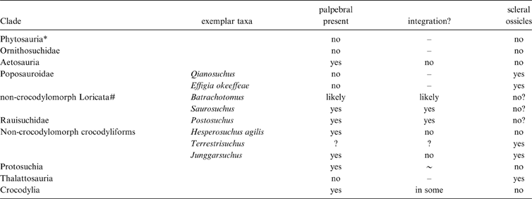
Our description of the morphology, position and connectivity of the crocodylian palpebral, reinforced with recent work on the development of the element (Vickaryous & Hall Reference Vickaryous and Hall2008), strongly suggests that the osseous elements dorsal to the orbit in extinct crocodylomorphs are homologous to those of living crocodylians (Table 1). Furthermore, these same similarities between Crocodylia and extinct Crocodylomorpha are also present in the closest relatives of crocodylomorphs, the non-crocodylomorph pseudosuchians, suggesting that osseous elements in non-crocodylomorph pseudosuchians and the palpebral of Crocodylia are also homologous. Therefore, palpebrals are not restricted to Crocodylomorpha, but are also present in their closest relatives. Yet, the origin of the palpebral is difficult to understand, given the discontinuous distribution among early pseudosuchian groups and the questionable homology of the elements in aetosaurs. Osseous elements in the dorsal portion of the orbit have not been identified in stem archosaurs (e.g. Euparkeria capensis, Ewer Reference Ewer1965; Vancleavea campi, Nesbitt et al. Reference Nesbitt, Stocker, Small and Downs2009), in any early bird-line archosaur (=Ornithodira, =Avemetatarsalia), phytosaurs, or in the early diverging pseudosuchian members such as Ornithosuchidae or Gracilisuchus stipanicicorum (Romer Reference Romer1972). At least some members of Aetosauria do possess palpebrals (see description above), but the homology of these elements remain unclear. The palpebrals of crocodylomorphs and aetosaurs share many similarities, but there are differences as well. The three ossified elements in Aetosaurus ferratus are likely ossified from the eyelid, given the position in the orbit as in Crocodylia. The external morphology is similar to that of osteoderms like those of crocodylomorphs. However, there are no crocodylomorphs with three independent palpebral elements (the palpebral of Osteolaemus is composed of two or three ossifications, but the separate ossifications still form a single palpebral). This may not be a problem, given that all of the Aetosaurus specimens are juveniles (Schoch Reference Schoch2007), and that later in ontogeny the three elements may form a single palpebral. The immaturity of the skeleton of Aetosaurus may also explain why the three elements have little to no contact with the circumorbital bones of the skull. Moreover, in the larger specimens of Aetosaurus, it appears that the palpebrals are touching the prefrontal, as in all other pseudosuchians with preserved palpebrals. Because of these similarities, we argue that these elements in aetosaurs represent homologous elements with Crocodylia.
Distribution of osseous skeletal elements in the orbit of pseudosuchians clades.
* Most archosaur phylogenies (Sereno Reference Sereno1991; Juul Reference Juul1994; Brusatte et al. Reference Brusatte, Benton, Desojo and Langer2010) have found Phytosauria as the basal-most pseudosuchian group, but recently Nesbitt (Reference Nesbitt2011) found them as the sister-taxon to Archosauria.
# does not include Rauisuchidae.
The other group (depending on the hypothesis of relationships, see Fig. 11A) is the “rauisuchians” of Brusatte et al. (Reference Brusatte, Benton, Desojo and Langer2010), or the non-crocodylomorph loricatans of Nesbitt (Reference Nesbitt2011). The three taxa with confirmed palpebrals, Saurosuchus, Polonosuchus and Postosuchus, share a general external morphology, position and connectivity with early crocodylomorphs such as Hesperosuchus. However, the non-crocodylomorph loricatan taxa have a unique articulation with the circumorbital bones not sampled in Crocodylomorpha, in which there is a cranial-like, interdigitating suture between the palpebral and the circumorbital bones.
(A) The evolution of skeletal elements in the orbit of early pseudosuchians, based on the relationships of Nesbitt (Reference Nesbitt2011) (left) and Brusatte et al. (Reference Brusatte, Benton, Desojo and Langer2010) (right). Skeletal elements in the orbit are highlighted in yellow/gray. (B) The evolution of skeletal elements in the orbit of Crocodyliformes, based on the relationships of Clark et al. Reference Clark, Xing, Forster and Wang2004, Pol et al. (Reference Pol, Turner and Norell2009), Turner & Sertich (Reference Turner and Sertich2010) and Pritchard et al. (Reference Pritchard, Turner, Allen and Norellin press). The distribution of scleral ossicles and the palpebral among pseudosuchians is complicated, even when considering skeletal elements are lost easily in taphonomic processes. Abbreviations: ?=unknown condition; ?*=likely a genuine absence; P=palpebral; S=scleral ossicles / sclerotic ring. The dotted lines represent uncertainty in phylogenetic position.
From these data, it is likely that the palpebral evolved in close relatives of Crocodylomorpha. The differences among the palpebral in early pseudosuchian groups such as Aetosauria and “rauisuchians” and crocodylomorphs may have been the result of early experimentation with a novel element before it became somewhat more standardised in basal Crocodylomorpha and Crocodyliformes. The palpebral was present by the Late Triassic in aetosaurs and “rauisuchians” and, given phylogenetic estimates of Pseudosuchia, the feature was likely present in the last common ancestor of aetosaurs and crocodylomorphs at the end of the Early Triassic (Butler et al. Reference Butler, Brusatte, Reich, Nesbitt, Schoch and Hornung2011; Nesbitt Reference Nesbitt2011; Nesbitt et al. Reference Nesbitt, Liu and Li2011). Furthermore, the palpebral may also be useful for determining relationships in early Archosauria. Nesbitt (Reference Nesbitt2011) employed three characters that focused on the palpebral, one character examining the presence or absence of the palpebral (character 147), one character addressing the size of the elements (character 148), and one character addressing the connectivity of the elements with the circumorbital bones (character 149). Even though we do not have the ability to examine developmental sequences in extinct pseudosuchians, the origin of the palpebral attests to the importance of the fossil record when examining seemingly novel features in living groups of vertebrates.
2.2. The evolution of osseous skeletal elements in the orbit of pseudosuchians
The evolution of scleral ossicles and the palpebral within Pseudosuchia is complicated and likely intertwined throughout the evolutionary history of the group. For example, there is only one example of a pseudosuchian, Junggarsuchus sloani, bearing both a palpebral and scleral ossicles, whereas most pseudosuchians lack scleral ossicles. Both the palpebral and scleral ossicles are lost or “gained” throughout the history of Pseudosuchia.
The palpebral originated outside Crocodylomorpha and is not a crocodylian invention. Clearly, the palpebral is plesiomorphic for Crocodylomorpha, given the distribution of the element within Pseudosuchia (Fig. 11A). The general morphology (osteoderm-like, concave ventrally-convex dorsally), position within the orbit (in the upper eyelid), and the attachment location (prefrontal) of the palpebral is rather conservative across Pseudosuchia, whereas the number of total palpebral elements, size of the palpebral and the integration with circumorbital bones varies across Crocodylomorpha. In the earliest forms, a large single palpebral occupies the dorsal portion of the orbit, and this condition appears to be inherited from their closest relatives in Loricata. At the taxonomic level of Crocodyliformes, two palpebrals, an anterior and posterior element, are present and occupy most of the dorsal portion of the orbit. Most members of Crocodyliformes outside the Crocodylia retain two palpebral elements, whereas all members of Crocodylia only bear one element (Fig. 11B). The single palpebral in Crocodylia is hypothesised to be homologous to the anterior palpebral of non-crocodylian crocodyliforms, given the similarities in size and attachment position.
Although the position of the palpebral is conserved in crocodyliforms, the degree of integration with the skull roof varies across Crocodylomorpha. The integration of the palpebral with the circumorbital bones within Crocodylomorpha is not to the same degree as in non-crocodylomorph loricatans (i.e., a cranial suture is present), but several crocodyliforms such as Sebecus, Simosuchus, and the crocodylians, Paleosuchus and Osteolaemus show high degrees of integration. Furthermore, taxa with better integrated palpebral with the circumorbital bones have large palpebrals that span the entire dorsal margin of orbit. Interestingly, the extinct (e.g. Simosuchus, Georgi & Krause Reference Georgi and Krause2010; Sertich & Groenke Reference Sertich and Groenke2010) and extant (Osteolaemus, Kofron Reference Kofron1992) taxa with this arrangement of palpebrals have been inferred to have a more terrestrial ecology than their more aquatic cousins.
No member of Crocodylia possess scleral ossicles. Nevertheless, scleral ossicles are present in early pseudosuchians and even within Crocodylomorpha. Therefore, the evolution of scleral ossicles within the orbit of pseudosuchians is complicated. The only non-crocodylomorph pseudosuchian clade to possess scleral ossicles is the highly disparate poposauroids. Interestingly, the earliest diverging member of this clade (Nesbitt Reference Nesbitt2011), Qianosuchus, has been hypothesised to have a marine ecology (Li et al. Reference Li, Wu, Cheng, Sato and Wang2006). However, other members of this clade, the shuvosaurids, also have scleral ossicles, but are inferred to be a terrestrial clade without clear aquatic adaptations (Nesbitt & Norell Reference Nesbitt and Norell2006; Nesbitt Reference Nesbitt2007). Among non-crocodyliform crocodylomorphs, Terrestrisuchus and Junggarsuchus are the only members to possess a sclerotic ring. Terrestrisuchus lacks any clear aquatic adaptations in the skeleton and is from a terrestrial assemblage (Whiteside & Marshall Reference Whiteside and Marshall2008). Within Crocodyliformes, sclerotic rings are present, but are restricted to a number of closely related marine forms (e.g. Dakosaurus andiniensis (Gasparini et al. Reference Gasparini, Pol and Spalletti2006), Geosaurus giganteus and Cricosaurus suevicus). No other crocodyliforms possess a sclerotic ring.
Scleral ossicles are rare among pseudosuchians, but in most cases are restricted to marine forms (e.g. Qianosuchus and Thalattosuchia) and may have helped stabilise the eye under pressure (Curtis & Miller Reference Curtis and Miller1938). Yet, scleral ossicles are present in a few non-marine taxa (e.g. Terrestrisuchus and Shuvosauridae) and are present in many non-marine amniotes. Given the distribution of these features among pseudosuchians (Fig. 11), it appears that multiple gains of scleral ossicles were occurring throughout pseudosuchian evolution. Interpreting the absence of scleral ossicles as multiple independent losses in the numerous lineages without them is a considerably less parsimonious scenario. Yet, the morphology, composition and location within the orbit of scleral ossicles in pseudosuchians are indistinguishable from those of other amniote taxa that retain the feature. Therefore, we conclude that scleral ossicles were likely lost early in pseudosuchian history, possibly near the origin of the clade. However, members of Pseudosuchia retained the ability to develop scleral ossicles. It is not clear if members of Crocodylia still retain this ability to develop scleral ossicles.
2.3. Further considerations of skeletal elements in the orbit and the use of the anatomical term palpebral
The term “palpebral” has previously been applied to any bony element in the dorsal portion of the orbit in members of Reptilia including: squamates, crocodylians, pterosaurs, birds and ornithischian dinosaurs (Romer Reference Romer1956; Coombs Reference Coombs1972; Lee Reference Lee1997; Clark et al. Reference Clark, Sues and Berman2000; Mayr Reference Mayr2005; Wang et al. Reference Wang, Kellner, Zhou and Campos2007; Maidment & Porro Reference Maidment and Porro2010). The use of the same term to describe somewhat similar features across a broad range of taxa implies homology, but the homology of these features has never been explicitly tested. Furthermore, a number of proposed synonyms are used interchangeably with palpebral, including the terms “supraorbital,” and “supraciliary”, for any element in the dorsal portion of the eye. Given recent breakthroughs in understanding the palpebral of crocodylians (Vickaryous & Hall Reference Vickaryous and Hall2008), it is clear that not all osseous elements in the dorsal portion of the orbit develop the same way. For example, the palpebral of crocodylians forms from metaplastic ossification, like that of osteoderms (Vickcaryous & Hall 2008), but this is different from the formation of supraorbitals in osteichthyan fishes, where the bones form from a cartilaginous precursor (Hall Reference Hall2005). Although examining the development of the skull bones of an extinct taxon is impossible, we can utilise Patterson's (Reference Patterson, Joysey and Friday1982) requirements for homology (similarity and congruence tests) to clarify the term palpebral. We propose restricting the term “palpebral” to the element that develops in a similar fashion to that of an osteoderm and resides in the upper eyelid (as in crocodylians). This is a considerably more refined definition of palpebral, but is similar to the original definition proposed by Peters (Reference Peters1964). Furthermore, it is our view that the terms “supraorbital,” and “supraciliary” should also be restricted to a particular group.
With this restricted definition of palpebral, it is clear that the term palpebral should not be applied to the structures across the orbits of ornithischians. In a comprehensive contribution examining the homology of the ornithischian “palpebral” across the entire clade, Maidment & Porro (Reference Maidment and Porro2010) explicitly test the homology of the “palpebral(s)” of some ornithischian taxa with that of the variously named supraorbital(s) of other clades using Patterson's (Reference Patterson, Joysey and Friday1982) homology tests. In short, they find that the “palpebral(s)” and supraorbital(s) of ornithischian clades are homologous. Their extensive and careful assessment of ornithischian “palpebral(s)” across the clade is welcomed, but in the end, the term “palpebral” is not appropriate for the orbital bone(s), although the first application of the term palpebral to that of the structure in some ornithischians was based on the inferred homology of the palpebral to those of crocodylians (Gregory & Mook Reference Gregory and Mook1925). Using the same methodology for determining homology, we can reject the homology of the ornithischian “palpebral” and crocodylian palpebral, given that the element fails Patterson's (Reference Patterson, Joysey and Friday1982) tests one (the morphology and connectivity differ) and three (the common ancestors of the two clades do not possess a “palpebral”). As a general recommendation, we advocate that ornithischian “palpebral” be renamed to further prevent future confusion.
3. Acknowledgements
We thank Sankar Chatterjee, Hans-Dieter Sues, Thomas Sulej, Patricia Holroyd, Wann Langston and Rainer Schoch for providing access to specimens under their care. Discussions with Randall Irmis, Christopher Brochu, Eric Wilberg, Eugenia Gold and Michael Vickaryous helped improve the manuscript. Wann Langston contributed to the discussion of the paper, provided useful publication, and access to rare specimens; his advice set us on the right track more than once. Critical reviews by James Clark and Christopher Brochu were instrumental to completing this paper. Jeffery Martz provided pictures of Aetosaurus. We thank James Clark for access to a cast of Junggarsuchus and pictures of the skull prior to full preparation.