Callous unemotional (CU) traits in childhood and adolescence characterize a relatively homogenous subgroup of youths with conduct problems who engage in severe and persistent antisocial and aggressive behaviors (Frick et al., Reference Frick, Stickle, Dandreaux, Farrell and Kimonis2005; Rowe et al., Reference Rowe, Maughan, Moran, Ford, Briskman and Goodman2010; Kahn et al., Reference Kahn, Byrd and Pardini2013) and who are at particularly high risk of developing psychopathic traits in adulthood (Vasey et al., Reference Vasey, Kotov, Frick and Loney2005; Salekin, Reference Salekin2006; Burke et al., Reference Burke, Loeber and Lahey2007). CU traits include limited empathy and remorse and reduced displays of emotion, and are most commonly assessed using the Inventory of Callous Unemotional Traits (ICU), which is comprised of three subfactors: callousness, uncaring, and unemotionality (Frick and Ray, Reference Frick and Ray2015). The emergence of these traits and subsequent behavior problems has been consistently linked to dysfunction in the amygdala (Marsh et al., Reference Marsh, Finger, Mitchell, Reid, Sims, Kosson, Towbin, Leibenluft, Pine and Blair2008, Reference Marsh, Finger, Fowler, Jurkowitz, Schechter, Yu, Pine and Blair2011a; Finger et al., Reference Finger, Marsh, Blair, Majestic, Evangelou, Gupta, Schneider, Sims, Pope, Fowler, Sinclair, Tovar-Moll, Pine and Blair2012; Viding et al., Reference Viding, Sebastian, Dadds, Lockwood, Cecil, De Brito and McCrory2012; Herpers et al., Reference Herpers, Scheepers, Bons, Buitelaar and Rommelse2014; Lozier et al., Reference Lozier, Cardinale, VanMeter and Marsh2014; Breeden et al., Reference Breeden, Cardinale, Lozier, VanMeter and Marsh2015). Although it has been posited that amygdala dysfunction in CU youths may stem from major anatomical aberrations, no evidence of atypical amygdala volume in children with CU traits has yet emerged. This may be considered surprising in light of consistent findings of reduced amygdala volume in adults with psychopathy or CU traits (Yang et al., Reference Yang, Raine, Narr, Colletti and Toga2009; Pardini et al., Reference Pardini, Raine, Erickson and Loeber2014; Vieira et al., Reference Vieira, Ferreira-Santos, Almeida, Barbosa, Marques-Teixeira and Marsh2015). In the present study, we explored whether a relationship between CU traits and amygdala volume would emerge in a moderately large sample of children and adolescents in which CU traits and externalizing behaviors were simultaneously modeled as continuous variables. We also considered the relationship between the three independent CU subfactors and amygdala volume.
Theories of the development of CU traits frequently focus on deficits in affective and reinforcement learning processes that rely on the amygdala (Blair, Reference Blair2013). Impaired amygdala functioning in CU youths is believed to impede their ability to learn to avoid behaviors like aggression and making threats, in part because the amygdala is important for recognizing and responding appropriately to signs of others’ distress (Marsh, Reference Marsh2016). Without appropriate signaling in the amygdala, CU youths fail to develop appropriate guilt and empathy in response to others’ distress, and so persist in behaviors like aggression and violence that would normally be inhibited by these emotional responses (Kochanska, Reference Kochanska1993; Frick and Morris, Reference Frick and Morris2004; Blair, Reference Blair2005, Reference Blair2013; Marsh, Reference Marsh2016; Seara-Cardoso et al., Reference Seara-Cardoso, Sebastian, Viding and Roiser2016). These theories are reinforced by consistent findings of reduced amygdala responsivity to fearful facial expressions in high CU youths (Marsh et al., Reference Marsh, Finger, Mitchell, Reid, Sims, Kosson, Towbin, Leibenluft, Pine and Blair2008; Jones et al., Reference Jones, Laurens, Herba, Barker and Viding2009; Viding et al., Reference Viding, Sebastian, Dadds, Lockwood, Cecil, De Brito and McCrory2012; White et al., Reference White, Marsh, Fowler, Schechter, Adalio, Pope, Sinclair, Pine and Blair2012; Lozier et al., Reference Lozier, Cardinale, VanMeter and Marsh2014) as well as aberrant functional connectivity between the amygdala and other regions implicated in emotion processing (Marsh et al., Reference Marsh, Finger, Fowler, Jurkowitz, Schechter, Yu, Pine and Blair2011a; Finger et al., Reference Finger, Marsh, Blair, Majestic, Evangelou, Gupta, Schneider, Sims, Pope, Fowler, Sinclair, Tovar-Moll, Pine and Blair2012; Aghajani et al., Reference Aghajani, Klapwijk, van der Wee, Veer, Rombouts, Boon, van Beelen, Popma, Vermeiren and Colins2016).
Despite a robust literature examining amygdala activity in CU youths, relatively few structural magnetic resonance imaging (MRI) studies have examined the association between CU traits and brain structure. Reduced amygdala volume has repeatedly been found in both studies of youths with conduct disorder (Sterzer et al., Reference Sterzer, Stadler, Poustka and Kleinschmidt2007; Huebner et al., Reference Huebner, Vloet, Marx, Konrad, Fink, Herpertz and Herpertz-Dahlmann2008; Fairchild et al., Reference Fairchild, Hagan, Walsh, Passamonti, Calder and Goodyer2013; Wallace et al., Reference Wallace, White, Robustelli, Sinclair, Hwang, Martin and Blair2014; Rogers and De Brito, Reference Rogers and De Brito2016) and studies of adults with psychopathic traits, who are distinguished from other antisocial populations primarily by their elevated CU traits (Yang et al., Reference Yang, Raine, Narr, Colletti and Toga2009; Cope et al., Reference Cope, Ermer, Nyalakanti, Calhoun and Kiehl2014; Pardini et al., Reference Pardini, Raine, Erickson and Loeber2014). These findings strongly implicate reduced amygdala volume in the development of antisociality and CU traits, but four studies of children and adolescents have not yet found an association between amygdala volume and CU traits (De Brito et al., Reference De Brito, Mechelli, Wilke, Laurens, Jones, Barker, Hodgins and Viding2009; Wallace et al., Reference Wallace, White, Robustelli, Sinclair, Hwang, Martin and Blair2014; Cohn et al., Reference Cohn, Viding, McCrory, Pape, van den Brink, Doreleijers, Veltman and Popma2016; Sebastian et al., Reference Sebastian, De Brito, McCrory, Hyde, Lockwood, Cecil and Viding2016) (although Cohn et al. found that increased CU traits were associated with reduced amygdala gray matter concentration). This could be interpreted to mean that CU traits are associated with amygdala volume only in adulthood, but not childhood or adolescence. Alternately, methodological considerations may have concealed a relationship between amygdala volume and CU traits in youths. For example, although CU traits are continuously distributed and more accurately assessed using continuous analyses (Guay et al., Reference Guay, Ruscio, Knight and Hare2007; Moffitt et al., Reference Moffitt, Arseneault, Jaffee, Kim-Cohen, Koenen, Odgers, Slutske and Viding2008; Lozier et al., Reference Lozier, Cardinale, VanMeter and Marsh2014), three of the four studies of volumetric differences in CU youths employed primarily group-based approaches (De Brito et al., Reference De Brito, Mechelli, Wilke, Laurens, Jones, Barker, Hodgins and Viding2009; Wallace et al., Reference Wallace, White, Robustelli, Sinclair, Hwang, Martin and Blair2014; Sebastian et al., Reference Sebastian, De Brito, McCrory, Hyde, Lockwood, Cecil and Viding2016). In addition, Wallace et al. and De Brito et al. compared youths with both CU traits and conduct problems to healthy control youths, an approach that hinders the dissociation of correlates of CU traits and conduct problems more generally (De Brito et al., Reference De Brito, Mechelli, Wilke, Laurens, Jones, Barker, Hodgins and Viding2009; Wallace et al., Reference Wallace, White, Robustelli, Sinclair, Hwang, Martin and Blair2014). Sebastian et al. compared groups of youths with conduct problems and both low and high levels of CU traits to healthy controls (Sebastian et al., Reference Sebastian, De Brito, McCrory, Hyde, Lockwood, Cecil and Viding2016). However, similar to the prior two studies, their use of a primarily group-based approach lacked the power of a continuous analysis, and may have been affected by suppressor effects (Sebastian et al., Reference Sebastian, McCrory, Cecil, Lockwood, De Brito, Fontaine and Viding2012).
Moreover, these studies all examined CU traits as a unitary construct, although, increasingly, assessments of CU traits have focused on the three subfactors comprising CU traits (callous, uncaring, and unemotional traits) (Kimonis et al., Reference Kimonis, Frick, Munoz and Aucoin2008a, Reference Kimonis, Frick, Skeem, Marsee, Cruise, Munoz, Aucoin and Morrisb). Examining the independent associations between callous, uncaring, and unemotional traits with aberrant amygdala volume may clarify the relevance of amygdala development to the emergence of CU traits. Given the key role of the amygdala in emotional processing, it may be primarily the unemotional component of CU traits that is associated with reduced amygdala volume, a finding that would implicate the amygdala in global affective deficits in CU youths. By contrast, associations with the callous and/or uncaring components of CU traits would suggest that the amygdala may play a more complex role in interpersonal empathy and caring. Examination of associations between unemotional traits and amygdala volume may also provide important insight into the validity of assessments of unemotional traits, the nature of which have been subject to recent concerns (Henry et al., Reference Henry, Pingault, Boivin, Rijsdijk and Viding2016; Cardinale and Marsh, Reference Cardinale and Marsh2017).
The current study therefore examined associations between amygdala gray matter volume and externalizing behaviors as well as callous, uncaring, and unemotional traits in a sample of youths with varying levels of conduct problems and CU traits. Through a series of multiple linear regression analyses, we investigated how total ICU scores and the three subfactor scores correspond to both aberrant structural development of the amygdala and the emergence of externalizing behaviors, and whether CU traits mediate the relationship between reduced amygdala volume and externalizing behavior severity. We hypothesized that CU traits would be associated with increased externalizing behavior problems and decreased bilateral amygdala volume. Furthermore, we predicted that these relationships would be driven by the callous and uncaring subscales of the ICU.
Methods
Participants
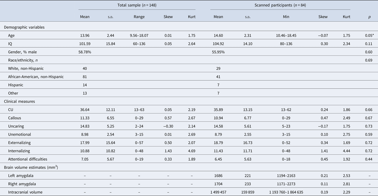
One hundred forty-eight children, aged 9–18 (M = 13.96, s.d. = 2.44, % male = 59.46), were recruited from Washington, DC and surrounding regions through referrals, advertisements, and fliers seeking both healthy children and children with conduct problems. All participants and their parents first completed an initial visit during which demographic and clinical measures were completed along with IQ testing using the Kauffman Brief Intelligence Test (Kaufman and Kaufman, Reference Kaufman and Kaufman2004). Participants reported a wide range of scores on our clinical measures, confirming that our sample included both healthy youths and youths with elevated conduct problems and varying CU traits, as well as psychiatric symptoms including externalizing behaviors, internalizing behaviors, and attentional difficulties (Table 1). Consistent with our recruitment effort to specifically target both healthy children and children with elevated conduct problems, 77 participants reported clinical levels of externalizing behavior as assessed by an age and gender standardized externalizing symptomology score on the Child Behavior Checklist (CBCL) that placed them above the 98th percentile (Achenbach, Reference Achenbach1991).
Descriptive statistics

p Values are reported for comparisons of total sample v. scanned participants.
Of participants who completed the initial visit, 93 were eligible for and consented to participate in an MRI scan. Participants were excluded from MRI scanning for: history of head trauma or neurological disorder, symptoms of pervasive developmental disorder, IQ <80, or MRI contraindications such as claustrophobia or metallic implants including braces or permanent retainers. The MRI sample consisted of children aged 10–17 (M = 13.98, s.d. = 2.36, % male = 59.14) and varied widely in externalizing behavior, including 46 participants with clinically significant externalizing scores. The MRI sample did not differ from the full sample in terms of externalizing and CU scores or any other clinical or demographic measures, with the exception of a trend-level difference in age between the full sample and the scanned sample (Table 1). All participants were native English speakers. Written informed assent and consent were obtained from children and parents before testing. Approval for all procedures was obtained from the Georgetown University Institutional Review Board. The authors assert that all procedures contributing to this work comply with the ethical standards of the relevant national and institutional committees on human experimentation and with the Helsinki Declaration of 1975, as revised in 2008.
Clinical measures
Inventory of Callous Unemotional Traits
The ICU was used to assess CU traits (Kimonis et al., Reference Kimonis, Branch, Hagman, Graham and Miller2008a, Reference Kimonis, Frick, Skeem, Marsee, Cruise, Munoz, Aucoin and Morrisb). The ICU was completed separately by parents and participants. Scores on the ICU were calculated by summing the highest item response from either the child or parent version (Jones et al., Reference Jones, Laurens, Herba, Barker and Viding2009; Sebastian et al., Reference Sebastian, McCrory, Cecil, Lockwood, De Brito, Fontaine and Viding2012; Viding et al., Reference Viding, Sebastian, Dadds, Lockwood, Cecil, De Brito and McCrory2012; Lozier et al., Reference Lozier, Cardinale, VanMeter and Marsh2014; Breeden et al., Reference Breeden, Cardinale, Lozier, VanMeter and Marsh2015). This scoring approach follows the recommended scoring practices for the parent scale of the ICU (Frick and Hare, Reference Frick and Hare2001), and has been shown to reduce susceptibility to social desirability biases and optimize accuracy across multiple contexts (Piacentini et al., Reference Piacentini, Cohen and Cohen1992; Frick et al., Reference Frick, Cornell, Bodin, Dane, Barry and Loney2003).
Child Behavior Checklist
The CBCL is a parent report-based assessment of behavioral and emotional problems in children and adolescents (Achenbach, Reference Achenbach1991). Externalizing and internalizing syndrome scales were calculated for each participant. Attentional difficulties were also measured using the attention difficulties syndrome scale. The use of the CBCL to assess the severity of various clinical symptoms in community samples has been demonstrated to be reliable and valid (Biederman et al., Reference Biederman, Faraone, Doyle, Lehman, Kraus, Perrin and Tsuang1993; Warnick et al., Reference Warnick, Bracken and Kasl2008).
Image acquisition and analysis
Three-dimensional anatomical images were acquired using a 3.0 Tesla Siemens (Erlangen, Germany) TIM Trio. High-resolution T1-weighted images were collected for each participant (TR = 1900 ms, TE = 2.52 ms, TI = 900.0 ms, 1.0 mm3 voxels, 176 slices, matrix = 246 × 256, field of view = 250 mm2). Prior to analyses, all images were visually inspected for motion artifacts. Any potential motion artifacts were examined by three independent evaluators and only scans for which all three evaluators reached agreement were included in the dataset. Thirty-nine participants completed more than one anatomical scan; the scan with the fewest motion artifacts and clearest contrast was selected for these participants. Data from nine participants could not be analyzed due to excessive motion artifacts in all completed anatomical scans, resulting in a final sample size of 84 participants. Images were collected using an eight-channel phased-array head coil for 16 participants and using a 12-channel phased-array head coil for the remaining 68 participants. For all analyses investigating neural volume, a dummy coded variable for use of the 8 v. 12-channel was included as a covariate (Breeden et al., Reference Breeden, Cardinale, Lozier, VanMeter and Marsh2015). Because participants completed the scan during a separate visit, we also included age at the time of the scan in addition to age at the time of the initial visit for all analyses investigating neural volume.
Anatomical images were analyzed using FreeSurfer version 5.3.0. Automated segmentation of subcortical regions occurred during the first stage of the FreeSurfer cortical reconstruction process (Fischl et al., Reference Fischl, Salat, Busa, Albert, Dieterich, Haselgrove, van der Kouwe, Killiany, Kennedy, Klaveness, Montillo, Makris, Rosen and Dale2002, Reference Fischl, Salat, van der Kouwe, Makris, Segonne, Quinn and Dale2004; Fischl, Reference Fischl2012). During this stage, neuroanatomical labels are automatically assigned to each voxel based on probabilistic information acquired through an a priori knowledge of spatial relationships acquired through a manually labeled training set. This classification technique is robust to anatomical variation typical in pediatric populations through the use of a non-linear registration procedure. Segmentation occurs following three automated strategies to disambiguate voxel labels, which assess the prior probability of the tissue class occurring at an atlas location, and given the tissue class, the likelihood of the image and the probability of the local spatial configuration. The resulting subcortical segmentation, has been shown to be reliable (Morey et al., Reference Morey, Selgrade, Wagner, Huettel, Wang and McCarthy2010) and comparable to manual segmentation (Fischl et al., Reference Fischl, Salat, Busa, Albert, Dieterich, Haselgrove, van der Kouwe, Killiany, Kennedy, Klaveness, Montillo, Makris, Rosen and Dale2002; Morey et al., Reference Morey, Petty, Xu, Hayes, Wagner, Lewis, LaBar, Styner and McCarthy2009; Grimm et al., Reference Grimm, Pohlack, Cacciaglia, Winkelmann, Plichta, Demirakca and Flor2015; Schoemaker et al., Reference Schoemaker, Buss, Head, Sandman, Davis, Chakravarty, Gauthier and Pruessner2016). Following segmentation, 42 subcortical regions were identified and labeled using both subject-independent probabilistic atlases and subject-specific measured variables for each subject. All images were visually inspected following segmentation. Volume estimates for all subcortical regions, including the left amygdala and right amygdala, as well as total intracranial volume were extracted and exported for analysis in STATA (Table 1; online Supplementary Table S2).
Results
For all analyses, variables were mean centered, and known correlates of amygdala volume and/or externalizing behaviors were entered as covariates. For analyses of clinical symptomology, gender, IQ, and age at initial visit were included as covariates. For analyses of brain volume, we included total intracranial volume, age at time of scan, and headcoil as additional covariates. Robust standard errors were used to account for heteroscedasticity of the experimental variables and to control for sibling effects. All analyses were repeated with dummy variables coding for the 18 sibling groups present in these data (n = 41). Findings were not affected by the inclusion of these covariates. Therefore, for ease of interpretation of results, we report the findings from analyses excluding sibling status as a covariate.
ICU scores and clinical symptomology
The internal consistency of total ICU, α = 0.90, callous subscale, α = 0.84, and uncaring subscale, α = 0.87 were acceptable, whereas the unemotional subscale showed relatively low internal consistency, α = 0.58. Intercorrelations among scores on the ICU were all large (online Supplementary Table S1).
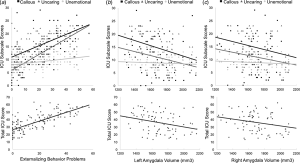
Results of a multiple linear regression analysis across all participants (n = 148) predicting externalizing behaviors from ICU scores confirmed that as total ICU scores increased, externalizing behaviors increased, β = 0.73, t(143) = 11.73, p < 0.001 (Fig. 1). This association remained significant after controlling for attentional difficulty and internalizing behavior scores, β = 0.31, t(141) = 4.55, p < 0.001. Next, a multiple regression with all three ICU subscales predicting externalizing behavior problems found that scores on the callous, β = 0.53, t(141) = 4.56, p < 0.001, and uncaring, β = 0.34, t(141) = 3.30, p = 0.001, subscales were independently associated with increased externalizing, whereas unemotional subscale scores were not, β = −0.07, t(141) = −1.13, p = 0.26 (Fig. 1). Associations between externalizing and the callous, β = 0.21, t(139) = 3.03, p = 0.003, and uncaring, β = 0.20, t(139) = 3.19, p = 0.002, subscales persisted when controlling for attentional difficulties and internalizing behaviors, whereas the unemotional subscale scores remained non-significant, β = −0.08, t(139) = −1.37, p = 0.17.
Scatter plots for the associations between total scores, callous, uncaring, and unemotional subscale scores on the ICU with (a) externalizing behaviors and (b) left and (c) right amygdala volumes.

We repeated all of the above analyses examining the relationship between ICU scores and clinical symptomologies restricted to only those participants who qualified for inclusion in MRI scanning (n = 84). All patterns of significant findings persisted when analyses were limited to this sample (online Supplementary Text S1), supporting the reliability of the identified patterns.
Amygdala volume
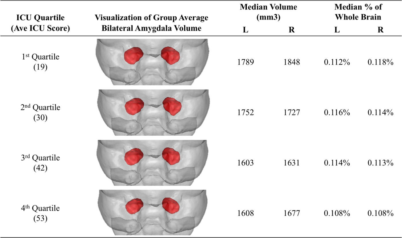
We next investigated associations between amygdala volume, scores on the ICU, and externalizing symptoms using a region of interest (ROI) approach, consistent with the approaches used in previous studies of neural correlates of CU traits and psychopathy (Sebastian et al., Reference Sebastian, McCrory, Cecil, Lockwood, De Brito, Fontaine and Viding2012; Lozier et al., Reference Lozier, Cardinale, VanMeter and Marsh2014; Pardini et al., Reference Pardini, Raine, Erickson and Loeber2014; Wallace et al., Reference Wallace, White, Robustelli, Sinclair, Hwang, Martin and Blair2014; Breeden et al., Reference Breeden, Cardinale, Lozier, VanMeter and Marsh2015; Vieira et al., Reference Vieira, Ferreira-Santos, Almeida, Barbosa, Marques-Teixeira and Marsh2015; Sebastian et al., Reference Sebastian, De Brito, McCrory, Hyde, Lockwood, Cecil and Viding2016). Again, analyses included age at scanning, headcoil type, and total intracranial volume as covariates in addition to age at time of initial visit, gender, and IQ. Results of separate multiple linear regression analyses revealed that total ICU scores were associated with decreased left, β = −0.36, t(76) = −3.21, p = 0.002, and right, β = −0.27, t(76) = −2.64, p = 0.01, amygdala volume (Fig. 2).
Visualizations of average amygdala volumes for each ICU total score quartile rendered within the mean brain volume of all subjects. Shown in anterior view with the right hemisphere displayed on the right.

Similar results were obtained for callous [left: β = −0.32, t(76) = −3.00, p = 0.004; right: β = −0.24, t(76) = −2.40, p = 0.02] and uncaring [left: β = −0.35, t(76) = −3.07, p = 0.003; right: β = −0.24, t(76) = −2.34, p = 0.02] subscale scores. Unemotional subscale scores were associated with right amygdala volume at a trend level, β = −0.17, t(76) = −1.97, p = 0.05, but not left amygdala volume, β = −0.18, t(76) = −1.64, p = 0.11 (Fig. 1).
To assess the specificity of these findings, we conducted parallel analyses examining associations between CU traits and subcortical volume estimates for the nucleus accumbens, caudate, hippocampus, pallidum, putamen, thalamus, and ventral diencephalon (DC). Across all of these subcortical regions, no significant associations (all p > 0.10) were found with ICU total (online Supplementary Table S3) or subscale scores (online Supplementary Table S4), or with externalizing behaviors (online Supplementary Table S5), with one exception: decreased right ventral DC volume was associated with increased externalizing behaviors, β = −0.18, t(76) = −2.18, p = 0.03, and uncaring traits, β = −0.17, t(76) = −2.18, p = 0.03, but no other measure of CU traits. Of note, neither ICU scores nor externalizing behaviors were associated with total intracranial volume, all p > 0.10.
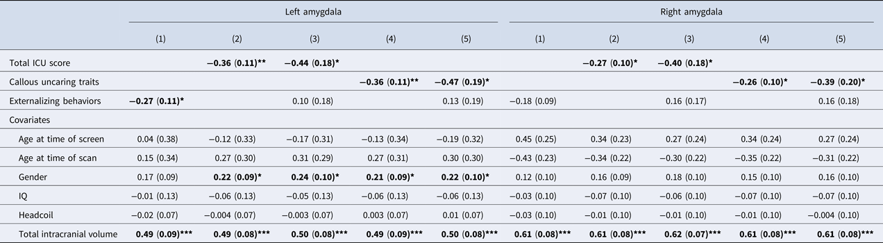
Multiple linear regression analyses in which externalizing behavior problems were entered as predictors of left and right amygdala volumes found that externalizing problems were associated with decreased left, β = −0.27, t(76) = −2.54, p = 0.01, but not right, β = −0.18, t(76) = −1.88, p = 0.06, amygdala volume. When both externalizing behaviors and total ICU scores were included simultaneously in the model, CU traits remained predictors of both left, β = −0.44, t(75) = −2.42, p = 0.02, and right, β = −0.40, t(76) = −2.21, p = 0.03, amygdala volume, but the relationship between externalizing behaviors and amygdala volume was rendered non-significant. Consistent with this pattern of findings, mediation analyses across all participants using the SPSS PROCESS macro revealed a significant indirect effect of amygdala volumes on externalizing behaviors through CU traits (online Supplementary Fig. S1), such that the observed direct statistical relationship between decreased left and right amygdala volumes with increased externalizing behaviors was explained by the statistical relationship between each of these variables with CU traits (left: Sobel Z = −3.50, p = .001; right: Sobel Z = −2.60, p = 0.01).
Following persistent findings that the unemotional subscale was not closely associated with either externalizing behaviors or amygdala volumes, we created a composite callous-uncaring score by summing responses to items comprising only the callous and uncaring subscales (scale reliability was acceptable, α = 0.90). Callous-uncaring scores predicted right and left amygdala volumes, even after accounting for externalizing scores (Table 2). Mediation analyses that included callous-uncaring composite scores revealed that callous-uncaring traits, specifically, mediated the statistical relationship between decreased amygdala volume and increased externalizing behaviors (left: Sobel Z = −3.56, p < 0.001; right: Sobel Z = −2.55, p = 0.01).
Multiple regression models predicting left and right amygdala volumes

*p < 0.05, **p < 0.01, ***p < 0.001. We report five independent regression models for the prediction of left and right amygdala volume separately. Each regression model is reported in its own column as indicated by the numbers 1–5. Standardized betas with standard error in parentheses. Significant effects in bold.
Age and gender as moderators
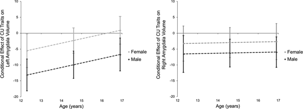
We next examined whether age or gender moderated the relationship between CU traits and amygdala volume. Moderation analyses were conducted using the SPSS PROCESS macro. We selected a model such that both age and gender were entered as moderators of the relationship between CU traits and amygdala volume. Results revealed that both age, β = 0.19, t(73) = 2.10, p = 0.04, and gender, β = −0.31, t(73) = −2.86, p = 0.01, were moderators, such that the conditional effect of CU traits on predicting amygdala volume is greater at younger ages and in male participants (Fig. 3). Whereas in male participants, the conditional effect of CU traits on amygdala volume is significant across all age ranges but greatest at younger ages; within female participants, the conditional effect of CU traits on right amygdala volume is non-significant at all ages and only in younger females is the conditional effect of CU traits on left amygdala volume significant. Of note, IQ, age, externalizing behaviors, and CU traits were not significantly different across male and female participants in our sample. Whereas age was unrelated to IQ, gender, or CU traits, we observed a significant bivariate correlation between age and externalizing behaviors, r(84) = 0.26, p = 0.02.
Age and gender moderate the relationship between CU traits and amygdala volume. The conditional effect of CU traits on amygdala volume plotted as a function of age separately for male and female participants. Note: 95% CI depicted as error bars at the mean age and ±1s.d.

Discussion
These findings provide the first evidence linking CU traits to reduced amygdala gray matter volume in youths. Across a mixed-gender sample of children with varying levels of externalizing behavior and CU traits, we found that variation in amygdala volume is associated with levels of both callous-uncaring traits and antisocial and externalizing behaviors, even after accounting for variation in children's age, sex, cognitive abilities, and total intracranial volume. In our sample, the volume of left amygdala was associated with engagement in externalizing behaviors (e.g. aggression, theft, rule-breaking) and the volume of both left and right amygdala was associated with CU traits including limited empathy, remorse, and guilt. Multiple regression analyses revealed, however, that when externalizing behaviors and CU traits were modeled simultaneously, only CU traits remained associated with amygdala volume. Moreover, the relationship between amygdala volume and CU traits primarily reflected callous and uncaring subscale scores (both of which were also robustly associated with externalizing behavior), supporting a role for the amygdala in interpersonal empathy and caring. Alone, unemotional traits as measured by the ICU were unrelated to externalizing behaviors or amygdala volume. Comparable patterns were not observed in parallel analyses of subcortical volume, suggesting that findings were specific to the amygdala rather than limbic structures generally.
Together, these findings suggest that aberrant development of the amygdala, particularly relative reductions in bilateral amygdala volume as a proportion of total intracranial volume, may play a role in the emergence of CU traits and subsequent externalizing behavior. Structural amygdala abnormalities in high CU youths may lead to functional impairments in stimulus reinforcement learning and empathy, two processes that typically promote the avoidance of externalizing behaviors that cause distress in others (Marsh, Reference Marsh2016; Seara-Cardoso et al., Reference Seara-Cardoso, Sebastian, Viding and Roiser2016). Among callous and uncaring youths, developmental deficits in the amygdala may stunt the development of empathy and guilt, leading to engagement in increased externalizing behaviors such as aggression (Kochanska, Reference Kochanska1993; Frick and Morris, Reference Frick and Morris2004; White et al., Reference White, Cruise and Frick2009). The results of our moderator analyses suggest a developmental trajectory for the association between amygdala volume and CU traits such that the association between reduced amygdala volume with elevated CU traits was greatest at younger ages within our sample. This is consistent with the hypothesis that the amygdala plays a key role in moral development in childhood and early adolescence, such that anatomical abnormalities during this period are particularly important. Gender also emerged as a significant moderator. While we observe similar patterns in both genders, the association between CU traits and amygdala volume was stronger and more consistent in males. Future studies should investigate younger developmental periods, as the associations between amygdala volume and CU traits in female children may be stronger at even younger ages given evidence that brain maturation occurs earlier in females in comparison to males (Giedd et al., Reference Giedd, Blumenthal, Jeffries, Castellanos, Liu, Zijdenbos, Paus, Evans and Rapoport1999; Lenroot et al., Reference Lenroot, Gogtay, Greenstein, Wells, Wallace, Clasen, Blumenthal, Lerch, Zijdenbos, Evans and Thompson2007).
Our findings contrast with those of four previous investigations that have observed no correspondence between amygdala gray matter volume and CU traits (De Brito et al., Reference De Brito, Mechelli, Wilke, Laurens, Jones, Barker, Hodgins and Viding2009; Wallace et al., Reference Wallace, White, Robustelli, Sinclair, Hwang, Martin and Blair2014; Cohn et al., Reference Cohn, Viding, McCrory, Pape, van den Brink, Doreleijers, Veltman and Popma2016; Sebastian et al., Reference Sebastian, De Brito, McCrory, Hyde, Lockwood, Cecil and Viding2016). However, the present investigation benefited from several alternate analytical approaches that may explain this disparity. We employed continuous analyses of CU traits rather than group-based analyses, in keeping with emerging trends in evaluating CU traits (and psychopathology more generally) (Guay et al., Reference Guay, Ruscio, Knight and Hare2007; Moffitt et al., Reference Moffitt, Arseneault, Jaffee, Kim-Cohen, Koenen, Odgers, Slutske and Viding2008; Lozier et al., Reference Lozier, Cardinale, VanMeter and Marsh2014). Given that CU traits are highly correlated with externalizing behaviors, groups defined by CU traits may also be characterized by high levels of externalizing behaviors, making it more difficult to isolate variables that are specifically associated with CU traits. Furthermore, lack of group differences in amygdala volume could result from suppressor effects arising from the strong positive correlation between externalizing behaviors and CU traits but inverse associations of CU traits and externalizing behaviors with various aspects of neural development (Sebastian et al., Reference Sebastian, McCrory, Cecil, Lockwood, De Brito, Fontaine and Viding2012; Viding et al., Reference Viding, Sebastian, Dadds, Lockwood, Cecil, De Brito and McCrory2012; Lozier et al., Reference Lozier, Cardinale, VanMeter and Marsh2014). Our findings are consistent with the existence of suppressor effects for the associations between CU traits, externalizing behaviors, and amygdala volume. Examined separately, both externalizing behaviors and CU traits were associated with decreased amygdala volume. But when entered together in a multiple regression, the suppressor effect became evident through the emergence of CU traits as associated with decreased amygdala volume while externalizing behaviors were (non-significantly) associated with increased amygdala volume.
In addition, the current study employed FreeSurfer to extract measures of subcortical volume, whereas three of the four previous studies used voxel-based morphometry (VBM) in Statistical Parametric Mapping (SPM) (De Brito et al., Reference De Brito, Mechelli, Wilke, Laurens, Jones, Barker, Hodgins and Viding2009; Cohn et al., Reference Cohn, Viding, McCrory, Pape, van den Brink, Doreleijers, Veltman and Popma2016; Sebastian et al., Reference Sebastian, De Brito, McCrory, Hyde, Lockwood, Cecil and Viding2016). There is some evidence that the use of different analytic techniques can produce different results for the examination of subcortical gray matter structures (Heinen et al., Reference Heinen, Bouvy, Mendrik, Viergever, Biessels and de Bresser2016; Katuwal et al., Reference Katuwal, Baum, Cahill, Dougherty, Evans, Evans, Moore and Michael2016; Popescu et al., Reference Popescu, Schoonheim, Versteeg, Chaturvedi, Jonker, Xavier de Menezes, Garre, Uitdehaag, Barkhof and Vrenken2016), which could be due to fundamental methodological differences or their varied statistical requirements. The main aim of VBM is to characterize differences in the local composition of brain tissues (at the voxel level) while discounting gross anatomical and positional differences (Mechelli et al., Reference Mechelli, Price, Friston and Ashburner2005) accomplished through spatial normalization to a template space. Each voxel is then assigned a value indicating the concentration of a given tissue class (i.e. gray matter), which is then statistically analyzed using mass-univariate testing. By contrast, FreeSurfer's subcortical segmentation pipeline labels each voxel as being part of a particular brain region based on anatomical priors. The resulting metrics are not at the voxel level but rather the volume of the segmented subcortical structure. While VBM is highly applicable for data-driven analyses, variations in local gray matter (i.e. within the medial temporal lobe) may be anatomically imprecise and difficult to interpret. FreeSurfer was chosen for the current study given our anatomically specific hypotheses, desire for strong interpretability and detailed statistical modeling procedure. One previous study employed similar analyses in FreeSurfer as the current study (Wallace et al., Reference Wallace, White, Robustelli, Sinclair, Hwang, Martin and Blair2014). However, the group-level statistical models primarily employed a group-based approach and did not account for covariation between CU traits and conduct problems.
Our findings are also consistent with recent concerns about the validity of the unemotional subscale of the ICU (Roose et al., Reference Roose, Bijttebier, Decoene, Claes and Frick2010; Byrd et al., Reference Byrd, Kahn and Pardini2013; Kimonis et al., Reference Kimonis, Branch, Hagman, Graham and Miller2013; Hawes et al., Reference Hawes, Byrd, Henderson, Gazda, Burke, Loeber and Pardini2014; Waller et al., Reference Waller, Wright, Shaw, Gardner, Dishion, Wilson and Hyde2015; Henry et al., Reference Henry, Pingault, Boivin, Rijsdijk and Viding2016). Among the studies that have investigated associations between externalizing behaviors and subfactors of CU traits, callous and uncaring traits generally demonstrate stronger associations with externalizing behaviors than do unemotional traits (Essau et al., Reference Essau, Sasagawa and Frick2006; Kimonis et al., Reference Kimonis, Frick, Skeem, Marsee, Cruise, Munoz, Aucoin and Morris2008b; Ciucci et al., Reference Ciucci, Baroncelli, Franchi, Golmaryami and Frick2014; Gluckman et al., Reference Gluckman, Hawes and Russell2016) and may emerge from distinct etiologies (Henry et al., Reference Henry, Pingault, Boivin, Rijsdijk and Viding2016), which, as our findings suggest, may influence the growth of the amygdala during adolescence. We found no association between the unemotional subscale and either externalizing behaviors or amygdala volume, suggesting that the unemotional subscale of the ICU may fail to capture the affective deficits underlying CU traits. This could be due to poor psychometric properties of the scale, such as poor internal reliability (α = 0.58 in our sample) and small correlations with total ICU scores. Alternatively, unemotionality as a construct may fail to capture the nature of affective deficits underlying CU traits, which are not uniformly associated with deficits in all aspects of emotion (Cardinale et al., Reference Cardinale, Breeden, Robertson, Lozier, VanMeter and Marsh2018). Whereas high CU youths frequently report and exhibit decreased experience of fear (Kimonis et al., Reference Kimonis, Frick, Munoz and Aucoin2008a; Muñoz et al., Reference Muñoz, Frick, Kimonis and Aucoin2008; Jones et al., Reference Jones, Happe, Gilbert, Burnett and Viding2010; Marsh et al., Reference Marsh, Finger, Shechter, Jurkowitz, Reid and Blair2011b), reports and experiences of, for example, disgust and happiness may be relatively unaffected (Marsh and Blair, Reference Marsh and Blair2008; Marsh et al., Reference Marsh, Finger, Shechter, Jurkowitz, Reid and Blair2011b; Dawel et al., Reference Dawel, O'Kearney, McKone and Palermo2012).
The current study is limited in its ability to draw causal conclusions regarding reduced amygdala volume and the emergence of CU traits and externalizing behavior problems due to the cross-sectional design of this study. Future longitudinal work assessing amygdala volume at various stages in childhood, as well as the trajectory of CU traits and externalizing behavior problems across childhood, adolescence, and into adulthood, would better allow for a more direct investigation of the causal role of the amygdala in the development of CU traits and externalizing behavior. In addition, the current study is limited in that it only investigated associations with subcortical structure. Previous work has linked CU traits to structural abnormalities in cortical regions such as the ventromedial prefrontal cortex, insula, and anterior cingulate cortex (Wallace et al., Reference Wallace, White, Robustelli, Sinclair, Hwang, Martin and Blair2014; Sebastian et al., Reference Sebastian, De Brito, McCrory, Hyde, Lockwood, Cecil and Viding2016). As such, future investigation of the association between dimensionally assessed CU traits and subsequent externalizing behaviors with measures of cortical thickness, surface area, and curvature using surface-based methods is necessary to acquire a full understanding of neuroanatomical deficits underlying the development of CU traits.
Despite this limitation, these findings provide the first evidence for volumetric abnormalities in the amygdala associated with CU traits in childhood and adolescence. Our study, along with findings linking psychopathy and decreased amygdala volume in adulthood (Pardini et al., Reference Pardini, Raine, Erickson and Loeber2014), supports theories that CU traits reflect underlying neuroanatomical deficits during development (Blair et al., Reference Blair, Peschardt, Budhani, Mitchell and Pine2006; Blair, Reference Blair2013) and provides the framework for further investigation of abnormal amygdala growth as a key causal pathway for the development of CU traits and conduct problems. This has implications for the potential identification of biomarkers for CU traits early in development and suggests that early interventions aimed at fostering healthy amygdala development may reduce the emergence of CU traits and conduct problems in youths.
Supplementary material
The supplementary material for this article can be found at https://doi.org/10.1017/S0033291718001927.
Acknowledgements
None.
Financial support
This research was supported by NIH/NICHD (A.A., R03 HD 064906-01); The Georgetown-Howard Universities Center for Clinical and Translational Science (GHUCCTS) (J.V.M., NIH/NCATS 1KL2RR031974-01); Intellectual and Developmental Disabilities Research Center (IDDRC) at Children's National Medical Center (J.V.M., NIH/NICHD 2P30HD040677-11).