I. INTRODUCTION
Tailoring one-dimensional (1D) micro-/nanostructures as building blocks for functional materials and devices was the key for specific applications in micro- and nanoscale electromechanical systems, optoelectronics, field emitters, and light emission diodes.Reference Wang and Song1–Reference Zeng, Cai, Li, Hu and Liu4 Among them, tubular structure was of special interest due to its confinement effect and potentials for fluid path, fuel cells, hydrogen storage, field emitters, etc.Reference Ajima, Yudasaka, Suenaga, Kasuya, Azami and Iijima5–Reference Liu, Fan, Liu, Cong, Cheng and Dresselhaus7 Many methods had been developed to synthesize tubular structure including vapor phase deposition, template-assisted, hydrothermal, and wet chemical methods.Reference Yan, Liu, Liu, Wu, Gao and Xue8–Reference Xi, Song, Xu, Yang, Gao, Hu and Wang14 However, in these methods, the prepared process was complex and obtained structures often were amorphous or polycrystalline, which largely restricted their applications as high efficient devices. Therefore, a challenging issue was to develop a feasible method to synthesize high-quality single-crystalline tubular structure.
In the growth of crystal, high temperature often favors to form product with high crystallization. So, the chemical vapor deposition method might be a feasible route to prepare high-quality tubular structure.
So, the chemical vapor deposition method might be a feasible route to prepare high-quality tubular structure. In vapor route, the growth of crystal was composed of four sequential steps: (i) transportation of the reagent’s species in the vapor phase; (ii) collision and absorption of reagent’s species onto the surface of the nucleus; (iii) transference of absorbed reagent’s species along the surface of nucleus; and (iv) precipitation, incorporation, and crystal growth.Reference Hao, Meng, Wang, Ye and Zhang15 In the growth process, the vapor pressure was considered as crucial factor determining the growth behavior of reagent’s species.Reference Yan, Zhang, Zeng and Zhang16, Reference Hsu and Tam17 Previous abundant reports revealed that some redundant Zn clusters were frequently found in ZnO structure; the coexistence of Zn and ZnO often induced the growth of novel structure.Reference Wang, Kong and Zuo18, Reference Gao and Wang19 For example, some Zn clusters were found on the tip of ZnO nanocomb reported by Wang et al.,Reference Wang, Kong and Zuo18 which resulted in the formation of comb-like structure. Inspired by this point, a synthesized route was proposed to prepare hollow structure, namely, a Zn-rich ZnO nanostructure first grew up under high Zn vapor pressure, which was largely higher than that of oxygen, and followed by second-volatilization of redundant Zn clusters under high temperature resulted in the formation of hollow structure.
In this article, the ZnO was selected as researched object. ZnO, a direct band gap semiconductor with a wide band gap (Eg = 3.4 eV) and a large exciton binding energy of 60 meV,Reference Wang and Song1 has been broadly studied for use in light-emitting diodes and lasers,Reference Bao, Zimmler, Capasso, Wang and Ren20, Reference Zimmler, Bao, Capasso, Muller and Ronning21 photodetectors,Reference Soci, Zhang, Xiang, Dayeh, Aplin, Park, Bao, Lo and Wang22 electrical generators,Reference Xu, Qin, Xu, Wei, Yang and Wang23 photovoltaics,Reference Yang, Wolcott, Wang, Sobo, Fitzmorris, Qian, Zhang and Li24 transparent electrodes,Reference Lee, Han, Jung, Shin, Lee, Noh, Lee, Cho, Lee, Kim and Shin25 chemical and biological sensors,Reference Yang, She, Wang, Ou, Zhang, Lee and Lee26 etc. In our experiments, ZnO microtubes were successfully prepared using mixed powder of Zn, ZnO, and carbon as source. The growth process was discussed at length, and a two-step growth model composed of deficient-oxidation and second-volatilization was proposed to explain the formation of tubular structure. Our experimental method developed a feasible route to prepare hollow structures via controlling reagent’s vapor pressure and growth temperature, and the growth discussions were also helpful to deeply comprehend the relationship between microcosmic growth behavior and regulative experimental parameters.
II. EXPERIMENTAL SECTION
The experiment (defined as experiment I) was performed in a conventional horizontal tube furnace. A mixed powder of Zn, ZnO, and carbon with molar ratio of 1:2:2 (1.0 g) was used as source. A cleaned silicon wafer (5 × 10 mm2) was placed downstream with distance 9 cm from the center of source for collecting products. Before heating, a 100 sccm Ar (mixed with 1% O2) flow was introduced into the reacting chamber for 30 min to purge the air, and then, the furnace was rapidly heated to 1000 °C in 11 min and kept at this temperature for 60 min. After cooling, a layer of white products was found deposited onto the silicon substrate. The obtained products were characterized with x-ray diffraction (XRD) spectra [Philips X’pert PRO, Cu Kα(0.15419 nm) radiation], field emission scanning electron microscope (SEM; Sirion 200 FEG).
III. RESULTS AND DISCUSSIONS
A. Characterizations of morphology and structure
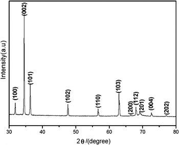
The XRD pattern (Fig. 1) showed that all diffraction peaks could be indexed to the hexagonal wurtzite phase of ZnO (JCPDS Card No. 89-0510) with lattice parameters a = 0.3248 nm and c = 0.5205 nm. The strong intensities relative to the background signal further verified the high purity and high crystallinity of the obtained ZnO structures.
XRD pattern of obtained products in experiment I.

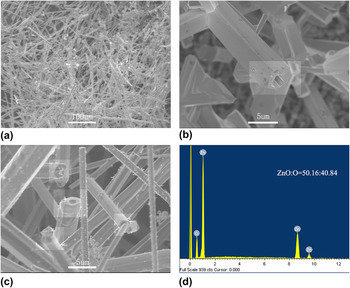
Figure 2 showed the SEM images of obtained product. The low-magnification image [Fig. 2(a)] demonstrated that bulk microrods deposited onto the substrate. The high-magnification images [Figs. 2(b) and 2(c)] revealed that these microrods possessed tubular structures with diameter of several micrometers. Further detailed observation indicated that the exterior configuration of these microtubes was hexagonal, but the interior configuration of hole was irregular.
SEM images of prepared products in experiment I: (a) low-magnification SEM image; (b, c) high-magnification SEM images, and (d) EDS focusing on the side plane of ZnO microtube.

B. Growth mechanism
As to these microtubes synthesized in our experiment, the distinct character was irregular configuration of hole, which was different with conventional regular hexagonal hole. This character indicated that the growth mechanism might be different with conventional one-step growth process. Generally, one-step growth process usually led to formation of tubular structure with regular hexagonal exterior and interior configuration. Obviously, these microtubes prepared in our experiment could not be well explained by the one-step growth mechanism.
Based on the point in Sec. I, the vapor pressure and growth temperature were two crucial factors determining the growth behavior of reagent’s species. In following sections, vapor pressure and growth temperature were all analyzed, and a two-step growth model was proposed to explain the formation of ZnO microtube.
1. Analysis of vapor pressure
In our experiment, Zn vapor reacted with oxygen to form ZnO. The reaction was as follows: Zn + O2→ ZnO. The oxygen mainly originated from the oxygen in the carrying gas. Its supply kept constant during the whole reaction. The Zn vapor came from two sources: direct evaporation of metal Zn powder and indirect carbon thermal reduction reaction of ZnO and carbon powder.
As to the Zn vapor provided by Zn powder, the evaporating rate was determined by the heating temperature. In our experiment (reacting temperature: 1000 °C), the high evaporating rate resulted in high Zn vapor pressure, which was higher than that of oxygen.Reference Yin, Chen, Dai, Liu, Zhang, Wang, Wu and Chai27 As the reaction proceeded, the Zn powder was largely consumed and would be exhausted after some time. This point could be confirmed by the EDS result of residue after reaction. It indicated that there was no residual Zn powder in the source. So, the supply of Zn vapor evaporated from Zn powder would disappear in the latter reacting stage. As a result, the Zn vapor pressure supplied by the Zn powder was unstable during the whole reaction: a high vapor pressure at the initial stage and no contribution at the latter stage.
As to the Zn vapor supplied by the carbon thermal reduction reaction of ZnO and carbon powder, it was gentle and steady.Reference Yan, Zhou, Xue and Zhang28 The consumption of ZnO in the whole reaction was not much, which could be validated by the abundant residue of ZnO powder in the source after reaction. So, the partial Zn vapor pressure coming from this part was low and constant during the whole reaction, which was smaller than that of oxygen.
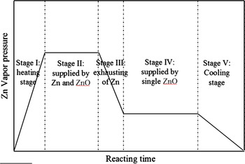
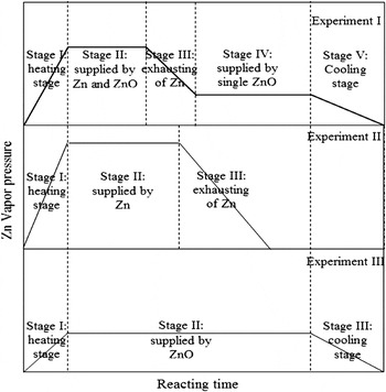
From above analysis, the changing curve of Zn vapor pressure during whole reaction could be divided into five stages, namely stage I: heating stage, stage II: supplied by Zn and ZnO, stage III: exhausting of Zn, stage IV: supplied by single ZnO, and stage V: cooling stage. At the initial reacting stage (before the Zn powder was exhausted), the supply of Zn vapor coming from both the rapid evaporation of Zn powder and the gentle carbon thermal reduction reaction of ZnO and carbon. Thus, the Zn vapor pressure prefers to remain high. At the latter reacting stage (after Zn powder was used up), the Zn vapor pressure would remain low due to the single supply from the gentle carbon thermal reduction of ZnO and carbon powder. The changing curve of Zn vapor pressure during experiment I was described Fig. 3.
Changing curve of reagent’s vapor pressure during whole reaction (Fig. 4).

2. Analysis of growth temperature
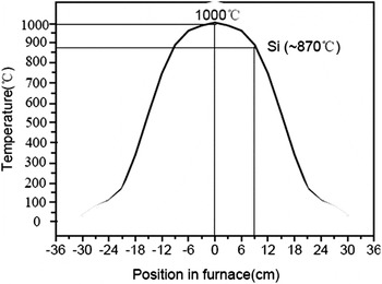
It was well known that the growth temperature was crucial factor to determine the migration probability of reagent’s species. Under high temperature, the reagent’s species had large opportunities transferring to preferential absorbed sites. The distributing curve of temperature in our horizontal tube furnace was presented in Fig. 4. From this distributing curve, it could be seen that the temperature of substrate was about 870 °C, which was much higher than the melting point 420 °C of Zn.
Distributing curve of temperature in the furnace in experiment I; the temperature of substrate was 870 °C.

3. Growth model
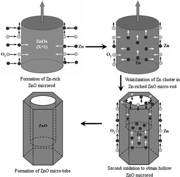
Concluded from the chemical bonding theory of single crystal growth,Reference Xue, Li, Liu, Sun and Chen29–32 a two-step growth model was proposed as shown in Fig. 5. At initial reacting stage, the Zn vapor pressure was higher than that of oxygen, which favored to form Zn-rich ZnO microrod; these redundant Zn species could coexist in ZnO lattice as interstitial Zn.Reference Wei, Lei and David33, Reference Zhang, Zhang, Xu, Wang, Chen and Yu34 It could be deduced that there were abundant structural defects (oxygen vacancies and Zn interstitials) in the formed ZnO microrod.
Growth model of microtube composed of four steps: (i) formation of Zn-rich ZnO microrod; (ii) second-volatilization of redundant interstitial Zn species along lateral and axial directions; (iii) formation of sunken tip in the Zn-rich ZnO microrod; and (iv) formation of microtube after reaction.

At the latter stages, the Zn powder was used up and the Zn vapor pressure was supplied by the single carbothermal reduction reaction of ZnO and carbon, which was lower than that of oxygen. The following lateral and axial growth of Zn-rich ZnO microrod was as follows.
As to the lateral growth, the Zn species had two sources. The first source was these collided Zn species from the vapor phase. Some of them transferred to the growing tip, some of them incorporated into the side crystal plane maintaining lateral growth, and the rest dissociated and got into the vapor phase again. The second source was these interstitial Zn coexisted in the Zn-rich ZnO microrod.Reference Zhang, Zhang, Xu, Wang, Chen and Yu34 Under high growth temperature of 870 °C, these interstitial Zn would gain enough energy to migrate via oxygen vacancies. Some of them transferred along axial direction to growing tip, while the others transferred along lateral direction via oxygen vacancies to outside plane maintaining lateral growth. After some time, the enough oxygen would lead to full oxidation of outside shell and formed approximately stoichiometric ZnO; this point was approved by the EDS focusing on the side plane as shown in Fig. 2(d); it could be seen that the atomic ratio of Zn and O was 50.16:49.84, and it was approximately equal to 1:1. After that, the lateral migration of interstitial Zn would stop, thanks to the absence of oxygen vacancies in outside shell. In addition, it was worth pointing out that the lateral growth would induce the formation of hexagonal exterior configurations terminated by equivalent index <0110> planes with low surface energy.Reference Yan and Zhou35
As to the axial growth, the Zn species also had two sources. One was colloided Zn species from vapor phase; these Zn species was composed of direct collided Zn species on the top plane and transferred Zn species from the side planes. The other was these interstitial Zn species coexisted in the Zn-rich ZnO microrod. Based on above analyses, though the lateral transference of interstitial Zn species would stop when stoichiometric ZnO outside shell formed, the axial transference would continued via oxygen vacancies existed in Zn-rich core. Obviously, second-volatilization of interstitial Zn would result in destroy of crystalline lattice.Reference Gao and Wang19 If the interstitial Zn in ZnO microrod was enough abundant, their volatilization would result in collapse of core and form hollow tubular structure. In our experiment, the Zn vapor pressure was far higher than that of oxygen at initial reacting stage as shown in Fig. 3. So there was abundant interstitial Zn coexisted in formed ZnO microrod. As a result, the second-volatilization of abundant interstitial Zn species under high temperature would result in the formation of hollow tubular structure.
C. Validation of growth mode
In our proposed growth mode, the Zn vapor pressure and growth temperature were two crucial factors determining the formation of tubular structure. To prove our growth model, four another experiments (defined as experiment II, III, IV, and V) were further conducted to observe reagent’s growth behavior under different Zn vapor pressure and growth temperature with higher and lower value, respectively.
1. Growth analysis under different Zn vapor pressure
Comparing with experiment I, experiments II and III were designed to analyze the growth behavior of reagent’s species under higher and lower Zn vapor pressure, respectively. Just as described above, the Zn powder would provide higher Zn vapor pressure than that of ZnO and carbon at same heating temperature. Herein, pure Zn powder and mixed powder of ZnO and carbon (molar ratio 1:1) were adopted as source in experiments II and III to obtain higher and lower Zn vapor pressure, respectively.
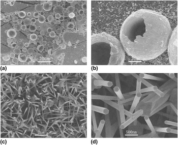
In experiment II, it was found that the Zn powder was completely exhausted after reaction, which indicated that the Zn vapor pressure would gradually decrease down to zero in latter reacting stages. Figures 6(a) and 6(b) exhibited the SEM images of obtained products in experiment II. From Fig. 6(a), it could be seen that abundant microballs deposited onto the substrate with diameter about 10 μm. Magnified image [Fig. 6(b)] indicated that these microballs possessed hollow structure.
(a) Low- and (b) high-magnification SEM images of products adopting pure Zn powder as source; (c) low- and (d) high-magnification SEM images of products adopting ZnO and carbon powder as source.

In experiment III, abundant residue of ZnO and carbon was found after reaction, which indicated that the supply of Zn vapor pressure from ZnO and carbon was persistent during whole reaction. Figures 6(c) and 6(d) presented SEM images of products. From Fig. 6(c), it could be seen that nanorod arrays deposited onto the substrate with diameter about 200 nm. The magnified image [Fig. 6(d)] indicated that these nanorods possessed perfect ZnO hexagonal configuration.
According to above analyses in Sec. III. B. 1, the change of Zn vapor pressure in three experiments I, II, and III was presented in Fig. 7. In experiment II, the pure Zn powder had higher vapor pressure than that of mixed powder of Zn, ZnO, and carbon in experiment I, and the Zn powder would be exhausted due to its quick evaporation after some time. So, the change of Zn vapor pressure was composed of three parts, namely stage I: heating stage, stage II: reacting stage, and stage III: exhausting stage. In experiment III, the mixed powder of ZnO and carbon provided persistent and low Zn vapor pressure via gentle and steady carbothermal reduction reaction. So, the change of Zn vapor pressure also had three parts, namely stage I: heating stage, stage II: reacting stage, and stage III: cooling stage.
Changing curves of reagent’s vapor pressure during whole reaction in experiments I, II, and III.

Based on our proposed growth mode, the growth behavior of reagent’s species was discussed in experiments II and III as follows.
In experiment II, higher Zn vapor pressure preferred to form Zn-rich ZnO structure with higher proportion of interstitial Zn, and latter second-volatilization and oxidation would result in formation of hollow structure. It was worth noting that the obtained structure was ball shaped instead of 1D structure. The formation of ball-shaped structure could be ascribed to isotropic growth under high vapor pressure.Reference Yan, Zhou, Zhang, Zhang and Hu36, Reference Hao, Meng, Ye, Zhang and Zhang37
In experiment III, the Zn vapor pressure was lower than that of oxygen, so there was no redundant interstitial Zn in the formation ZnO 1D nanorod. The growth of 1D nanorod could be ascribed to anisotropic growth under low vapor pressure.Reference Yan, Zhou, Zhang, Zhang and Hu36, Reference Hao, Meng, Ye, Zhang and Zhang37
From above experimental results, it could be seen that formation of Zn-rich structure was important to obtain hollow structure.
2. Growth analysis under different heating temperature
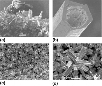
Comparing with experiment I, experiments IV and V were designed to analyze the growth behavior of reagent’s species under higher and lower growth temperatures. Accordingly, the heating temperature was set to 1150 and 850 °C, respectively. Figures 8(a) and 8(b) exhibited SEM images of products in experiment IV. Figure 8(a) presented that abundant microtubes formed with larger diameter about 10 μm. Magnified image [Fig. 8(b)] indicated that some small-sized nanorods grew up along inside wall of hole. Figures 8(c) and 8(d) showed SEM images of products in experiment V. From Fig. 8(c), it could be seen that abundant microrods were synthesized with smaller diameter about 1 μm. The magnified image [Fig. 8(d)] further indicated that there was a small hole on tip of some microrods.
(a) Low- and (b) high-magnification SEM images of products with heating temperature increased to 1150 °C; (c) low- and (d) high-magnification SEM images of products with heating temperature decreased to 850 °C.

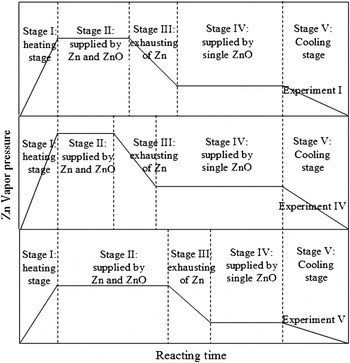
According to above analyses in Sec. III. B. 1, the change of Zn vapor pressure in three experiments I, IV, and V was presented in Fig. 9. Obviously, high heating temperature would accelerate evaporation of Zn powder and carbothermal reduction reaction of ZnO and carbon, which resulted in high Zn vapor pressure. In contrast, low heating temperature resulted in low Zn vapor pressure. So, their changing curves of Zn vapor pressure in three experiments had similar trend with different value.
Changing curves of reagent’s vapor pressure during whole reaction in experiments I, IV, and V.

Based on proposed growth mode, the growth behavior of reagent’s species in experiments IV and V was discussed as follows.
In experiment IV, the higher Zn vapor pressure would result in formation of Zn-rich ZnO microrod with higher proportion of interstitial Zn. The second-volatilization of these interstitial Zn would result in formation of tubular structure. In addition, the larger diameter could be ascribed to quicker lateral growth under the higher Zn vapor pressure,Reference Yan and Zhou38 and the second-volatilization of the large-sized Zn-rich core would lead to formation of large-sized hole. This point could be validated by the magnified SEM image [Fig. 8(b)]; it could be seen that the size of the hole was about 5–6 μm. As a result, oxygen could directly transfer into the hole and reacted with the second-evaporated interstitial Zn, which induced the growth of small-sized nanorods along inside wall.
In experiment V, the lower Zn vapor pressure led to formation Zn-rich ZnO microrod with less interstitial Zn. Furthermore, lower growth temperature also results in decrease of migration probability of interstitial Zn species coexisted in ZnO microrod. As a result, low migration probability led to incomplete second-volatilization, which favored to form small and irregular hole on the tip of some microrods.
From above experimental results, it could be concluded that full evaporation of interstitial Zn under enough high temperature was crucial for the formation of hollow structure.
Through these four experiments, the growth behavior of reagent’s species under different Zn vapor pressure and the growth temperature were fully discussed. These experimental results further indicated that the Zn vapor pressure and growth temperature were two crucial factors in the growth of tubular structure, and the rationality of our proposed two-step growth model could be well approved.
IV. CONCLUSIONS
In conclusion, ZnO microtubes were well prepared using mixed powder of Zn, ZnO, and carbon as source via chemical vapor deposition method. The growth process was well discussed, the high Zn vapor pressure and high growth temperature were considered as two crucial factors determining formation of hollow structure, and a two-step growth model was proposed. In the growth process, the high Zn vapor pressure led to formation of Zn-rich ZnO structure with abundant interstitial Zn; the high growth temperature induced to second-volatilization of these interstitial Zn and formed hollow structure. Four another experiments were designed to analyze the growth behavior under different Zn vapor pressure and growth temperature. These experimental results further approved proposed two-step growth model. Our growth analyses were helpful in comprehending the microcosmic growth behavior of reagent species in the vapor growth route, and our experimental method provided a feasible route to prepare hollow building blocks for functional nanodevices.
ACKNOWLEDGMENTS
This work was financially supported by the Foundation for New Teachers of Education (Grant No. 20090133120004) and the Science Foundation of Shandong Province (Grant No. ZR2010AQ014).