Introduction
Chaenotheca (Th. Fr.) Th. Fr. is a genus among the calicioid lichens that has attracted a fair amount of attention. It has recently been placed in Coniocybomycetes M. Prieto & Wedin (Prieto et al. Reference Prieto, Baloch, Tehler and Wedin2013). Chaenotheca was revised by Tibell (Reference Tibell1980) and has subsequently been included in a number of regional revisions: Australasia (Tibell Reference Tibell1987), South America (Tibell Reference Tibell1996, Reference Tibell1998), North America (Peterson & Rikkinen Reference Peterson and Rikkinen1999; Selva & Tibell Reference Selva and Tibell1999; Selva Reference Selva2010, Reference Selva2013, Reference Selva2014; McMullin & Arsenault Reference McMullin and Arsenault2016) and Northern Europe (Tibell Reference Tibell and Ahti1999). It has been noted that some Chaenotheca species occur in habitats with long forest continuity and high species diversity (Tibell Reference Tibell1992; Selva Reference Selva and Davis1996; Maloof Reference Maloof2016). A key to the European species was provided by Tibell (Reference Tibell and Ahti1999) but this key did not include C. servitii Nádv, although this species was described in some detail in Tibell (Reference Tibell1980) and further notes are given below. Several species have been included in Red Lists of countries in both the Northern and Southern Hemispheres. Many taxonomic problems still remain in the genus, and additional species can undoubtedly be found in underexplored areas. Thus a rich diversity has recently been discovered in the Himalayas (unpublished results of the first author) and most of these species have still not been described.
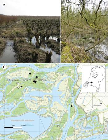
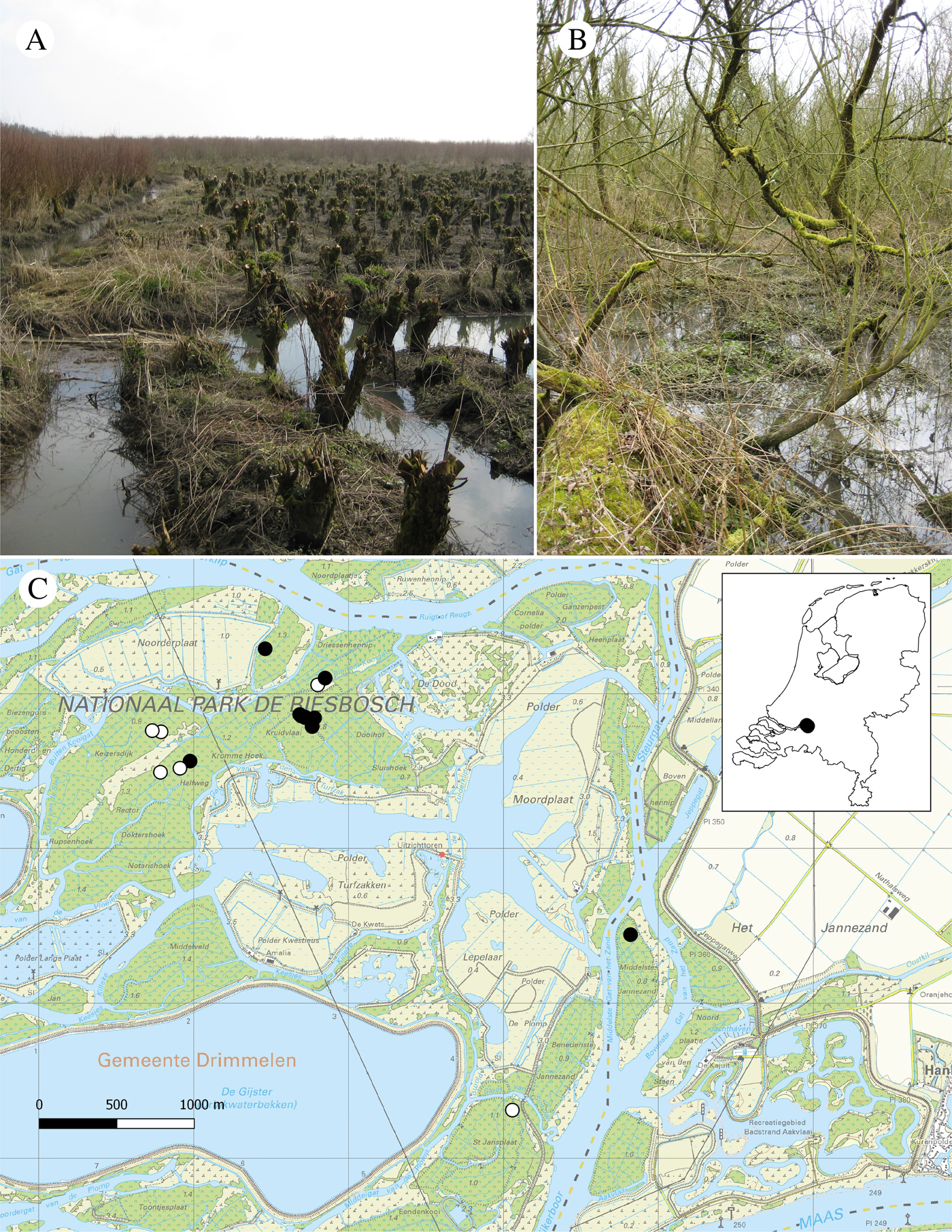
In the Netherlands, willow coppices (in Dutch: ‘grienden’) were for many centuries a common form of land use in the freshwater tidal areas of the Meuse and Rhine Rivers. In an extremely fertile and wet environment, willows (mainly Salix alba L. or Salix × rubens - S. alba × S. fragilis L.) were planted on narrow beds of clay drained by numerous ditches (Fig. 1A). Every three or four years, branches were harvested from these trees. In recent times most of these coppices have been abandoned and developed into semi-natural willow forests (Fig. 1B). Large areas of such woods occur in the Biesbosch National Park near the city of Dordrecht. In the past there was a tidal amplitude of c. two metres. After the closure of the Volkerak and Haringvliet estuaries by dams in 1969 and 1970 an amplitude of mostly 20–30 cm, and locally to 80 cm, remained. The greater part of the park is accessible only by boat.
Freshwater tidal habitats in the Biesbosch National Park. A, willow coppices, location: ‘Sterling’, 2009; B. Salix woodland, c. 50 years old, location: ‘Ottergriend’, 2009; C, distribution of Chaenotheca biesboschii in the Biesbosch. Filled circles = vouchered specimens included in this paper; open circles = field observations or photographs. In colour online.
The willow forests of the Biesbosch have been intensively surveyed and have proved to be rich in bryophytes and lichens (e.g. van der Pluijm Reference van der Pluijm1995, Reference van der Pluijm2000). In the 1980s and 1990s most of the interesting discoveries, including several species new to the Netherlands, were pioneer epiphytes on ‘young’, 12–20-year old branches of the former coppices. Thus the moss Orthotrichum rogeri Brid. was found in several locations (van der Pluijm Reference van der Pluijm1991). It is virtually endemic to Europe and is included in Annex II of the EU Habitats Directive. In the Biesbosch near Sliedrecht, Timmia megapolitana Hedw. occurs on mud-covered stems subjected to daily flooding (van der Pluijm Reference van der Pluijm1993). Recently, in Western Europe outside of the Biesbosch it has been found only in a freshwater tidal area in Norfolk, England (Porley & Ellis Reference Porley and Ellis2002). Most willow forests in the National Park date from c. 1970 but the so-called Grienden van de Dood (‘Coppices of Death’) in the central area of the Brabantse Biesbosch, go back to 1950. With the aging of the forest, variation in habitats has increased and solitary old trees, fallen trees that regenerate by new vertical stems, rotting logs, pit-and-mound topography from wind-thrown trees, and gaps in the canopy can now be found in the woods. Changes in the floristic composition of the woodlands are still ongoing. Since 2010, several ‘old-woodland’ lichens have even established themselves such as Phaeographis dendritica (Ach.) Müll. Arg., P. smithii (Leight.) B. de Lesd., Graphis inustuloides Lücking, Sporodophoron cretaceum (Hue) Ertz & Frisch and Arthonia cinnabarina (DC.) Wallr. (van der Pluijm Reference van der Pluijm2015; van der Pluijm & van Dort Reference van der Pluijm and van Dort2016).
Calicioid lichens were searched for on a field trip in 2016 to the ‘Grienden van de Dood’, and an intriguing Chaenotheca was discovered by J.-J. Spaargaren and M. Langbroek. On later excursions additional material was collected in several other locations in the Biesbosch. The aims of this paper are to ascertain the identity and relationships of this unidentified Chaenotheca, and to provide data on its ecology and local distribution.
Material and Methods
Material
Material was collected from a total of nine localities in the Biesbosch area (The Netherlands, c. 51°45′N, 4°49′E; Fig. 1C). There are nine filled circles on Fig. 1C representing nine vouchered localities: Doolhof (5), Palen Hennip (1), Driessen Hennip (1), Middelste Jannezand (1) and also Rector (1). However, only eight filled circles are discernible since two in Doolhof are overlapping. In addition, there are six open circles on the map that were not documented with a collection.
For the anatomical observations, mature apothecia were sectioned longitudinally with a freezing microtome. A 5% solution of gum arabic was used as an embedding medium. Forty mature spores were randomly selected and measured.
DNA extraction and sequencing
The holotype specimen had been stored for four weeks at room temperature. Twelve apothecia, lacking any visible symptoms of fungal infection, were selected, placed in 1·5 ml Eppendorf tubes and a small droplet of ultrapure water was added. After 2 h the Eppendorf tube was immersed in liquid nitrogen at −196 °C and the material was crushed with a small pestle. The DNeasy Plant Mini Kit (Qiagen) was used for isolating total DNA following the manufacturer's instructions. Diluted (1:10) or undiluted DNA was used for PCR amplifications with AccuPower® PCR PreMix (Bioneer) adding 3 µl of DNA extraction, 1·5 µl of each primer (10 µM) and water to a total volume of 20 µl.
Primers used for the ITS region were ITS1F (Gardes & Bruns Reference Gardes and Bruns1993) and ITS4 (White et al., Reference White, Bruns, Lee, Taylor, Innis, Gelfand, Sninsky and White1990). Thermal cycling parameters were: initial denaturation for 4 min at 95 °C, followed by 35 cycles of 1 min at 94 °C, 1 min at 54 °C, 45 s at 72 °C, and a final elongation for 5 min at 72 °C. Amplification products were visualized on 0·5% agarose gels stained with GelRed. The product was cleaned with Illustra™ ExoProStar (Thermo Fisher Scientific), following the protocol of the manufacturer. The DNA amplification product was sequenced in both directions by using the above mentioned primers also as sequencing primers (5 µM). Sequencing, automated reaction clean up and visualization were carried out by Macrogen Inc. (www.macrogen.com). The sequence chromatograms were inspected for quality and base-calling accuracy with Chromas v. 2.6.5 (http://technelysium.com.au/wp/chromas/), and finally edited to contigs using BioEdit v. 7.0.5 (http://www.mbio.ncsu.edu/bioedit/bioedit.html).
Alignments and phylogenetic analyses
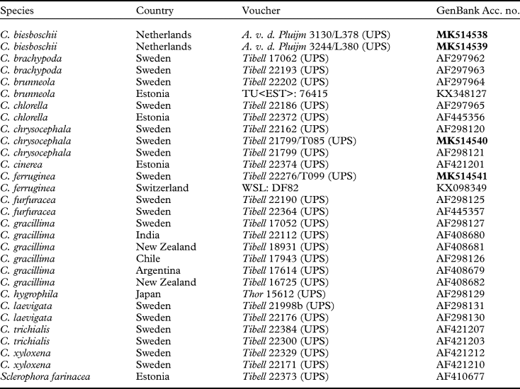
The two ITS sequences of the taxon discovered in the Biesbosch, together with two other newly generated sequences were aligned along with 26 sequences downloaded from GenBank (Table 1) using MAFFT v.7 (Katoh & Standley Reference Katoh and Standley2013; Katoh et al. Reference Katoh, Rozewicki and Yamada2017) on an online server (http://mafft.cbrc.jp/alignment/server/). An alignment was created utilizing the L-INS-I iterative refinement method and using the default settings (gap opening penalty = 1·53 and offset value = 0·00). The alignment was additionally edited using AliView (Larsson Reference Larsson2014). Sclerophora farinacea was chosen as outgroup based on the phylogeny presented by Prieto et al. (Reference Prieto, Baloch, Tehler and Wedin2013). The ITS data matrix contained 495 unambiguously aligned sites. The best-fit model of DNA evolution for the ITS data matrix was estimated with TOPALi v2.5 (Milne et al. Reference Milne, Wright, Rowe, Marshal, Husmeier and McGuire2004) using the Akaike Information Criterion (AIC) implemented in ‘Model Selection’. Phylogenetic relationships and confidence were inferred using a Bayesian approach. Additional support values were estimated with the program for Maximum-likelihood based phylogenetic inference, RAxML, using Maximum Likelihood bootstrapping (MLbs).
Specimens used in an ITS-based phylogenetic analysis of Chaenotheca species together with their GenBank Accession numbers. New sequences are in bold.

Using the AIC, the Bayesian analysis employed the GTR + G model. Two analyses of two parallel runs were carried out for 10 M generations using MrBayes v3.2.6 (http://mrbayes.sourceforge.net/). Each run included four chains and trees were sampled every 1000 generations. All runs converged on the same average likelihood score and topology. A burn-in fraction of 25% was discarded from each run.
RAxML v7.7.1 (Stamatakis et al. Reference Stamatakis, Hoover and Rougemont2008) as RAxML BlackBox (https://embnet.vital-it.ch/raxml-bb/) was used for the MLbs analysis with 500 bootstrap replicates and a single model of molecular evolution (GTRCAT). Bayesian posterior probabilities (PP) ≥95% and MLbs ≥70% were considered to be significant.
Phylogenetic trees were visualized using FigTree v1.4.3 (Rambaut Reference Rambaut2012). Adobe Illustrator CC 2017.0.2 was used for artwork.
Results
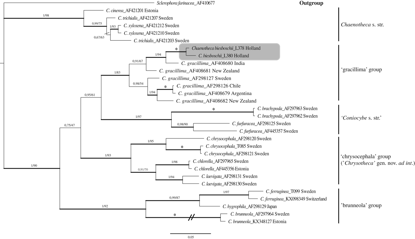
Analyses were based on a total of 29 ITS sequences representing 13 species of Chaenotheca (Table 1), with Sclerophora farinacea as outgroup (Fig. 2). Chaenotheca biesboschii, in a phylogeny of the ITS region along with 27 other Chaenotheca sequences, is most closely related to C. gracillima (Vain.) Tibell (Fig. 2). Several strongly supported monophyletic groups were discovered (Fig. 2) and were given provisional names and/or ad int. names, such as the ‘brunneola’ group (PP = 1, MLbs = 92), the ‘chrysocephala’ group (‘Chrysotheca’ gen. nov. ad int., PP = 1, MLbs = 93), Coniocybe s. str. (PP = 1, MLbs = 97), the ‘gracillima’ group (PP = 1, MLbs = 83) and Chaenotheca s. str. (PP = 1, MLbs = 98). The provisional names for ‘Chrysotheca’ and ‘Coniocybe’ are used, in anticipation of the recognition of a new genus and an emendation of Coniocybe Ach. (S. Tibell, G. Hillman & L. Tibell, unpublished data). Problems at the species level were also revealed, such as the relationships within the ‘gracillima’ group and within Chaenotheca s. str., the full resolution of which, however, are outside the aim of this paper. In the phylogenetic analysis, C. biesboschii belongs in the ‘gracillima’ group, and interestingly is closest to a specimen of C. gracillima from India.
Phylogenetic relationships among 29 sequences (Table 1) representing 13 species of Chaenotheca based on a Bayesian analysis of an ITS dataset. The tree was rooted using Sclerophora farinacea. The two support values associated with each internal branch correspond to PP and MLbs proportions, respectively. Branches in bold indicate a support of PP ≥95% and MLbs ≥70%. An asterisk on a bold branch indicates that this node has a support of 100% for both support estimates. The branch with the double slash is shortened. The informal name ‘Chrysotheca’ is being used as a place-holder for the recognition of a new genus and here indicated with an ad int. name. Similarly, ‘Coniocybe s. str.’ is used for a provisional recognition of an emendation of Coniocybe. Chaenotheca biesboschii is highlighted by a shaded box.

Taxonomic treatment
Chaenotheca biesboschii Tibell & van der Pluijm sp. nov.
MycoBank No.: MB 828546
Chaenotheca biesboschii has an insignificant thallus, yellow to brown pruinose apothecia, spherical spores and Stichococcus as photobiont. It is somewhat similar to C. gracillima but differs in having shorter and usually not flexuous stalks, and slightly larger spores. It is also similar to C. servitii but differs in having sturdier apothecia with much wider capitula and by having a swollen, ring-shaped excipulum rather than the gradually and evenly thickened, conical excipulum of C. servitii.
Type: The Netherlands, N-Brabant, Werkendam, Biesbosch, Middelste Jannezand, 51°44′7·69″N, 4°50′56·29″E, on wood of decorticated stump of Salix, 2 m above the ground, 8 January 2017 A. v. d. Pluijm 3244 (DNA-extraction L380, UPS—holotype; L—isotype; GenBank Accession no. MX514539).
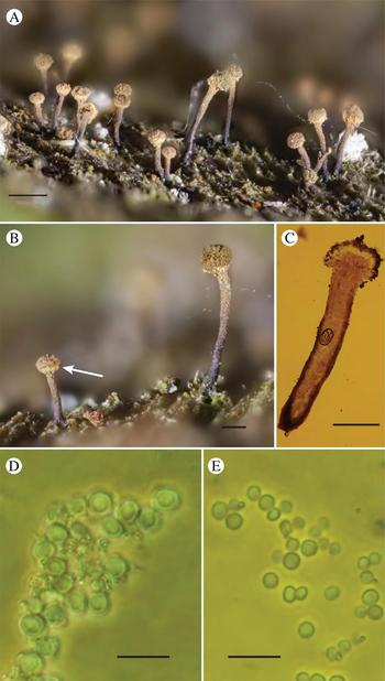
(Fig. 3)
Thallus immersed, smooth to superficial and minutely granular, glaucous green. Apothecia 0·9–1·4 mm high; capitulum 0·2–0·3 mm diam.; stalk 0·04–0·07 mm diam., when young the capitulum on the lower side has a ring-like thickening and is covered by a yellow pruina; when mature the stalk is rusty brown. The stalk surface at maturity cracks to form small, irregular to polygonal projections rendering an impression of a rusty pruina, although there is no deposition of a true crystalline pruina. Asci formed singly, well stalked. Spores spherical, 3·5–5·5 µm diam., ornamented by irregular cracks, medium brown. Photobiont: Stichococcus.
A–D, morphology and anatomy of Chaenotheca biesboschii (holotype): A, mature and young apothecia on thin thallus; B, apothecia (note yellow thickening at the base of the capitulum of the young apothecium (arrow) and the prominent brown pruina of the mature apothecium to the right); C, longitudinal section of apothecium with thickened excipulum; D, released, mature spores. E, released, mature spores of C. gracillima, at the same magnification and in the same (mixed) preparation as in D (Tibell 23030, UPS). Scales: A = 0·5 mm; B & C = 0·2 mm; D & E = 10 μm.

Etymology
Named after the Biesbosch National Park, origin of the holotype.
Habitat and distribution
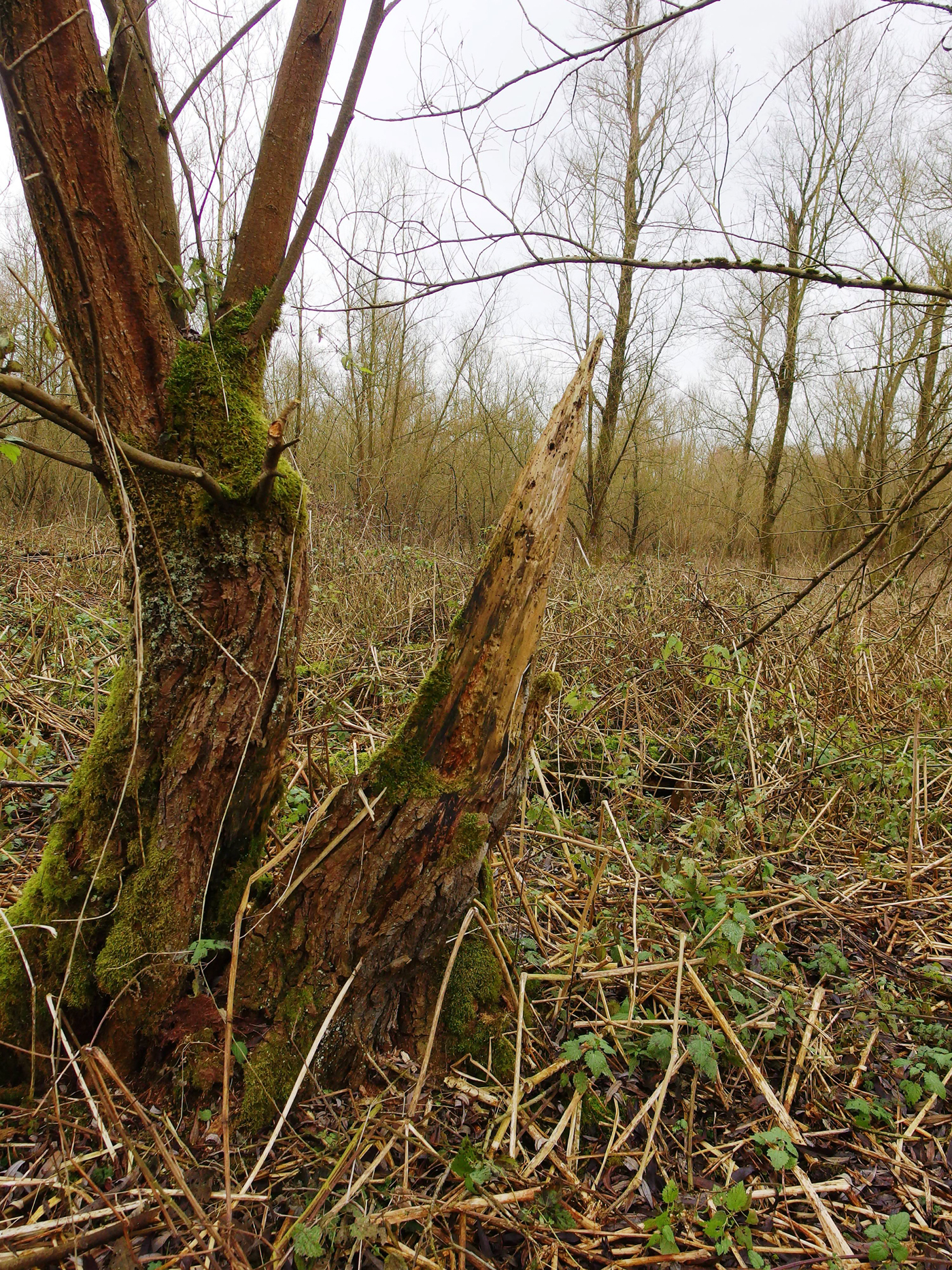
Found on decorticated wood of Salix in willow forests in freshwater tidal areas of rivers (Fig. 4). For its distribution, see Fig. 1C.
Selected additional specimens examined. The Netherlands: Biesbosch, Doolhof, 51°44′52·47″N, 4°49′9·42″E, 1·3–1·5 m high on soft rotten stump of Salix, L. B. Sparrius 9215 & A. v. d. Pluijm 3130, DNA-extraction L378 (hb. van der Pluijm).
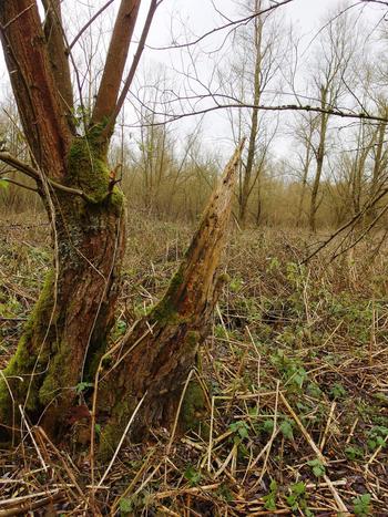
Habitat of Chaenotheca biesboschii: decorticated stump of Salix in willow forest, Biesbosch National Park, location: ‘Doolhof’, 2016. In colour online.
Discussion
Taxonomic position
Chaenotheca biesboschii might be mistaken for C. gracillima, and in a phylogenetic analysis of the ITS region it was most closely related to this species (Fig. 2), more specifically to a specimen from India. The wide sample of sequences tagged C. gracillima in the analysis, however, is heterogeneous, and C. gracillima as conceived here might be a complex including a number of hitherto unrecognized species. In the following discussion, C. gracillima pertains to the genotype suggested to be most closely related to the type (from Finland). Morphologically, C. biesboschii differs from C. gracillima in having: shorter and usually not flexuous stalks; apothecia with a ratio between the height and stalk width of 8–35 compared to 24–40 in C. gracillima; initially a yellow pruina on the young capitula; larger spores (2·5–3·5 µm in C. gracillima). The two ITS sequences obtained from C. biesboschii (gathered from locations 2·5 km apart, Table 1) proved to be of the same haplotype and were compared with sequences of C. gracillima originating from Argentina, Chile, India, New Zealand and Sweden. The latter collection (Tibell 17052, AF298127) was obtained from a location rather close to the type locality and is thus probably genetically quite similar to the type of C. gracillima. In a pairwise comparison there was a difference of 27 SNPs and four indels between the types of C. biesboschii and C. gracillima from Sweden (Tibell 17052). In an analysis of all available sequences, including those from extra-European areas, the sequences of C. gracillima differ and are part of two moderately supported groups, one of which contains C. biesboschii. Chaenotheca gracillima as conceived here may thus represent separate entities in a complex of species (Fig. 2) and further extensive work is required to clarify this situation.
Chaenotheca biesboschii is morphologically similar to C. servitii, but in the material studied (Tibell Reference Tibell1980), the latter differed in having taller apothecia (1·6–2·5 mm high) with flexuous stalks with a ratio between the height and stalk width of 27–42 as compared to the sturdier apothecia of C. biesboschii that have a ratio of 8–35, narrower capitula (0·10–0·15 mm wide) compared to C. biesboschii (0·2–0·3 mm) and smaller spores (3·8–4·2 µm). In addition, the thallus of C. biesboschii is more coarsely granular and the excipulum, although quite thick, is not evenly conical as it is in C. servitii, but is quite low and juts out abruptly; thus the capitulum on the lower side displays a ring-like thickening. Both C. servitii and C. biesboschii are lignicolous and occur on Salix, with the former found in the vicinity of Velké Kapušany in eastern Slovakia and the latter in coastal woods in tidal areas of the Atlantic zone. Chaenotheca servitii is known only from the type locality and the collection distributed in Nádvornik's exsiccate (Nádvorník, Caliciaceae exsiccatae No 6) and studied by Tibell (Reference Tibell1980) has been quite fragmented when distributed. This material was collected by Nádvorník at, or very close to, the type locality. A conventional isolation of DNA from this material is not feasible and further work on the molecular characterization of C. servitii has to await the discovery of additional material or the application of a more advanced methodology requiring less material.
Ecology and local distribution
In the woodlands of the Biesbosch many willow trees have succumbed to parasitic fungi such as Armillaria spp. Chaenotheca biesboschii was found on rotten lignum of partly decorticated but still standing trunks and also on thick branches of Salix in wet willow forests (Fig. 4), 0·5–2·5 m above the ground. The substratum is typically very soft. It is accompanied here by free-living algae but no other lichens or bryophytes are usually present. On a few occasions, however, Hypnum cupressiforme Hedw., Chaenotheca brachypoda (Ach.) Tibell, C. hispidula (Ach.) Zahlbr, Lecanora expallens Ach., Micarea misella (Nyl.) Hedl., Opegrapha vermicellifera (Kunze) J. R. Laundon, O. herbarum Mont. and O. vulgata (Ach.) Ach were found as accompanying species.
In 2016 and 2017, further excursions in search of C. biesboschii were undertaken by the third author. These led to the discovery of several new locations also included in Fig. 1C. Suitable habitats with dead wood today seem abundant but Chaenotheca biesboschii was found only in a limited number of localities. The woodland parcels called ‘Keizersdijk’, ‘Rektor’, ‘Halfweg’, ‘St.-Jansplaat’ and ‘Middelste Jannezand’ (Fig. 1C) were surveyed thoroughly, but so far C. biesboschii has mainly been found in the north-eastern parts. This might be explained by the predominantly westerly winds in the Netherlands and a preference of C. biesboschii for sheltered niches.
Chaenotheca biesboschii, overlooked or a recent establishment?
So far Chaenotheca biesboschii has been found at more than 15 locations in the Biesbosch (Fig. 1C). It is surprising that an undescribed species of Chaenotheca has turned up in the Netherlands, considering that most parts of Europe have been quite well investigated lichenologically. We find it unlikely that it has been overlooked previously, and there are some indications that it might be a recent arrival from outside the Netherlands. The woodlands of the Biesbosch are not older than c. 60 years. Before that, cultures (rush and reed fields, willow coppices and agricultural polders) dominated the area. In any case, forests of some age on fertile, clayish soils are a fairly recent phenomenon in the Netherlands. In the past coppice woods, arable lands, hay fields and meadows were the dominant land uses. Therefore the habitats in which C. biesboschii is found today were probably very scarce or not available at all in the Netherlands in previous centuries.
Furthermore, the Biesbosch has been intensively studied by the third author since 1983. The minute Chaenotheca species might possibly have been overlooked, although from 1969 onwards the Biesbosch area has also been visited by many other experienced Dutch lichenologists. In reports of their early excursions to the willow forests, no Chaenotheca was ever mentioned. The diversity of this genus in the area has been revealed only in very recent years. In 1992, C. trichialis (Ach.) Th. Fr. was the first to be discovered. Chaenotheca brachypoda and C. chlorella (Ach.) Müll. Arg. were found in 1998. Since then, C. brachypoda in particular has become very common in the Biesbosch, where it occurs in crevices of bark on the rain-sheltered side of trees. In 2012 the eye-catching C. furfuracea (L.) Tibell was collected for the first time from an upturned tree root of a windswept willow, and this species seems to have spread rapidly since then, also occurring on living trees. More recently in 2016, C. hispidula and C. stemonea (Ach.) Müll. Arg. were discovered (BLWG 2017).
Finally, the characteristics of dead wood and dead branches in older woods of the Biesbosch have changed significantly since c. 1990. This is indicated by the fact that mosses and lichens that prefer mildly acidic and oligotrophic habitats have declined dramatically here. Common species that were abundant or frequent on dead wood in the 1980s (van der Pluijm Reference van der Pluijm1995) have now almost completely disappeared, such as Dicranoweisia cirrata (Hedw.) Lindb., Ceratodon purpureus (Hedw.) Brid., Aulacomnium androgynum (Hedw.) Schwägr., Dicranum scoparium Hedw., D. montanum Hedw., Ptilidium pulcherrimum (Weber) Vainio, Lophocolea heterophylla (Schrad.) Dumort., Cladonia spp., Hypogymnia physodes (L.) Nyl., Platismatia glauca (L.) W. L. Culb. & C. F. Culb., Pseudevernia furfuracea (L.) Zopf and Usnea spp. In contrast, mosses such as Amblystegium serpens (Hedw.) Schimp., Brachythecium rutabulum (Hedw.) Schimp., Hypnum cupressiforme and Kindbergia praelonga (Hedw.) Ochyra are dominant today, indicating that dead wood of Salix has become more eutrophicated and now exhibits a higher pH. These local observations are in accordance with national trends. Decreased levels of SO2 and increased levels of NH3 in the atmosphere over previous decades, combined with increased temperatures, are probably causes of these changes (van Herk et al. Reference van Herk, Aptroot and van Dobben2002). This development also makes a relict status of Chaenotheca biesboschii on dead wood less likely.
We can, however, only speculate about the geographical origin of C. biesboschii. In recent decades in the Netherlands, many newly established lichens, or lichens that are increasing in frequency, originate from southern regions (van Herk et al. Reference van Herk, Aptroot and van Dobben2002; Aptroot & van Herk Reference Aptroot and van Herk2007). It is possible that the occurrence of C. biesboschii is the result of a similar rather recent dispersal. A recent example of a new, southern lichen in the Biesbosch is Rinodina biloculata (Nyl.) Sheard (van der Pluijm Reference van der Pluijm2017). Therefore, perhaps C. biesboschii has arrived from a poorly investigated warmer area. The spores of Chaenotheca species are extremely small and might possibly be able to disperse over very long distances (Tibell Reference Tibell1994).
Key to Chaenotheca in Europe
Note. This is a key to the morpho-species as traditionally conceived. Several of the species named below are place-holders for complexes showing genetic diversification that in some instances may or may not overlap and several additional species may well be described, but this requires detailed investigations at the population level.
1 Lower side of capitulum and stalk at least partly covered by a yellowish green, yellow-reddish brown or brown pruina ………2
Lower side of capitulum and stalk at least partly with a white pruina or without pruina ………12
2(1) Pruina of mature apothecia yellowish green or yellow ……… 3
Pruina of mature apothecia reddish brown to brown ……… 10
3(2) Photobiont Trentepohlia or trebouxioid ……… 4
Photobiont Stichococcus .………. 8
4(3) Thallus intensely yellow ……… C. chrysocephala (Ach.) Th. Fr.
Thallus greenish, grey or brownish or immersed ……… 5
5(4) Photobiont Trentepohlia, thallus immersed ………C. hispidula
Photobiont trebouxioid, thallus not immersed ………6
6(5) Spores ellipsoid to cylindrical ……… C. laevigata Nádv.
Spores spherical ……… 7
7(6) Thallus minutely granular, whitish grey, apothecia 0·8–1·5 mm high ………C. subroscida (Eitner) Zahlbr.
Thallus olive to brownish, thick, verrucose, apothecia 0·5–1·2 mm high ………C. phaeocephala (Turner) Th. Fr.
8(3) Spores partly ellipsoid, thallus verrucose to granular, greyish green C. chlorella
All spores spherical, thallus farinaceous, intensely green or immersed ……… 9
9(8) Apothecia 1·6–2·6 mm high, mazaedium pale brown, spores minutely verrucose at high magnification ……… C. furfuracea
Apothecia 0·4–1·4 mm high, mazaedium medium brown, spores smooth but with minute fissures at high magnification ………C. brachypoda
10(2) Young capitula with a reddish brown pruina below, spores 2·5–3·5 µm diam. ……… C. gracillima
Young capitula with yellow pruina below, spores larger ………11
11(10) Capitula with collar-like swelling at the base, spores 3·5–5·5 µm diam ………C. biesboschii
Capitula with a conical, gradually thickened excipulum, spores 3·8–4·2 µm diam. ………C. servitii
12(1) Photobiont with tiny (< 5 µm) cylindrical cells forming chains (Stichococcus) ……… 13
Photobiont with larger (> 6 µm) spherical cells (trebouxioid or Dictyochloropsis) ……… 17
13(12) Thallus farinaceous ……… 14
Thallus immersed or episubstratic, not farinaceous ………15
14(13) Thallus Pd+ yellowish red, capitulum with a cobwebby felt of hyphae below, spores medium brown, 4·0–5·0 µm diam ……… C. stemonea
Thallus Pd−, capitulum without cobwebby hyphae on lower side, spores pale brown to hyaline, 2·5–3·0 µm diam ………C. gracilenta (Ach.) Mattsson & Middelb.
15(13) Lower part of stalk pale, excipulum edge irregularly split ………C. cinerea (Pers.) Tibell
Lower part of stalk black, excipulum edge not split………16
16(15) Thallus thin to thick, verrucose to squamulose, hyphae of the excipulum periclinal ……… C. trichialis
Thallus immersed, hyphae of the outer part of the excipulum anticlinal ……… C. xyloxena Nádv.
17(12) Thallus distinct, grey, with yellow, K+ red spots …... C. ferruginea (Turn.) Migula
Thallus immersed or episubstratic, without yellow spots ……… 18
18(17) Thallus immersed, photobiont Dictyochloropsis (cells > 10 µm diam.) ……… C. brunneola (Ach.) Müll. Arg.
Thallus episubstratic, granular, photobiont trebouxioid (cells < 10 µm diam.). 19
19(18) Asci irregular in shape, without stalks, formed in chains ………C. sphaerocephala Nádv.
Asci with stalks, not formed in chains ……… C. hygrophila Tibell
Author ORCIDs
Leif Tibell, 0000-0002-8629-7989.
Arno van der Pluijm wishes to thank Theo Muusse (Staatsbosbeheer) and Arie van der Ree for transportation by boat, and Laurens Sparrius for composing the distribution map. Georg Hillman is thanked for contributing Fig. 3A & B.