Introduction
Larval stage of genus Echinococcus is the causing agent for the zoonotic infection which is life-threatening known as Echinococcosis. After two decades of data accumulation on epidemiology, geographical data and biochemical data on Echinococcus spp and the analysis of nuclear and mitochondrial gene sequences as well as the phylogenetic relationship of species, limitations and contradiction of strains/genotypes system, the taxonomic revision of E. granulosus was reviewed as a result of which the recent taxonomic reshuffle consists of Echinococcus granulosus sensu lato which is further divided in to Echinococcus granulosus sensu stricto (G1–G3), Echinococcus equinus (G4), Echinococcus ortleppi (G5), Echinococcus canadensis (G6/G7 and G8–G10) causes cystic echinococcosis (CE) (Romig et al., Reference Romig, Ebi and Lato2015). CE is transmitted between two hosts, the intermediate hosts including herbivorous and omnivorous mammals and carnivorous i.e. dogs and other canids usually serve as definitive hosts, however, humans are considered as accidental host and serve as intermediate host (Galeh et al., Reference Galeh, Spotin, Mahami-Oskouei, Carmena, Rahimi, Barac, Ghoyounchi, Berahmat and Ahmadpour2018).
There is substantial data of prevalence of genotypes present in different countries, helping in the updating of information regarding genetic variations and geographical spread of Echinococcosis infection in livestock. Currently, there are three Echinococcus species which are E. granulosus s.s., E. equinus and E. canadensis (G6/G7) present in Turkey. Studies have shown the presence of E. granulosus s.s. being the prevailing species in Turkey (Simsek et al., Reference Simsek, Kaplan and Ozercan2011; Kesik et al., Reference Kesik, Simsek, Kilinc and Koroglu2019a, Reference Kesik, Kilinc, Simsek and Gulb). Previous records show that the CE has not been eliminated in Turkish population as 14 789 cases were reported during the period 2001–2005 (Yazar et al., Reference Yazar, Ok, Hokelek, Polat, Yilmaz, Ozbilge, Ustun, Koltas, Ertek, Sakru, Alver, Cetinkaya, Koc, Demirci, Aktas, Parsak, Ozerdem, Sakman, Cengiz, Ozer, Keklik, Yemenici, Turan, Dastan, Kaya, Tamer, Kilimcioglu, Turk, Sinirtas, Evci, Kilicturgay, Mutlu and Artis2008) and several new CE cases were recorded (Bakal et al., Reference Bakal, Simsek and Kazez2015; Sevinc et al., Reference Sevinc, Karahan, Simsek, Bakdik, Aksoy and Soydan2016).
Previous epidemiological studies were performed to assess the molecular and genetic variations of Echinococcus spp. in domestic animals from Iran, the eastern neighbour of Turkey. Available molecular evidence has been shown two species that infected livestock in Iran which includes E. granulosus s.s. and E. canadensis (G6 genotype) (Khademvatan et al., Reference Khademvatan, Majidiani, Foroutan, Hazrati Tappeh, Aryamand and Khalkhali2019). Besides, E. granulosus s.s and E. canadensis (G7 genotype) were prevalent in Greece where the west neighbour of Turkey (Roinioti et al., Reference Roinioti, Papathanassopoulou, Theodoropoulou and Simsek2016). Recent investigations have indicated the circulation of species E. canadensis (G6-G7) in Greece, Italy and Spain also in Central and Eastern Europe. However, G6 genotype of E. canadensis was found in two human isolates (Simsek et al., Reference Simsek, Kaplan and Ozercan2011).
Considering the previous studies and the present data about E. granulosus s.l., the objectives of the current study are the identification, molecular characterization and genotyping of the Echinococcus spp. in sheep and cattle samples collected from the slaughterhouse of Elazig, Turkey.
Material and methods
Sample collection
Hydatid cyst samples have been collected from EL-KAS slaughterhouse in Elazig province of Turkey between February and July 2019. In our study, 128 hydatid cyst isolates have been collected from lungs and liver of 85 animals (cattle, n = 66 and sheep, n = 19). The hydatid cysts were dissected and the germinal layers were collected into Eppendorf tubes containing 70% ethanol. Besides, the cysts collected from the same organs were stored in different Eppendorf tubes in order to check any nucleotide differences in the same organ hydatid cysts. All cyst samples were stored at −20°C till further use.
Genomic DNA isolation
Genomic DNA was isolated from teach individual cyst samples. The germinal layers were crushed by the surgical blade and washed at least 5 times with 1X PBS solution in order to eliminate the traces of ethanol. Then the gDNA extraction kit (RTA Genomic DNA Isolation Kit from tissue, Turkey) protocol has been followed.
Polymerase chain reaction (PCR) of target gene
Amplification of mt-CO1 gene having a fragment of 446 bp was performed using JB3 (5´-TTTTTTGGGCATCCTGAGGTTTAT-3´), and JB4.5 (5´-TAAAGAAAGAACATAATGAAAATG-3´) primer set (Bowles et al., Reference Bowles, Blair and McManus1992). The conditions used in the PCR reaction were: pre-denaturation step at 95 ˚C for 5 min following the 35 cycles (50 s at 94°C, 50 s at 45°C, 50 s at 72°C) and a final extension step (10 min at 72°C). Of the total, 1.4% stained ethidium bromide agarose gel was used in order to check the band quality in gel electrophoresis and electrophoretically separated PCR products were visualized under UV. The PCR bands with good results were then removed and were purified by using of QIAquick Gel Extraction Kit (Qiagen, Hilden, Germany). ABI PRISM Sequence Detection System was used to obtain sequences.
For the confirmation of E. canadensis (G6/G7) in a sheep isolate a long mt-CO1 gene primer sets also were used for PCR and following the sequence analyses were performed. A partial mt-CO1 gene (875 bp) was amplified by using the following primer pairs F/CO1 (5′-TTGAATTTGCCACGTTTGAATGC-3′) and R/CO1 (5′-GAACCTAACGACATAACATAATGA-3′) (Nakao et al., Reference Nakao, Sako, Yokoyama, Fukunaga and Ito2000). Amplification was performed according to the following temperature profiles: initial denaturation step of 10 min at 94°C and 30 cycles of 30 s at 94°C, 45 s at 52°C, 1 min at 72°C followed by a final extension step of 10 min at 72°C.
Data analysis by using bioinformatic tools
FinchTV 1.4.0 (Geospiza Inc., Seattle Washington, USA) (http://www.geospiza.com) has been used for viewing the sequencing data. ‘BLAST’ has been used to compare the sequences of our samples with the database and to find the genotype of each sample. The sequence ends were trimmed by comparing the published sequences using ‘BLAST’ search. The trimmed sequences were then added to ‘CLC Sequence viewer 8’ (Knudsen et al., Reference Knudsen, Knudsen, Flensborg, Sandmann, Heltzen, Andersen, Dickenson, Bardram, Steffensen, Mansted, Lauritzen, Forsberg, Thanbichler, Bendtsen, Gorlitz, Rasmussen, Tordrup, Vaerum, Ravn, Hachenberg, Fisker, Dekker, Schultz, Hein and Sinding2007). The alignment was performed with the published reference and out-group sequences retrieved from NCBI PubMed. The trimmed sequences were then added to MEGA X (Kumar et al., Reference Kumar, Stecher, Li, Knyaz and Tamura2018). For the sequence alignment and subsequent analysis of several output formats including PHYLIP, NEXUS and FASTA have been used which were generated by using ClustalW. The evolutionary history was investigated by using Maximum Likelihood method. Hasegawa-Kishino-Yano and Tamura-Nei model to create phylogenetic trees (Tamura and Nei, Reference Tamura and Nei1993). A discrete Gamma distribution was used to model evolutionary rate differences among sites (5 categories (+G, parameter = 0.1727)) (Kumar et al., Reference Kumar, Stecher, Li, Knyaz and Tamura2018).
Haplotype analysis
PopART- 1.7 (Population Analysis with Reticulate Trees) software was used in order to detect the haplotype diversities (Leigh et al., Reference Leigh, Bryant and Steel2015). Haplotypes were generated using Minimum Spanning Networks, and the relationships in between these haplotypes were demonstrated. The data were then uploaded to DnaSP 6 (Rozas et al., Reference Rozas, Ferrer-Mata, Sánchez-DelBarrio, Guirao-Rico, Librado, Ramos-Onsins and Sánchez-Gracia2017). Haplotype (h), haplotype diversity (Hd), nucleotide diversity (π), and the neutrality indices including Tajima's D (Tajima, Reference Tajima1989) and Fu's Fs are the indices of population diversity (Fu, Reference Fu1997).
Results
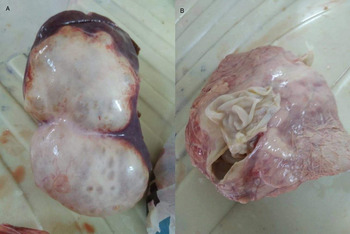
In our study, 85% of the hydatid cysts have been found in the lungs, whereas 15% were from the liver. After excluded the calcified and caseified cysts a total of 85 samples (cattle, n = 66 and sheep, n = 19) with clean germinal membranes were used for molecular analyses in the current study (Fig. 1). Multiple organ occurrences have been found in cattle 5, where cysts were present in both lungs and liver.
Showing the hydatid cysts with germinal layer, A: Sheep liver hydatid cysts contained cysts fluid, B: Dissected lung hydatid cysts and showing the germinal layer.

A total of 85 samples were sequenced. The genetic tree was generated through alignment of the sequences showing the phylogenetic relationship of samples with their reference sequences and out-groups retrieved from BLAST search. The results have shown the predominance of E. granulosus s.s. in all cyst samples except one sheep sample (Sample no: S2) which has shown resemblance with the E. canadensis (G6/G7) (Fig. 2). It has been confirmed with the using of long mt-CO1 sequence as well (Fig. 3). The detection of E. canadensis (G6/G7) in sheep is reported for the first time in Turkey. We have observed mutations (C→T and T→C substitution) at different positions within the sequences of E. granulosus s.s. Thus, we performed alignments on samples which were taken from the same cattle.
Comparison of phylogenies of different isolates using partial mt-CO1 (446 bp) sequences. The phylogenetic tree was constructed using the Maximum Likelihood method and HKY + G model. Evolutionary analyses were conducted in MEGA X. The GenBank accession numbers and the species name were given for each reference sequences as follow: KT382540 (E. granulosus s.s.), MN990735 (E. granulosus s.s.), KR920701 (E. granulosus s.s.), JX854028 (E. granulosus s.s.), NC021144 (E. felidis), KY766905 (E. equinus), AB461420 (E. multilocularis), KT382535 (E. ortleppi), AB235848 (E. canadensis, G8-10), MK321260 (E. canadensis, G6/G7), KX010854 (E. canadensis, G6/G7). The circle indicates the sequence of E. canadensis (G6/G7) from the present study.

Phylogenetic confirmation of E. canadensis (G6/G7) isolate using partial long mt-CO1 (875 bp) sequence. The phylogenetic tree was constructed using the Maximum Likelihood method and TN93 + G model. Evolutionary analyses were conducted in MEGA X. The GenBank accession numbers and the species name were given for each reference sequences as follow: KX010854 (E. canadensis, G6/G7), MH791328 (E. canadensis, G8-10), MN058591 (E. ortleppi), KP161207 (E. equinus), MK774655 (E. granulosus s.s.), EF558356 (E. felidis), AB461419 (E. multilocularis). The circle indicates the sequence of E. canadensis (G6/G7) from the present study.

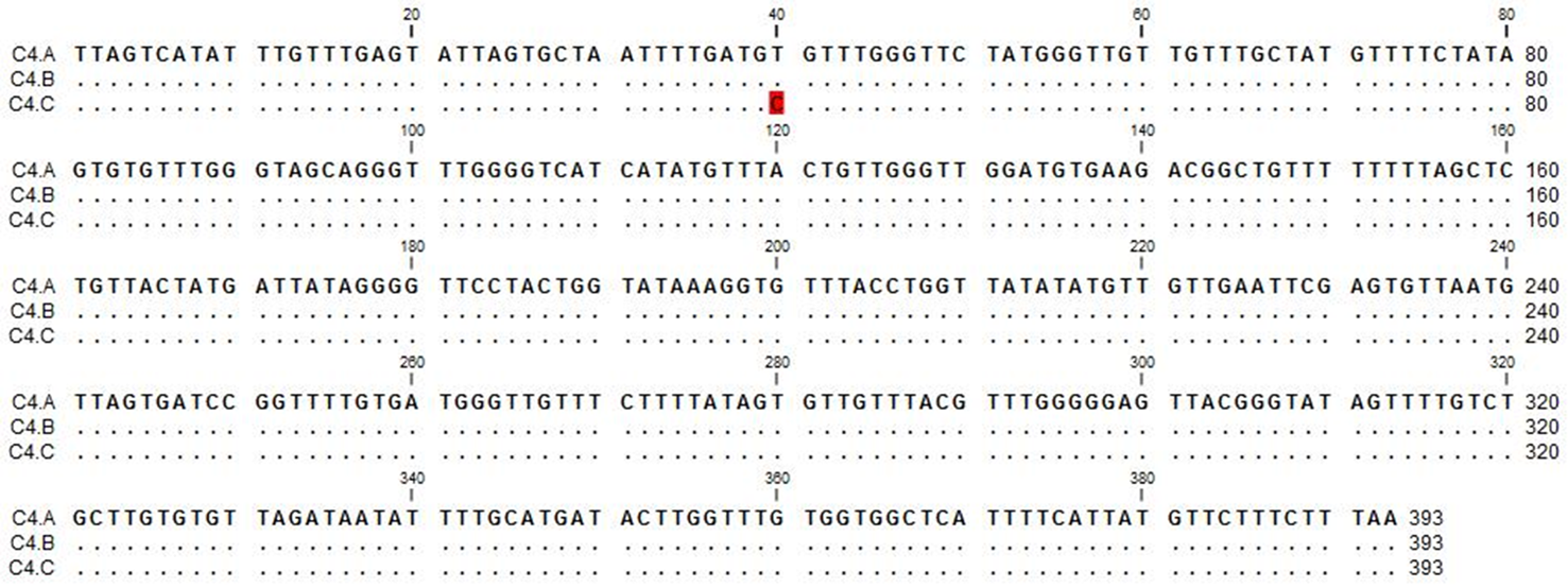
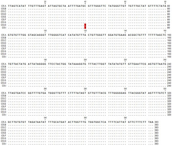
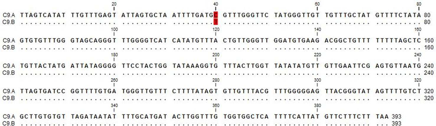
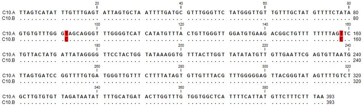
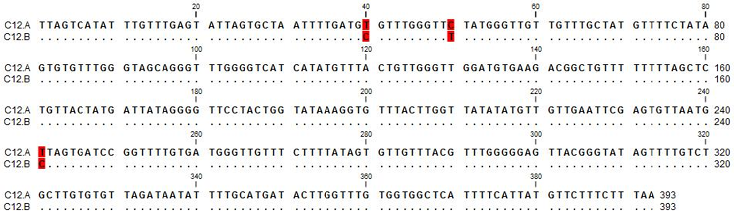
Three lung isolates which were collected from the same cattle (Cattle 4) were sequenced then a single nucleotide polymorphism in 40th nucleotide in a sample (C4.C) where there was T→C substitution occurred as shown in Fig. 4. We detected six liver (C5.A-F) and three lungs (C5.G-I) cysts in Cattle 5. However, although no nucleotide differences have been observed in the liver samples of Cattle 5, there was single nucleotide polymorphism (C→T) in 40th nucleotide of two lung cysts (C5.H and C5.I) as shown in Fig. 5. Two samples (C9.A and C9.B) have been collected from the lung of Cattle 9. The isolates have shown C→T substitution at 40th nucleotide as presented in Fig. 6. The samples of Cattle 10 (C10.A and C10.B) have been obtained from the lung and the sequence has shown the mutations at two points, T→C substitutions at nucleotide 92 and 158 as shown in Fig. 7. However, samples of Cattle 12 (C12.A and C12.B) which were collected from the lung showed mutations at three points in the sequences, T→C substitution at 40th and 241th nucleotides and C→T substitution at 50th nucleotide as shown in Fig. 8.
Sequence comparison of lung hydatid cysts of Cattle 4. C4.C has T→C substitution in 40th nucleotide.

Sequence comparison of six liver (C5.A-F) and three lung (C5.G-I) hydatid cysts of Cattle 5. C5.H and C5.I indicate the single nucleotide polymorphism (C→T) in 40th nucleotide of two lung cysts.

Sequence comparison of two lung samples (C9.A and C9.B) of Cattle 9. The C→T substitution at 40th nucleotide is shown.

C10.A and C10.B indicate the lung samples sequences of Cattle 10. Sequences show the mutations at two points (T→C substitutions at nucleotide 92 and 158).

C12.A and C12.B show the lung sample sequences of Cattle 12. Mutations at three points in the sequences were highlighted (T→C substitution at 40th and 241th nucleotides and C→T substitution at 50th nucleotide).

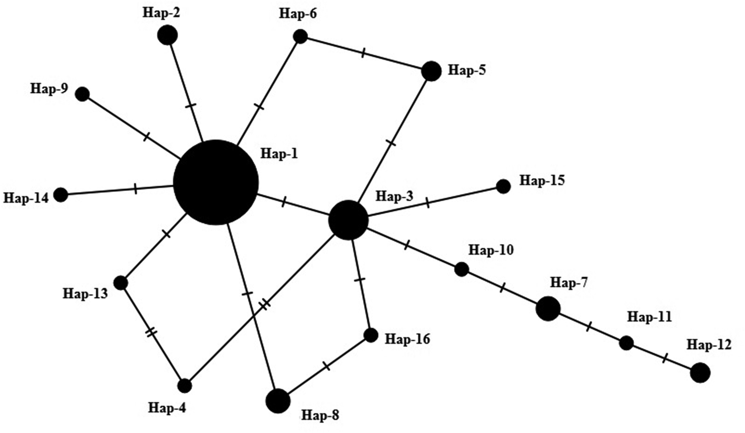
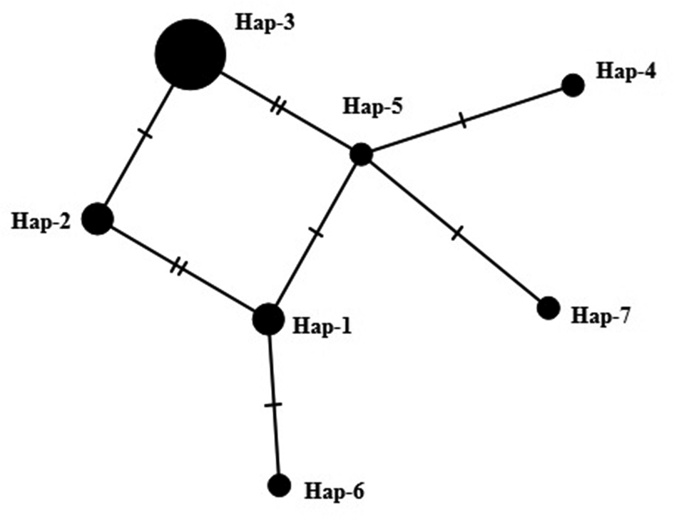
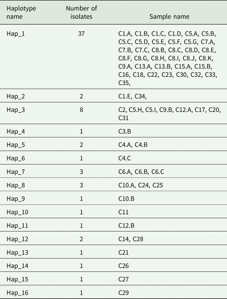
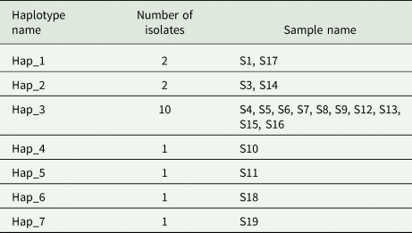
We generated the haplotype analyses in order to show genetic variability within the cattle and sheep samples separately. A total of 16 haplotypes of E. granulosus s.s. have been detected within 66 cattle isolates and the results were presented in Fig. 9. The haplotype network of cattle isolates had 16 haplotypes arranged within a star-like configuration with the main haplotype, the haplotypes that were separated from 1–6 mutations comprised 56.1% (Hap_1: 37/66) of the total isolates (Table 1). A total of 12 polymorphic sties has been detected in the CO1 gene sequences. The analysis showed that the overall nucleotide diversities were 0.00319 ± 0.00046 and haplotype diversities were 0.672 ± 0.063 for the mt-CO1 sequences. The overall neutrality indices were negative for the mt-CO1 (Tajima's D −1.42122, P > 0.10; Fu's Fs −11 722, P = 0.000). Besides, the haplotype analysis of 18 sheep isolates showed 7 different haplotypes of E. granulosus s.s. with one or two mutations and the results were described in Fig. 10. The haplotype network analysis of sheep isolates showed 7 haplotypes with the main haplotype which was distinct from others by 1–4 mutational steps comprised 55.5% (Hap_3: 10/18) of the total isolates (Table 2). A total of 6 polymorphic sites has been detected in CO1 gene sequences of 18 sheep isolates. The analysis showed that the overall nucleotide diversities were 0.00432 ± 0.00082 and haplotype diversities were 0.693 ± 0.114 for the mt-CO1 sequences. The overall neutrality indices were negative for CO1 gene (Tajima's D −0.08506, P > 0.10; Fu's Fs −1.942, P = 0.082).
The appearance of mt-CO1 haplotypes of cattle isolates of E. granulosus s.s. The size of the circles is related to the haplotype frequency. Small circles indicate additional mutational areas. The numbers in the figure indicate the number of mutations.

The appearance of mt-CO1 haplotypes of sheep isolates of E. granulosus s.s. The size of the circles is related to the haplotype frequency. Small circles indicate additional mutational areas. The numbers indicate the number of mutations.

Haplotypes of the cattle isolates detected in this study using partial mt-CO1 gene (446 bp) sequences

Haplotypes of the sheep isolates detected in this study using partial mt-CO1 gene (446 bp) sequences

Discussion
Echinococcosis which is a life-threatening zoonotic infection, recognized as 17 neglected tropical diseases in the world by WHO, the causing agent of the infection is the larval stage of cestode species of genus Echinococcus. It is responsible for significant economic as well as medical losses each year (Agudelo Higuita et al., Reference Agudelo Higuita, Brunetti and McCloskey2016).
The current study showed the predominance infection in lungs 85% as compared to liver 15%. Our study has shown some similarities with the previous studies. A recent study in Oman performed on slaughtered goats showed the predominance of infection in lungs 53.4% (Al-Kitani et al., Reference Al-Kitani, Baqir, Hussain and Roberts2014), another study conducted in Oman showed the high prevalence in lungs 82.9% (Al-Kitani et al., Reference Al-Kitani, Baqir, Hussain and Robert2015). Moreover, in a recent study conducted in Turkey, 23 out of 30 hydatid cysts were found in the lungs (Kesik et al., Reference Kesik, Simsek, Kilinc and Koroglu2019a; Reference Kesik, Kilinc, Simsek and Gulb). The predominance of infection in lungs is that the E. granulosus s.l. oncospheres migrate to the lungs and lungs possess a great number of capillaries sites than any other organ (Kebede et al., Reference Kebede, Hagos, Girna and Lobago2009).
For the molecular classification and to determine of the E. granulosus genotypes, mitochondrial as well as nuclear genomes have been used, however, for phylogenetic analysis of E. granulosus among closely related species, mt-DNA has been more efficient as compared to nuclear genomes because of the large datasets and rapid sequence evolution (Nakao et al., Reference Nakao, McManus, Schantz, Craig and Ito2006). The mt-CO1 gene can be used as a significant evolutionary marker for the distinction of intra- and interspecific variants (Nakao et al., Reference Nakao, Lavikainen, Yanagida and Ito2013a, Reference Nakao, Yanagida, Konyaev, Lavikainen, Odnokurtsev and Zaikovb). Generally, E. granulosus s.s. is considered as predominant specie in CE infections worldwide (Alvarez Rojas et al., Reference Alvarez Rojas, Romig and Lightowlers2014; Kinkar et al., Reference Kinkar, Laurimäe, Acosta-Jamett, Andresiuk, Balkaya, Casulli, Gasser, van der Giessen, González, Haag, Zait, Irshadullah, Jabbar, Jenkins, Kia, Manfredi, Mirhendi, M'rad and Rostami-Nejad2018a, Reference Kinkar, Laurimäe, Acosta-Jamett, Andresiuk, Balkaya, Casulli, Gasser, van der Giessen, González, Haag, Zait, Irshadullah, Jabbar, Jenkins, Kia, Manfredi, Mirhendi, M'rad and Rostami-Nejadb).
Our findings have shown the occurrence of E. granulosus s.s. in 84 out of 85 samples which is in agreement with recent studies. In a recent study out of 12 sheep and 10 human isolates of E. granulosus, 19 isolates have shown the presence of E. granulosus s.s. (Snábel et al., Reference Snábel, Altintas, D'Amelio, Nakao, Romig, Yolasigmaz, Gunes, Turk, Busi, Hüttner, Sevcová, Ito, Altintas and Dubinský2009). Another study has been conducted on 120 isolates from two provinces (Elazig and Erzincan) of Turkey, 114 isolates have shown CE infection caused by E. granulosus s.s. through 12S rRNA-PCR while the other 6 samples have also shown the occurrence of E. granulosus s.s after the analysis of mt-CO1 gene (Kesik et al., Reference Kesik, Simsek, Kilinc and Koroglu2019a, Reference Kesik, Kilinc, Simsek and Gulb). Moreover, another study has been conducted on 58 isolates (3 sheep, 13 cattle and 42 humans) in Turkey, E. granulosus s.s. (G1 genotype) have been detected in all cattle and sheep samples however E. granulosus s.s. (G1 genotype) and E. canadensis (G7) have been characterized in human isolates (Eryildiz and Sakru, Reference Eryildiz and Şakru2012). The study conducted in Turkey on 70 FFPE tissue samples of humans, the results have shown the occurrence of E. canadensis (G6) genotype in two Echinococcus isolates (Simsek et al., Reference Simsek, Kaplan and Ozercan2011). According to recent studies genotype of E. canadensis (G6) has been circulating in Sudan (Omer et al., Reference Omer, Dinkel, Romig, Mackenstedt, Elnahas, Aradaib, Ahmed, Elmalik and Adam2010), although the prevalence of this genotype is comparatively low in cattle and sheep as compared to camel (Omer et al., Reference Omer, Dinkel, Romig, Mackenstedt, Elnahas, Aradaib, Ahmed, Elmalik and Adam2010; Rostami et al., Reference Rostami, Talebi, Babaei, Sharbatkhori, Ziaali, Rostami and Harandi2013). In the current study one sheep sample has shown the presence of E. canadensis (G6/G7 genotype) and 100% homology has been observed with reference sequence of E. canadensis (G6/G7). The occurrence of E. canadensis (G6/G7 genotype) in sheep is reported for the first time in Turkey using of both short (446 bp) and long (880 bp) mt-CO1 sequences. The occurrence of the species E. granulosus s.s. and E. canadensis (G6/G7) in sheep is an indication of the presence of two different cycles of E. granulosus s.l. in Turkey. Although Turkey has very low camel and pig populations, finding of E. canadensis (G6/G7) in sheep may indicate that the adult parasites may enter from the neighbour countries border with an infected dog. It is already known that E. granulosus s.s. is well adapted to sheep as intermediate hosts however occurrence of E. canadensis (G6/G7) in the region need further characterization to understand the transmission dynamics of this parasite. Besides, this should be supported by studies of human CE in Turkey.
Echinococcus spp. possess distinct reproduction system as the adult worms are hermaphrodites and larval form of the parasite develops during the asexual phase of reproduction, this novel process of reproduction may cause variations at genus and specie level because of self-fertilization following the amplification by asexual reproduction which results in mutations (Smyth and Smyth, Reference Smyth and Smyth1964). The study showed the presence of two different haplotypes in a single intermediate host (Casulli et al., Reference Casulli, Interisano, Sreter, Chitimia, Kirkova, Rosa and Pozio2012). In our study sequence analysis has also been performed on samples of the same cattle which showed the single nucleotide polymorphism within the samples of the same animals. In this study, three lung cysts from the same cattle were sequenced then a T→C substitution in 40th nucleotide in a sample (C4.C) was determined. We detected six liver and three lung cysts in an isolate (Cattle 5). Although no nucleotide differences have been observed in the liver samples, there was a single nucleotide polymorphism (C→T) in 40th nucleotide of two lung cysts. Similarly, two samples have been collected from the lung of Cattle 9 and the isolates have shown C→T substitution at 40th point. Also, the samples of Cattle 10 have been obtained from the lung and the sequences shown T→C substitutions at nucleotide 92 and 158. However, samples of Cattle 12 which were collected from the lung showed T→C substitution at 40th and 241th nucleotides and C→T substitution at 50th nucleotide. All these mutations mean the presence of different variants of E. granulosus s.s. within the same host and existence of different variants in one intermediate host shows the chances of out-crossing among different adult worms in the same definitive host.
We have also generated the haplotypes of E. granulosus s.s. by using the alignment of a different sequence of cattle and sheep origin separately. There were 16 haplotypes of E. granulosus s.s. found in 66 cattle samples with the mutations at either one or two points. However, in sheep samples 7 haplotypes of E. granulosus s.s. have been found with mutations either at one point or two. The study has reported extremely high haplotype diversity in G1 genotype as reported 171 haplotypes out of 212 samples have found (Kinkar et al., Reference Kinkar, Laurimäe, Acosta-Jamett, Andresiuk, Balkaya, Casulli, Gasser, van der Giessen, González, Haag, Zait, Irshadullah, Jabbar, Jenkins, Kia, Manfredi, Mirhendi, M'rad and Rostami-Nejad2018a, Reference Kinkar, Laurimäe, Acosta-Jamett, Andresiuk, Balkaya, Casulli, Gasser, van der Giessen, González, Haag, Zait, Irshadullah, Jabbar, Jenkins, Kia, Manfredi, Mirhendi, M'rad and Rostami-Nejadb). Based on shorter sequence lengths previous studies have reported excessive genetic variations of E. granulosus s.s. in different regions of the world (Casulli et al., Reference Casulli, Interisano, Sreter, Chitimia, Kirkova, Rosa and Pozio2012; Alvarez Rojas et al., Reference Alvarez Rojas, Ebi, Gauci, Scheerlinck, Wassermann, Jenkins, Lightowlers and Romig2016). The mt-CO1 gene is considered to be the most suitable gene in the investigation of genetic diversity of E. granulosus s.s. at intraspecific level (Kamenetzky et al., Reference Kamenetzky, Gutierrez, Canova, Haag, Guarnera, Parra, García and Rosenzvit2002; Haag et al., Reference Haag, Ayala, Kamenetzky, Gutierrez and Rosenzvit2004; Badaraco et al., Reference Badaraco, Ayala, Bart, Gottstein and Haag2008; Nakao et al., Reference Nakao, Li, Han, Ma, Xiao, Qiu, Wang, Yanagida, Mamuti, Wen, Moro, Giraudoux, Craig and Ito2010). Study has reported the presence of E. granulosus s.s. (G1 and G3) in Italy and Eastern European population and confirmed four haplotypes in Italy and 21 haplotypes in Eastern parts of Europe (Casulli et al., Reference Casulli, Interisano, Sreter, Chitimia, Kirkova, Rosa and Pozio2012).
In conclusion, 85 isolates (19 sheep, 66 cattle) were sequenced out of them 84 were characterized as E. granulosus s.s. and polymorphism within E. granulosus s.s. has been observed in not only samples of different animals but also the isolates collected from same cattle. Moreover, multiple organ infections in one cattle have also been detected in both lungs and liver, and samples of the cattle (both lungs and liver) also showed the C→T substitution. The presence of polymorphic sites within the samples of the same animal indicates the chance of crossing over between worms in definitive hosts; however more study is needed in this section. We have found 16 haplotypes of E. granulosus s.s. within cattle isolates and 7 haplotypes in sheep samples. However, we have found G6/G7 genotypes of E. canadensis in one of the sheep sample which is identified for the first time in sheep in Turkey. The previous study has shown the occurrence of E. canadensis (G6 genotype) in two human isolates, the presence of this E. canadensis (G6/G7) in sheep indicates that the genotype circulates both in humans as well as in livestock animals in this region.
Acknowledgements
The authors would like to thank all veterinarians for their kind help during sample collection.
Financial support
This study has been supported by a grant from Firat University Scientific Research Projects Unit (Project No: VF.19.05).
Conflict of interest
The authors declare that there is no conflict of interest or financial disclosure about this publication.
Ethical standards
Not applicable.