Despite some initial controversy concerning an increased prevalence of major malformations with assisted reproductive technology, a few large and recent investigationsReference Koivurova, Hartikainen, Gissler, Hemminki, Sovio and Järvelin1–Reference Allen, Wilson and Cheung3 have pointed to an increased risk for congenital malformations among children conceived by these techniques, although no differences have been noted for the different techniques, which include in vitro fertilization, intracytoplasmic injection of sperm, and in vitro maturation.Reference Buckett, Chian, Holzer, Dean, Usher and Tan4 Thus, prenatal screening for congenital structural abnormalities in pregnancies achieved by assisted reproductive technology is recommended, including appropriate use of biochemical and sonographic screening.Reference Allen, Wilson and Cheung3
Case report
A 38-days-old male infant was referred to our hospital because of respiratory distress and tachycardia, detected by his physician during a routine visit on the previous day. The patient was the first child of a caucasian couple, and the mother had become pregnant after intracytoplasmic injection of sperm, one of the procedures developed in the field of assisted reproductive technology. Both the pregnancy and delivery had been uneventful.
On physical examination, the infant had mild central cyanosis, was in respiratory distress, with subcostal and intercostal retractions, had a pulse rate of 140 beats per minute, respiratory rate of 65 breaths per minute, and pulse oximetry was 89% on room air. Chest examination revealed a systolic ejection murmur graded at II from VI at the upper left sternal border, with fixed splitting of the second heart sound. Systolic blood pressure, measured in the right arm, was 71 over 50 millimetres of mercury, with no gradient between the arms and legs. Pulmonary ventilation was clear, and no visceral enlargement was detected. Further physical examination was unremarkable.
The electrocardiogram showed regular sinus rhythm, at a rate of 150 beats per minute, a QRS axis of 105°, and right ventricle predominance, without other significant findings. Chest radiography showed a mild increase in the flow of blood to the lungs, along with mild cardiomegaly.
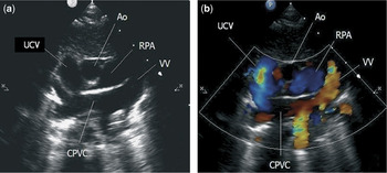
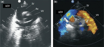
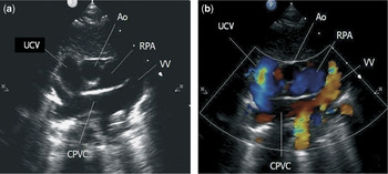
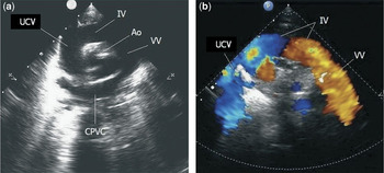
On echocardiography, both the cavities of the right-sided chambers and the superior caval vein were found to be dilated, the right ventricle diastolic diameter measured at 16 millimetres, the right atrial diameter at 29 millimetres, and the diameter of the superior caval vein at 11 millimetres. A defect was found in the floor of the oval fossa of 6 millimetres diameter, permitting right-to-left shunting. The ventricular septum was seen to move in paradoxical fashion. From the suprasternal notch, we noted that all 4 pulmonary veins entered a confluence posterior to the left atrium. This confluence communicated with a left vertical vein, which drained through a brachiocephalic vein into the right superior caval vein. No points of obstruction to venous flow were detected (Figs 1 and 2). The child was referred for surgical correction, which was successful, the postoperative course being uneventful.
The echocardiogram is shown (a) in the cross-sectional suprasternal short-axis view, and (b) with colour-flow Doppler interrogation. Abbreviations: UCV, superior caval vein; CPVC, common pulmonary venous confluence; VV, vertical vein; RPA, right pulmonary artery; Ao, aorta.
By tilting the transducer from the suprasternal short axis view (a), the entire anomalous supracardiac connection can be seen, resembling a vascular collar surrounding the transverse aorta. The addition of colour-flow Doppler (b) shows the flow through the anomalous supracardiac connection. UCV, superior caval vein; CPVC, common pulmonary venous confluence; VV, vertical vein; Ao, aorta; IV, brachiocephalic vein.
Discussion
Although congenital structural abnormalities are more often found in multiple pregnancies, assisted reproductive technology itself is supposed to play an important role. It has been hypothesized that the increase of chromosomal aberrations, in direct relation with the relatively advanced age of the infertile couples, and the possible induction of point mutations due to the exposure to physical and chemical teratogens, act as aetiopathogenic factors, though the possible underlying mechanisms are still unclear, and require further study.Reference Buckett, Chian, Holzer, Dean, Usher and Tan4 Whether assisted reproductive technology has a direct causative effect is still the subject of much debate.Reference Anthony, Buitendijk, Dorrepaal, Lindner, Braat and den Ouden5
A higher incidence of children born prior to term, and with low weight at birth, is also reported in those conceived by assisted reproductive technology, although multiplicity has been considered to be the main factor for these findings.Reference Koivurova, Hartikainen, Gissler, Hemminki, Sovio and Järvelin1
When considering the cardiovascular system, a higher incidence of congenital cardiac malformations has been well documented after assisted reproductive technology.Reference Koivurova, Hartikainen, Gissler, Hemminki, Sovio and Järvelin1, Reference Buckett, Chian, Holzer, Dean, Usher and Tan4, Reference McDonald, Murphy, Beyene and Ohlsson6, Reference Budziszewska, Włoch and Rozmus-Warcholińiska7 Lesions reported including atrial, ventricular, or atrioventricular septal defects, coarctation of the aorta, tetralogy of Fallot, tricuspid atresia, transposition, hypoplastic left heart syndrome, and other complex cardiopathies. In our patient, we found non-obstructive totally anomalous pulmonary venous connection to the superior caval vein. To the best of our knowledge, this malformation has not been previously described in a child conceived following intracytoplasmic injection of sperm.