Introduction
Neurocysticercosis (NCC), caused by encysted larvae of the tapeworm Taenia solium, is one of the most recurrent parasitic diseases of the human central nervous system (Fabiani and Bruschi, Reference Fabiani and Bruschi2013; Gripper and Welburn, Reference Gripper and Welburn2017). In 2012, the World Health Organization included NCC in the list of neglected tropical diseases (WHO, 2012). Most cases of NCC occur in less developed countries. However, the immigration of people from endemic areas to developed countries, and people travelling to areas where the disease is endemic, generated an increase in the incidence of NCC in the USA, Canada and European countries, among others (Sorvillo et al., Reference Sorvillo2011; Coyle et al., Reference Coyle2012).
Human taeniasis is acquired by the ingestion of T. solium metacestodes present in poorly cooked pork. The parasite develops the adult stage (tapeworm) in the intestine. Neurocysticercosis can occur when a human ingests eggs from faeces of a human parasite carrier. These eggs release oncospheres that can penetrate the intestinal mucosa and enter the bloodstream to migrate mostly to the central nervous system and the eyes (Gripper and Welburn, Reference Gripper and Welburn2017).
In the brain, the location of cysts can be parenchymal or extraparenchymal. Cysticerci mostly affect the cerebral hemispheres, mainly at the junction of grey and white matter. Less frequently, they can be observed in the ventricles, basal cisterns and subarachnoid spaces. In the posterior fossa, NCC usually involves the fourth ventricle, cerebellopontine angle cistern, cisterna magna and, infrequently, the cerebellum (Amaral et al., Reference Amaral2003; Rocca et al., Reference Rocca, Rosell and Alvarez2005). Although the cerebellar location is rare, some human cases have been reported (Zhu et al., Reference Zhu2003; Kim et al., Reference Kim2006, Reference Kim2010). The low blood flow in the cerebellum, in relation to the cerebrum, could explain the low frequency of cerebellar cysts (Kim et al., Reference Kim2010).
Generally, NCC has a long asymptomatic period, which extends from the first contact with the parasite until an intense inflammatory response begins in response to the degeneration of the larvae. In symptomatic NCC, neurological manifestations are non-specific and depend on characteristics of the parasite, such as number, size, location and stage of the larvae, and characteristics of the host, such as immune and inflammatory response. Although the reported signs and symptoms are varied, the majority of patients with cerebral parenchymal cysts have seizure disorders and the cysts located in extraparenchymal tissue produce intracranial hypertension (Del Brutto, Reference Del Brutto2012).
Symptomatology and treatment are associated with the stage of the cysticercus and with the immune response of the host to the parasite in the brain. In an attempt to understand the immune response and associated pathology in NCC, Cardona et al. (Reference Cardona1999) developed a mouse model using metacestodes of Mesocestoides corti, a related cestode to T. solium. The authors demonstrated that the distribution of M. corti metacestodes is similar to T. solium in the human brain. The aim of this work is to report the cerebellum as a new location for M. corti tetrathyridia in the murine model of NCC.
Materials and methods
In this study, animal procedures and management protocols were approved by the Institutional Animal Care and Use Committee (RD 148/15) of the Faculty of Exact and Natural Sciences, National University of Mar del Plata, Mar del Plata, Argentina and carried out in accordance with the revised form of The Guide for the Care and Use of Laboratory Animals (National Research Council US, 2011). Unnecessary animal suffering was avoided throughout the study. Animals were housed in a temperature-controlled (22 ± 1°C), light-cycled (12 h light/dark cycle) room, and food and water were given ad libitum.
Parasite material was maintained by serial passages in female CF-1 mice and Wistar rats, as described by Markoski et al. (Reference Markoski2003). To simulate human NCC, tetrathyridia were recovered from the peritoneum of infected animals, and 3–5 week old female CF-1 mice were injected intracranially with 20 μl of M. corti metacestodes (Alvarez et al., Reference Alvarez2010).
Before intracranial infection, animals were anaesthetized with a mixture of ketamine (50 mg/kg) and xylazine (5 mg/kg). Mice were given an intraperitoneal injection of tramadol analgesia (2 mg/kg) every 24 h for three days, starting the same day of infection. After 4 weeks of infection, mice were anaesthetized with a mixture of ketamine (100 mg/kg) and xylazine (10 mg/kg), and euthanized. Necropsies were carried out immediately and the brains were removed rapidly. To determine the location of the larvae, brains were fixed by immersing in 10% buffered formalin before dehydration in an ethanol series and ethyl acetate. Then, samples were embedded in paraffin. Horizontal histological sections of 12 μm were cut using a microtome (Arcano 1508) and stained with the routine technique of haematoxylin and eosin.
Results and discussion
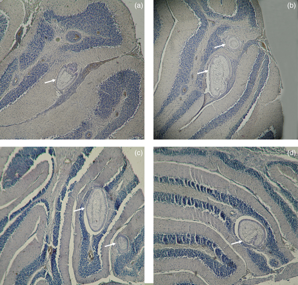
To determine the distribution of larvae in brain tissue, 13 infected mouse brains were analysed. Tetrathyridia were found in four locations: parenchyma, ventricles, meninges and in a previously unreported location, the cerebellum (fig. 1). Approximately 51% of metacestodes were observed in parenchyma, 35% in ventricles, 4% in meninges and the remaining 10% in the cerebellum. Seventy percent of the analysed brains presented larvae in this new location.
Light microscopy of histological sections of cerebellum obtained from mice with experimental NCC. Haematoxylin–eosin stain (120×). Larvae located in molecular layer on grey matter (a, b) or in the central white matter core (c, d) of cerebellum. Arrows indicate the tetrathyridia.

Similar to T. solium larvae in humans and in coincidence with the results reported by Cardona et al. (Reference Cardona1999) and Alvarez et al. (Reference Alvarez2010), tetrathyridia of M. corti quickly invade the brain. A few days post infection, the parasites reach the ventricular and subarachnoid spaces as well as the parenchyma.
After 3–5 weeks of infection, the parasites are distributed equally between parenchymal and extraparenchymal spaces, but eventually most larvae penetrate and invade the parenchymal tissue. This parallels the locations reported in humans with T. solium, the majority presenting parenchymal cysts (Gripper and Welburn, Reference Gripper and Welburn2017).
The presence of T. solium cysts in the cerebellum is rare, but it is a reported location in human NCC. A murine model that reproduces the human pathology is essential to evaluate the symptomatology and response to drug treatment in experimentally infected mice.
Financial support
This work was supported by PICT 2015 No. 0717 (ANPCyT, Argentina) and Universidad Nacional de Mar del Plata (Grants EXA769/16 and EXA871/18), Argentina.
Conflict of interest
None.
Ethical standards
The authors assert that all procedures contributing to this work comply with the ethical standards of the relevant national and institutional guides on the care and use of laboratory animals.