INTRODUCTION
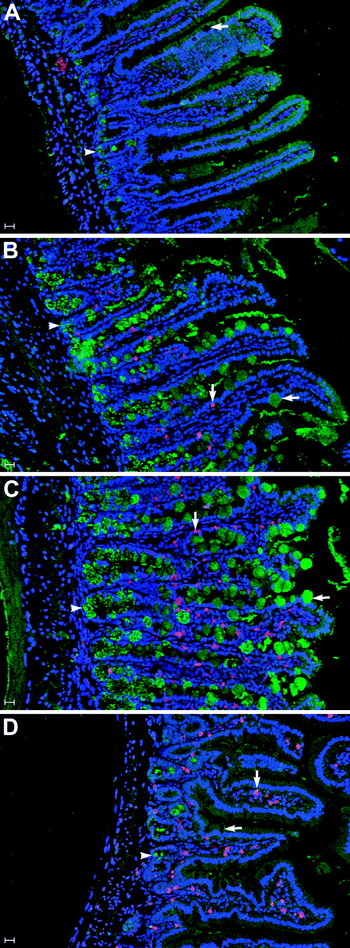
Much of our understanding of the processes involved in the immune expulsion of Trichinella spiralis derives from studies employing mice, in which adult worms are expelled from the gastrointestinal tract within 14–20 days of the establishment of a primary infection, depending on larval challenge dose and genetic background of the host (Miller and Jarrett, Reference Miller and Jarrett1971; Miller, Reference Miller1987, Reference Miller1996; Lawrence et al. Reference Lawrence, Paterson, Higgins, Macdonald, Kennedy and Garside1998; Artis et al. Reference Artis, Humphreys, Bancroft, Rothwell, Potten and Grencis1999; Dehlawi and Wakelin, Reference Dehlawi and Wakelin2002). It is these studies, conducted using mouse models, that we predominantly review in this article. T cell-mediated mucosal mast cell (MMC) and goblet cell hyperplasia, accompanied by the release of mediators, are characteristic features of gastrointestinal nematode infections (Nawa and Miller, Reference Nawa and Miller1978; Miller, Reference Miller1987, Reference Miller1996), and are induced by T. spiralis infection in rodents, pigs and humans (Miller and Jarrett, Reference Miller and Jarrett1971; Gustowska et al. Reference Gustowska, Ruitenberg, Elgersma and Kociecka1983; Lawrence et al. Reference Lawrence, Paterson, Higgins, Macdonald, Kennedy and Garside1998; Kamal et al. Reference Kamal, Wakelin, Ouellette, Smith, Podolsky and Mahida2001; Knight et al. Reference Knight, Pemberton, Robertson, Roy, Wright and Miller2004; Theodoropoulos et al. Reference Theodoropoulos, Hicks, Corfield, Miller, Kapel, Trivizaki, Balaskas, Petrakos and Carrington2005). MMC expand rapidly in the mucosa, predominantly within the epithelium, and release effector molecules such as histamine, cytokines and serine proteases such as mouse mast cell protease-1 (Mcpt-1) (Miller et al. Reference Miller, Huntley, Newlands, Mackellar, Lammas and Wakelin1988; Friend et al. Reference Friend, Ghildyal, Austen, Gurish, Matsumoto and Stevens1996; Vliagoftis and Befus, Reference Vliagoftis and Befus2005) (Fig. 1). Goblet cells enlarge and release mucus (Miller, Reference Miller1987; Miller et al. Reference Miller, Knight and Pemberton2006) as well as the novel peptides intelectin 2 and RELMβ (Artis et al. Reference Artis, Mei, Keilbaugh, He, Brenes, Swain, Knight, Donaldson, Lazar, Miller, Schad, Scott and Wu2004a; Pemberton et al. Reference Pemberton, Knight, Gamble, Colledge, Lee, Pierce and Miller2004a, Reference Pemberton, Knight, Wright and Millerb; Miller et al. Reference Miller, Knight and Pemberton2006) (Fig. 1).
Mucosal mast cell and goblet cell hyperplasia during Trichinella spiralis infection. Representative images of the jejunal mucosa of BALB/c mice before (A) and 7 (B), 14 (C) or 28 (D) days after infection with T. spiralis. Sections were probed with Mcpt-2 (red) and intelectin (green) specific antibodies, as markers for mucosal mast cells and goblet cells respectively. Nuclei were counterstained with SYBR Green I (blue). Goblet cells (horizontal arrows), paneth cells (arrowheads) and mucosal mast cells (vertical arrows) are highlighted. Note the pronounced goblet cell hyperplasia and peak ITLN expression on days 7–14 after infection, which is undetectable in uninfected jejunum and on day 28. Also note the predominantly intraepithelial location of mucosal mast cells on days 7–14 as opposed to day 28, and their absence in uninfected jejunum.

Trichinella spiralis-induced MMC and goblet cell hyperplasia is dependent on mucosal T cells (Kamal et al. Reference Kamal, Wakelin, Ouellette, Smith, Podolsky and Mahida2001) and, more specifically, on the Th2 cytokines IL-4 and IL-13 (Urban et al. Reference Urban, Schopf, Morris, Orekhova, Madden, Betts, Gamble, Byrd, Donaldson, Else and Finkelman2000; Helmby and Grencis, Reference Helmby and Grencis2002; McDermott et al. Reference McDermott, Humphreys, Forman, Donaldson and Grencis2005; Dehlawi et al. Reference Dehlawi, Mahida, Hughes and Wakelin2006; Scales et al. Reference Scales, Ierna and Lawrence2007). IL-4 and IL-13 induce protective responses against T. spiralis and Nippostrongylus brasiliensis through a common receptor, IL-4 receptor alpha (IL-4Rα), by activating signal transducer and activator of transcription 6 (STAT6) (Finkelman et al. Reference Finkelman, Shea-Donohue, Morris, Gildea, Strait, Madden, Schopf and Urban2004). STAT6 is a critical mediator for the activation and/or expression of many IL-4 responsive genes, including the IgE H chain and MHC Class II molecules. IL-4/IL-13- mediated protective effects against N. brasiliensis include increased mucus production as well as altered barrier function and smooth muscle contractility, and can occur independently of intestinal mastocytosis, whereas protective effects against T. spiralis include the induction of intestinal mastocytosis by T cells (Finkelman et al. Reference Finkelman, Shea-Donohue, Morris, Gildea, Strait, Madden, Schopf and Urban2004). T. spiralis-infected Stat-6−/− mice, which exhibit impaired IL-4 and IL-13 signalling, have reduced goblet cell and mast cell hyperplasia, and show a delayed expulsion of the worms (Urban et al. Reference Urban, Schopf, Morris, Orekhova, Madden, Betts, Gamble, Byrd, Donaldson, Else and Finkelman2000; Khan et al. Reference Khan, Blennerhasset, Ma, Matthaei and Collins2001a, Reference Khan, Vallance, Blennerhasset, Deng, Verdu, Matthaei and Collinsc). However, there are key differences in the regulation of expansion of these two cell types within the gastrointestinal mucosa, which will be discussed later in this review.
In accordance with the Th2 dependence of these responses, T. spiralis-induced goblet and mast cell hyperplasia can be abrogated by Th1-type cytokines. Goblet cell hyperplasia is downregulated by IL-12 (Khan et al. Reference Khan, Blennerhassett, Deng, Gauldie, Vallance and Collins2001b), while IL-27, which acts through the WSX-1 receptor and is structurally and functionally similar to IL-12, is a negative regulator of Th2 type responses and both goblet and mast cell hyperplasia in Trichuris muris infection (Artis et al. Reference Artis, Villarino, Silverman, He, Thornton, Mu, Summer, Covey, Huang, Yoshida, Koretzky, Goldschmidt, Wu, De Sauvage, Miller, Saris, Scott and Hunter2004b). More recently, IL-25 has been found to both upregulate IL-4 and IL-13 responses and downregulate Th1- type responses in this model (Owyang et al. Reference Owyang, Zaph, Wilson, Guild, McClanahan, Miller, Cua, Goldschmidt, Hunter, Kastelein and Artis2006). The Th1 cytokine IL-18 is a key negative regulator of mast cell hyperplasia but, interestingly, not of goblet cell hyperplasia in T. spiralis infection, and delays worm expulsion (Helmby and Grencis, Reference Helmby and Grencis2002). Conversely, in the presence of IL-2, IL-18 induced Th2-dependent intestinal mastocytosis in response to Strongyloides venezeulensis (see Sasaki et al. Reference Sasaki, Yoshimoto, Maruyama, Tegoshi, Ohta, Arizono and Nakanishi2005).
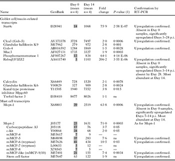
The rapid expansion of the MMC and goblet cell population in the intestinal epithelium, and increased production of their effectors, was highlighted by an analysis of the transcriptome of the jejunal epithelial compartment from uninfected and T. spiralis-infected BALB/c mice (Knight et al. Reference Knight, Pemberton, Robertson, Roy, Wright and Miller2004). MMC and goblet cell-derived transcripts were the most highly upregulated in the jejunal epithelium of T. spiralis-infected mice, and an analysis of a selection of these transcripts confirmed that upregulation was maximal 14 days after infection, corresponding with expulsion of the worms (Knight et al. Reference Knight, Pemberton, Robertson, Roy, Wright and Miller2004; summarized in Table 1). Upregulated transcripts included the MMC serine proteases mouse mast cell protease-1 (Mcpt-1) and -2 (Mcpt-2), and the goblet cell products, resistin-like moleculeβ (RELMβ), sialyl transferase 4c (Siat4c) and calcium-activated chloride channel 3 (CLCA-3; Gob-5). Parallel proteomic studies identified the novel goblet cell lectin intelectin-2 (ITLN-2), which was also highly inducible by T. spiralis infection but absent from the jejunum of uninfected mice (Pemberton et al. Reference Pemberton, Knight, Gamble, Colledge, Lee, Pierce and Miller2004a, Reference Pemberton, Knight, Wright and Millerb). Transcripts for Mcpt-1, ITLN-2, CLCA-3 and RELMβ, were also highly upregulated in the large intestine of BALB/c mice infected with T. muris (see Artis et al. Reference Artis, Mei, Keilbaugh, He, Brenes, Swain, Knight, Donaldson, Lazar, Miller, Schad, Scott and Wu2004a; Datta et al. Reference Datta, Deschoolmeester, Hedeler, Paton, Brass and Else2005; Artis, Reference Artis2006). These transcripts were absent or increased at significantly lower levels in T. muris-infected AKR mice, a strain susceptible to infection by this nematode (Artis et al. Reference Artis, Mei, Keilbaugh, He, Brenes, Swain, Knight, Donaldson, Lazar, Miller, Schad, Scott and Wu2004a; Datta et al. Reference Datta, Deschoolmeester, Hedeler, Paton, Brass and Else2005). An association between reduced Mcpt-1 expression and susceptibility to nematode infection has also been observed in response to the intestinal nematode Heligmosomoides polygyrus (see Menge et al. Reference Menge, Behnke, Lowe, Gibson, Iraqi, Baker and Wakelin2003).
Relative abundance of selected mast cell and goblet cell transcripts detected by expression profiling
(Summary of some of the key findings from an analysis of the transcriptome of the jejunal epithelial compartment from uninfected and Trichinella spiralis-infected BALB/c mice, using Affymetrix MG-U74AV2 oligonucleotide microarrays (Knight et al. Reference Knight, Pemberton, Robertson, Roy, Wright and Miller2004). Summarized are the findings for a selection of MMC- and goblet cell-derived transcripts, which were the most highly upregulated detected in the jejunal epithelium of T. spiralis-infected mice 14 days after primary challenge with 300 L3 by gavage. The mean signal level at day 0 (uninfected) and day 14 p.i. (post-infection), and fold change (mean Day 0 signal vs mean Day 14 signal), is shown. Greyed-out values were below the threshold level of detection (set at mean of 50). For comparative purposes, the effective fold-change includes instances where one of the values is below threshold of detection. P values are shown for signals which changed significantly on infection ((t-test with Welch correction; n=4 (P⩽0·05)). Upregulation of a selection of these transcripts was confirmed by RT-PCR analysis as indicated; where a complete temporal analysis was carried out using RNA samples collected on days 0–56 p.i. This is also summarized. Reproduced from Knight et al. (Reference Knight, Pemberton, Robertson, Roy, Wright and Miller2004), with kind permission from Infection and Immunity.)

In the present article, we review the factors which appear to control T. spiralis-induced MMC and goblet cell differentiation in the small intestine, and focus on some key MMC and goblet cell effector molecules which are considered to contribute to the expulsion of the adult worms and/or inhibition of larval development.
FACTORS CONTROLLING INTESTINAL MUCOSAL MAST CELL (MMC) HYPERPLASIA AND DIFFERENTIATION
Intestinal MMC are rare in uninfected mice, but are recruited into the jejunal epithelium in large numbers during T. spiralis infection, under regulation by TH2-type cytokines as described in the previous section. In BALB/c mice, MMC frequency typically peaks around the time of adult worm expulsion, approximately 14 days after infection, and returns to normal levels by around 8 weeks (Friend et al. Reference Friend, Ghildyal, Austen, Gurish, Matsumoto and Stevens1996, Reference Friend, Ghildyal, Gurish, Hunt, Hu, Austen and Stevens1998) (Fig. 1). Intra-epithelial MMCs are morphologically distinct from mast cells in non-mucosal sites and differ in their content of granule mediators, characteristically expressing mast cell proteases Mcpt-1 (Miller et al. Reference Miller, Huntley, Newlands, Mackellar, Lammas and Wakelin1988) and Mcpt-2, and to a lesser extent, Mcpt-9 (Friend et al. Reference Friend, Ghildyal, Austen, Gurish, Matsumoto and Stevens1996, Reference Friend, Ghildyal, Gurish, Hunt, Hu, Austen and Stevens1998). Intestinal MMC hyperplasia is accompanied by marked upregulation of Mcpt-1 and Mcpt-2 expression and the release of Mcpt-1 into the serum (Scudamore et al. Reference Scudamore, McMillan, Thornton, Wright, Newlands and Miller1997; Wastling et al. Reference Wastling, Scudamore, Thornton, Newlands and Miller1997).
The contribution of individual factors to initial mast cell precursor (MCp) recruitment, and subsequent MMC differentiation and proliferation once in the mucosa, has been investigated in vitro using cultured bone marrow precursors, and in vivo using gene deleted/transgenic mice, or via the administration of blocking antibodies. Stem cell factor (SCF) is a key regulator of T. spiralis-induced intestinal mastocytosis (Grencis et al. Reference Grencis, Else, Huntley and Nishikawa1993; Donaldson et al. Reference Donaldson, Schmitt, Huntley, Newlands and Grencis1996). SCF is produced by the small intestinal epithelium (Rosbottom et al. Reference Rosbottom, Knight, Mclachlan, Thornton, Wright, Miller and Scudamore2002a; Knight et al. Reference Knight, Brown, Wright, Thornton, Pate and Miller2007) and has both systemic and local effects on mast cell development. In vivo and in vitro studies have shown SCF to have multiple effects on mast cell biology, including directing migration (Tan et al. Reference Tan, Yazicioglu, Ingram, McCarthy, Borneo, Williams and Kapur2003; Pennock and Grencis, Reference Pennock and Grencis2004) and, in conjunction with IL-3 and IL-4, mast cell growth and proliferation (Tsuji et al. Reference Tsuji, Zsebo and Ogawa1991). The chemokine CCL2 (monocyte chemotactic protein-1), which induces migration of bone marrow-derived mast cells (Taub et al. Reference Taub, Dastych, Inamura, Upton, Kelvin, Metcalfe and Oppenheim1995), is upregulated in the small intestinal epithelium in nematode-infected mice and is expressed and released by MMC homologues in vitro (Rosbottom et al. Reference Rosbottom, Scudamore, von der Mark, Thornton, Wright and Miller2002b; Brown et al. Reference Brown, Knight, Wright, Thornton and Miller2003). More recently, mast cell-derived leukotiene-B4 (LTB4) has been shown to be a potent chemoattractant for immature mast cells and their progenitors (Weller et al. Reference Weller, Collington, Brown, Miller, Al-Kashi, Clark, Jose, Hartnell and Williams2005). MMC homologues have also been shown to bind the extracellular matrix protein laminin via integrin α7β1, which may, along with integrin-αEβ7 mediated adhesion to E-cadherin, facilitate their sequestration within the epithelial compartment (Rosbottom et al. Reference Rosbottom, Scudamore, von der Mark, Thornton, Wright and Miller2002b; Brown et al. Reference Brown, Knight, Pemberton, Wright, Pate, Thornton and Miller2004).
In vivo studies indicate roles for SCF, IL-3, IL-4 and IL-9 in the proliferation and/or survival of intestinal MMC (Abe et al. Reference Abe, Ochiai, Minamishima and Nawa1988; Madden et al. Reference Madden, Urban, Ziltener, Schrader, Finkelman and Katona1991; Grencis et al. Reference Grencis, Else, Huntley and Nishikawa1993; Faulkner et al. Reference Faulkner, Humphreys, Renauld, Van Snick and Grencis1997). Studies in our laboratory have also established a central role for the multifunctional cytokine TGF-β1 in the differentiation and function of MMC (Miller et al. Reference Miller, Wright, Knight and Thornton1999; Knight et al. Reference Knight, Wright, Brown, Huang, Sheppard and Miller2002; Wright et al. Reference Wright, Brown, Knight, Thornton, Kilshaw and Miller2002; Brown et al. Reference Brown, Knight, Wright, Thornton and Miller2003, Reference Brown, Knight, Pemberton, Wright, Pate, Thornton and Miller2004; Miller et al. Reference Miller, Knight and Pemberton2006; Pemberton et al. Reference Pemberton, Brown, Wright, Knight and Miller2006a). Crucially, expression of the MMC-specific β-chymases, Mcpt-1 and -2, and the αE subunit of the intraepithelial MMC integrin αEβ7 are strictly regulated by TGF-β1 (Miller et al. Reference Miller, Wright, Knight and Thornton1999; Brown et al. Reference Brown, Knight, Wright, Thornton and Miller2003, Reference Brown, Knight, Pemberton, Wright, Pate, Thornton and Miller2004; Knight et al. Reference Knight, Wright, Brown, Huang, Sheppard and Miller2002, Reference Knight, Brown, Wright, Thornton, Pate and Miller2007; Pemberton et al. Reference Pemberton, Brown, Wright, Knight and Miller2006a). TGF-β1 also induced morphological and granule ultrastructural changes similar to those observed in intestinal MMC in vivo (Miller et al. Reference Miller, Wright, Knight and Thornton1999; Knight et al. Reference Knight, Wright, Brown, Huang, Sheppard and Miller2002) and upregulates the expression of the α7 subunit of the laminin-binding integrin α7β1 (Rosbottom et al. Reference Rosbottom, Scudamore, von der Mark, Thornton, Wright and Miller2002b).
In vivo, TGFβ1 is secreted by many cell types, but the nascent protein is usually biologically inactive since it is complexed with latency-associated peptide (LAP) in the extracellular matrix. Latent TGF-β1-LAP complex can be activated by a diverse range of factors, including members of the αv integrin family (αvβ1, αvβ3, αvβ6 and αvβ8) (Mu et al. Reference Mu, Cambier, Fjellbirkeland, Baron, Munger, Kawakatsu, Sheppard, Broaddus and Nishimura2002; Annes et al. Reference Annes, Munger and Rifkin2003). Integrin αvβ6 is expressed exclusively by epithelial cells (Breuss et al. Reference Breuss, Gillett, Lu, Sheppard and Pytela1993; Brown et al. Reference Brown, Mcaleese, Thornton, Pate, Schock, Macrae, Scott, Miller and Collie2006). Thus, restricted expression of integrin αvβ6 to epithelial tissues, and studies in vitro and in null mice, support a role for this integrin as a key activator of TGF-β1 at mucosal surfaces (Huang et al. Reference Huang, Wu, Cass, Erle, Corry, Young, Farese and Sheppard1996, Reference Huang, Wu, Zhu, Pytela and Sheppard1998; Munger et al. Reference Munger, Huang, Kawakatsu, Griffiths, Dalton, Wu, Pittet, Kaminski, Garat, Matthay, Rifkin and Sheppard1999; Annes et al. Reference Annes, Munger and Rifkin2003; Ludlow et al. Reference Ludlow, Yee, Lipman, Bronson, Weinreb, Huang, Sheppard and Lawler2005). We have confirmed the presence of β6 integrin transcripts in enterocytes of the jejunum in normal and nematode-infected S129 mice, where it is co-expressed with TGFβ1 (Knight et al. Reference Knight, Wright, Brown, Huang, Sheppard and Miller2002). In β6−/− mice infected with (rat adapted) N. brasiliensis both MMC recruitment and Mcpt-1 expression were virtually abolished (Knight et al. Reference Knight, Wright, Brown, Huang, Sheppard and Miller2002). In contrast, infection of β6−/− mice with T. spiralis resulted in greatly enhanced MMC recruitment into the lamina propria with significantly reduced recruitment into the epithelial compartment, and greatly reduced expression of the integrin αEβ7 by the aberrantly located MMC (Brown et al. Reference Brown, Knight, Pemberton, Wright, Pate, Thornton and Miller2004). There is also co-ordinate reduction in expression of both Mcpt-1 and Mcpt-2 by gastrointestinal MMC in β6−/− mice and Mcpt-9 transcripts become undetectable (Knight et al. Reference Knight, Brown, Wright, Thornton, Pate and Miller2007). These studies support a central role for epithelially-expressed αvβ6 integrin in the activation of latent TGF-β1 in T. spiralis-induced MMC differentiation and function in vivo.
MUCOSAL MAST CELL SERINE PROTEASES INDUCED BY T. SPIRALIS INFECTION
There is clear evidence that MMC hyperplasia is necessary for the expulsion of adult T. spiralis from the intestine, but not for N. brasiliensis expulsion (Ha et al. Reference Ha, Reed and Crowle1983; Nawa et al. Reference Nawa, Ishikawa, Tsuchiya, Horii, Abe, Khan, Bing, Itoh, Ide and Uchiyama1994; Faulkner et al. Reference Faulkner, Humphreys, Renauld, Van Snick and Grencis1997). However, the key effector molecules responsible have not begun to be elucidated until relatively recently. The fact that MMC mediators are apparently more effective in dislodging adults of T. spiralis may be due to the intraepithelial niche of these parasites, as opposed to N. brasiliensis or H. polygyrus, which do not invade enterocytes. Mast cells in the intestinal mucosa and submucosa variously express the β-chymases Mcpt-1, Mcpt-2 and Mcpt-9, the α-chymase Mcpt-5, and the tryptases Mcpt-6 and Mcpt-7, depending on their location and the stage of infection (Friend et al. Reference Friend, Ghildyal, Austen, Gurish, Matsumoto and Stevens1996, Reference Friend, Ghildyal, Gurish, Hunt, Hu, Austen and Stevens1998; Miller and Pemberton Reference Miller and Pemberton2002). The most abundant proteases produced by MMC during T. spiralis infection are the MMC specific β-chymases, Mcpt-1 and Mcpt-2; for the purpose of this article, we focus on the potential roles of these serine proteases (Miller et al. Reference Miller, Huntley, Newlands, Mackellar, Lammas and Wakelin1988; Knight et al. Reference Knight, Pemberton, Robertson, Roy, Wright and Miller2004).
Role of MMC proteases; Mcpt-1 as a key effector molecule
Gastrointestinal nematode infections have long been associated with pronounced accumulation of fluid in the gut lumen, and this has been clearly demonstrated in both primary and secondary infection with T. spiralis (see McDermott et al. Reference McDermott, Bartram, Knight, Miller, Garrod and Grencis2003; Madden et al. Reference Madden, Yeung, Zhao, Gause, Finkelman, Katona, Urban and Shea-Donohue2004). Along with increased muscle contractility, this is thought to promote expulsion of the adult worms; sometimes referred to as “weep and sweep” (Madden et al. Reference Madden, Yeung, Zhao, Gause, Finkelman, Katona, Urban and Shea-Donohue2004). Increased fluid accumulation arises from a combination of increased epithelial secretion promoted by MMC mediators (e.g. prostaglandins and histamines) and enteric nerves, and increased paracellular epithelial permeability to macromolecules (Madden et al. Reference Madden, Yeung, Zhao, Gause, Finkelman, Katona, Urban and Shea-Donohue2004). Studies using Ussing chambers have shown that T. spiralis infection promotes both epithelial permeability and epithelial secretion, both of which are STAT6 dependent (Madden et al. Reference Madden, Yeung, Zhao, Gause, Finkelman, Katona, Urban and Shea-Donohue2004). Increased paracellular permeability may increase access of antibodies and other effector molecules to the niche of the parasites, but can also cause undesirable consequences such as diarrhoea, protein loss and electrolyte imbalance (Field, Reference Field2003). The intraepithelial location of MMCs at the time of worm expulsion may facilitate access of MMC serine proteases to epithelial apical junctional complex (AJC) proteins. A range of studies in the rat first demonstrated a role for MMC-derived, chymase-mediated increased epithelial paracellular permeability during nematode infection (Scudamore et al. Reference Scudamore, Thornton, McMillan, Newlands and Miller1995, Reference Scudamore, Jepson, Hirst and Miller1998). The rapid development of macromolecular leak in response to challenge of rats with N. brasiliensis was found to be associated with the translocation of the rat mucosal mast cell chymase II (RMCP-II), and suggested, for the first time, that mast cell chymases increase epithelial permeability via a paracellular route (Scudamore et al. Reference Scudamore, Thornton, McMillan, Newlands and Miller1995). These observations were confirmed in a series of experiments using polarized epithelial monolayers (MDCK; Madin-Darby canine kidney cell line) on porous supports. Basolateral, but not apical, exposure of the monolayers to varying concentrations of RMCP-II led to concentration- and time-dependent increases in electrical conductance and macromolecular permeability, with decreased immunostaining of the tight junction-associated proteins occludin and ZO-1 (Scudamore et al. Reference Scudamore, Jepson, Hirst and Miller1998). After prolonged exposure to RMCP-II (>12 h), gaps between adjacent epithelial cells could be identified (Scudamore et al. Reference Scudamore, Jepson, Hirst and Miller1998).
To address the in vivo functions of MMC-specific β-chymases, we generated transgenic mice that lack the gene for Mcpt-1, and backcrossed them onto a BALB/c background in order to investigate the responses during T. spiralis and N. brasiliensis infections (Wastling et al. Reference Wastling, Knight, Ure, Wright, Thornton, Scudamore, Mason, Smith and Miller1998; Knight et al. Reference Knight, Wright, Lawrence, Paterson and Miller2000). We demonstrated that the deletion of the Mcpt-1 gene was associated with significantly delayed expulsion of T. spiralis and increased deposition of muscle larvae, despite the presence of normal and sometimes increased numbers of MMCs (Knight et al. Reference Knight, Wright, Lawrence, Paterson and Miller2000). In contrast, neither worm fecundity nor worm burdens were altered in Nippostrongylus-infected Mcpt-1−/− mice. Additionally, MMC distribution was altered, with increased numbers of MMC in the submucosa during nematode infection. These data demonstrated, for the first time, a functional role for an MMC-derived effector molecule in the expulsion process. McDermott et al. (Reference McDermott, Bartram, Knight, Miller, Garrod and Grencis2003) provided further evidence that Mcpt-1, like RMCP-II, has a key role in the control of paracellular permeability. These workers investigated the influence of mast cells on intestinal epithelial permeability during T. spiralis infection using mast cell-depleted and Mcpt-1−/− mice. Ex vivo studies using Ussing chambers demonstrated that intestinal epithelial permeability is markedly elevated during T. spiralis infection, was maximal at the time of worm expulsion and was associated with degradation of the tight junction protein, occludin (McDermott et al. Reference McDermott, Bartram, Knight, Miller, Garrod and Grencis2003). The treatment with anti-c-kit antibody or use of IL-9 transgenic mice, with enhanced MMC hyperplasia, demonstrated that mast cells were directly responsible for this increase in epithelial permeability. This increased permeability failed to occur in Mcpt-1−/− mice and, in accordance to our previous study, was associated with delayed expulsion of the adult worms (McDermott et al. Reference McDermott, Bartram, Knight, Miller, Garrod and Grencis2003). Mast cells and Mcpt-1 were also associated with the increased intestinal permeability observed in oral allergen-induced diarrhoea (Brandt et al. Reference Brandt, Strait, Hershko, Wang, Muntel, Scribner, Zimmermann, Finkelman and Rothenberg2003; Pemberton et al. Reference Pemberton, Wright, Knight and Miller2006b).
Lawrence et al. (Reference Lawrence, Paterson, Higgins, Macdonald, Kennedy and Garside1998) demonstrated a potential role for Mcpt-1 in the regulation of nematode-induced epithelial turnover/remodelling. Villus atrophy and crypt hyperplasia, and an associated increase in numbers of mitotic figures and apoptosis in the crypt epithelium, were maximal during the commencement of worm expulsion in T. spiralis-infected BALB/c mice. These changes were significantly ameliorated in mast cell-deficient W/Wv and Mcpt-1−/− mice compared with wild-type controls, concurrently with reduced epithelial apoptosis and numbers of mitotic figures (Lawrence et al. Reference Lawrence, Paterson, Wright, Knight and Miller2004). These data indicate that mast cells contribute to epithelial turnover and remodelling through the action of Mcpt-1. Numerous in vitro studies indicate that mast cell proteases can degrade extracellular matrix proteins, either directly or via the activation of matrix metalloproteases (Miller and Pemberton, Reference Miller and Pemberton2002). Increased rates of epithelial turnover, under the control of IL-13, have been shown to promote expulsion of T. muris from the large intestine, and may point to a further common mechanism for nematode expulsion (Cliffe et al. Reference Cliffe, Humphreys, Lane, Potten, Booth and Grencis2005). Lawrence et al. (Reference Lawrence, Paterson, Wright, Knight and Miller2004) also demonstrated significantly reduced levels of TNF-α and nitric oxide in mast cell-deficient W/Wv and Mcpt-1−/− mice, compared to wild-type controls. These observations imply that mast cells, and Mcpt-1, contribute to induction of intestinal inflammation that is associated with nematodiasis, possibly by increasing paracellular exposure to pyrogens from the gut lumen. However, the relationship between intestinal pathology and immune expulsion of nematodes remains controversial, since immune expulsion occurs with minimal enteropathy in TNFR1 and inducible NO synthase (iNOS) deficient mice (Lawrence et al. Reference Lawrence, Paterson, Higgins, Macdonald, Kennedy and Garside1998, Reference Lawrence, Paterson, Wei, Liew, Garside and Kennedy2000).
Recently, we have shown that Mcpt-1 cleaves Group IVC phospholipase A2 (PLA2g4c) (Brown et al. Reference Brown, Knight, Thornton, Pate, Coonrod, Miller and Pemberton2008). This, calcium-independent, cytosolic phospholipase A2 is expressed de novo in response to T. spiralis infection, becoming one of the most highly expressed proteins in the intestinal epithelium of parasitized mice within days of worm establishment. However, intact PLA2g4c protein is only detectable in infected Mcpt-1−/− mice, suggesting that its activity is, at least partially, regulated by Mcpt-1−/−. Phospholipase A2 enzymes play a central role in the initiation, propagation and resolution of inflammation (Gilroy et al. Reference Gilroy, Newson, Sawmynaden, Willoughby and Croxtall2004) and we propose that Mcpt-1 may influence the allergic inflammatory response to T. spiralis through its ability to cleave PLA2g4c. PLA2g4c has been shown to contribute to the synthesis of both LTB4 and PGE2 (Mancuso et al. Reference Mancuso, Canetti, Gottschalk, Tithof and Peters-Golden2004), which are chemoattractants for mast cells and their progenitors (Weller et al. Reference Weller, Collington, Brown, Miller, Al-Kashi, Clark, Jose, Hartnell and Williams2005, Reference Weller, Collington, Hartnell, Conroy, Kaise, Barker, Wilson, Taylor, Jose and Williams2007). Induction of PLA2g4c expression by enterocytes, and its subsequent inactivation by Mcpt-1 and related chymases once sufficient numbers of mature MMC are resident within the intestinal mucosa, would represent an entirely novel mechanism for regulating pro-inflammatory eicosanoid synthesis during allergic inflammation.
The β-chymase Mcpt-2
Mcpt-2 appears to be closely related to Mcpt-1, and is co-expressed with Mcpt-1 both in intestinal MMC and in MMC homologues in vitro under the regulation of TGFβ1. However, Mcpt-2 appears to have a more widespread distribution in the gastrointestinal tract than Mcpt-1, so may be regulated by additional factors (Scudamore et al. Reference Scudamore, McMillan, Thornton, Wright, Newlands and Miller1997; Pemberton et al. Reference Pemberton, Brown, Wright, Knight, Mcphee, McEuen, Forse and Miller2003; Knight et al. Reference Knight, Brown, Wright, Thornton, Pate and Miller2007). Despite the presence of an active serine protease catalytic site, Mcpt-2 lacks chymase activity, and indeed no native substrate has been detected so far (Pemberton et al. Reference Pemberton, Brown, Wright, Knight, Mcphee, McEuen, Forse and Miller2003, Reference Pemberton, Wright, Knight and Miller2006b). Mcpt-2, unlike Mcpt-1, also does not appear to bind serine protease inhibitors (serpins) (Pemberton et al. Reference Pemberton, Wright, Knight and Miller2006b). During worm expulsion, Mcpt-2 is detectable in the peripheral bloodstream but at negligible levels compared to Mcpt-1, and is released at 8–10 fold lower levels into the gut lumen, during N. brasilensis and T. spiralis infection (Pemberton et al. Reference Pemberton, Brown, Wright, Knight, Mcphee, McEuen, Forse and Miller2003, Reference Pemberton, Wright, Knight and Miller2006b) and Wright (unpublished observations). Interestingly, Mcpt-1 and -2 are secreted at similar levels in TGFβ1-treated BMMC (Pemberton et al. Reference Pemberton, Wright, Knight and Miller2006b). Our recent studies explained these differences by demonstrating that Mcpt-2 is cleared much more rapidly from the bloodstream than Mcpt-1, presumably due to its inability to complex with serpins which stabilize Mcpt-1 in the blood (Pemberton et al. Reference Pemberton, Wright, Knight and Miller2006b). These results demonstrate that, despite their apparent similarity, Mcpt-2 and Mcpt-1 are clearly not functionally redundant; the role for Mcpt-2 is still speculative and a subject of future investigation.
GOBLET CELLS AND MUCUS PRODUCTION; GOBLET CELL AND MMC HYPERPLASIA ARE CONTROLLED BY DIFFERENT Th2 CYTOKINE REPERTOIRES
The association of goblet cell hyperplasia and mucus overproduction with intestinal parasite infection and mucosal allergic hypersensitivity has long been appreciated (Miller, Reference Miller1987). Mucus secretions, which can physically entrap worms, are considered to be important in effecting rejection of lumen-dwelling nematodes, such as N. brasiliensis (Miller et al. Reference Miller, Huntley and Wallace1981) and Haemonchus contortus (see Newlands et al. Reference Newlands, Miller and Jackson1990), and this is most evident in challenge infection of immune rats with T. spiralis (reviewed by Miller, Reference Miller1987). However, Bell et al. (Reference Bell, Adams and Ogden1984) also showed that rapid expulsion of T. spiralis could occur in the absence of physical entrapment in mucus.
Goblet cell hyperplasia following primary T. spiralis infection in NIH mice takes approximately 8 days to develop (Ishikawa et al. Reference Ishikawa, Wakelin and Mahida1997); the rapidity of the response can vary according to host strain. Through the use of numerous experimental models, Th2 cytokines have been shown to be necessary for the development of goblet cell hyperplasia. Both IL-4 and IL-13 contribute to airway goblet cell hyperplasia (Fallon et al. Reference Fallon, Jolin, Smith, Emson, Townsend, Fallon and Mckenzie2002; Kuperman et al. Reference Kuperman, Lewis, Woodruff, Rodriguez, Yang, Dolganov, Fahy and Erle2005), and a role for IL-9 has also been demonstrated (Townsend et al. Reference Townsend, Fallon, Matthews, Smith, Jolin and McKenzie2000). The activation of the IL-4/IL-13 receptor IL-4Rα, leading to the downstream activation of STAT6, is also essential for goblet cell hyperplasia to occur, and both Stat6 and intestinal smooth muscle cell IL-4Rα deficient mice infected with T. spiralis do not increase goblet cell numbers and fail to expel the parasite (Khan et al. Reference Khan, Blennerhasset, Ma, Matthaei and Collins2001a, Reference Khan, Vallance, Blennerhasset, Deng, Verdu, Matthaei and Collinsc; Horsnell et al. Reference Horsnell, Cutler, Hoving, Mearns, Myburgh, Arendse, Finkelman, Owens, Erle and Brombacher2007). However, recent data strongly indicate that, while mast cell hyperplasia is strongly influenced by IL-4, goblet cell hyperplasia is apparently much more dependent on IL-13 than IL-4, and the effects of these cytokines is influenced by host genetics (Dehlawi et al. Reference Dehlawi, Mahida, Hughes and Wakelin2006; Scales et al. Reference Scales, Ierna and Lawrence2007). Recently published findings (Dehlawi et al. Reference Dehlawi, Mahida, Hughes and Wakelin2006) demonstrated that, while C57/BL6 mice deficient in IL-4 had reduced numbers of MMC and increased worm survival, goblet cell hyperplasia was normal, although whether the goblet cells still produced the normal array of mediators was not known. Furthermore, Scales et al. (Reference Scales, Ierna and Lawrence2007) compared anti-worm and pathological responses to T. spiralis infection in mice deleted for IL-4 from different genetic backgrounds (C57/BL6 and BALB/c) as well as BALB/c IL-13 and IL-4Rα deleted BALB/c mice. These authors, like Dehlawi et al. (Reference Dehlawi, Mahida, Hughes and Wakelin2006), found both MMC hyperplasia and protective responses were compromised in IL-4−/− C57BL/6 mice in comparison to wild-type controls, but in IL-4−/− BALB/c mice, while MMC hyperplasia and Th2 cytokine responses were reduced, worm expulsion was apparently normal. BALB/c IL-13−/− mice and IL-4Rα−/− mice showed reduced worm expulsion and enteropathy in comparison to wild-types, but MMC hyperplasia was only compromised in BALB/c IL-4Rα−/− mice; goblet cell hyperplasia was not measured in this study. They suggested that this discrepancy between IL-4−/− C57BL/6 and IL-4−/− BALB/c mice was due to differences in the IL-4Rα gene; the two variants have different binding capacities to IL-4. IL-4 has been shown to have an increased ability to stimulate allergic responses when signalling via the BALB/c IL-4Rα compared to C57BL/6 IL-4Rα (Scales et al. Reference Scales, Ierna and Lawrence2007). If a similar phenomenon applied to IL-13 binding to IL-4Rα, IL-13 may compensate for IL-4 deletion in BALB/c mice but not C57BL/6. Potent sources of IL-4 and IL-13 cytokines within the epithelium include mucosal mast cells and resident lymphocytes. Indeed, IL-13 derived from intraepithelial NK cells in T. spiralis-infected immunodeficient SCID mice is sufficient to induce goblet cell hyperplasia (McDermott et al. Reference McDermott, Humphreys, Forman, Donaldson and Grencis2005). Thus, the goblet cell hyperplasia observed in T. spiralis infections is a stereotypic mucosal epithelial response to Th2 cytokines, but mainly controlled by IL-13.
As far as we can establish from the published literature on T. spiralis infection in knockout and immunodeficient mice, IL-4Rα dependent goblet cell hyperplasia is a requirement for efficient gastrointestinal nematode expulsion. While there is clear evidence that IL-4Rα expression on non-immune cells is required for parasite expulsion; this has been demonstrated for both intestinal epithelial cells and smooth muscle cells in the mucosa (Urban et al. Reference Urban, Noben-Trauth, Schopf, Madden and Finkelman2001; Finkelman et al. Reference Finkelman, Shea-Donohue, Morris, Gildea, Strait, Madden, Schopf and Urban2004; Horsnell et al. Reference Horsnell, Cutler, Hoving, Mearns, Myburgh, Arendse, Finkelman, Owens, Erle and Brombacher2007). The contribution of goblet cell hyperplasia alone in these models has not been determined. The T. spiralis-induced increased synthesis and release of a number of potential effector molecules from goblet cells into the intestinal lumen supports the evidence that goblet cell hyperplasia is causally related to parasite expulsion.
GOBLET CELL-DERIVED EFFECTOR MOLECULES INDUCED BY T. SPIRALIS INFECTION
Mucins
Only a relatively small number of potential effector molecules are known to be selectively expressed by goblet cells. Of these, the mucins are the most well known. Mucin monomers consist of a polypeptide backbone (5159 amino acids long in the case of human Muc2), and are highly glycosylated, primarily O-linked, via serine and threonine residues. Mucin monomers are linked by disulphide bridges to form the cross-linked viscoelastic mucus gel which protects mucosal surfaces. Small intestinal goblet cells synthesize predominantly mucin types Muc2 and Muc3 (secreted and membrane bound, respectively). T. spiralis infection increased expression levels of both transcripts by 2·9 and 1·6-fold, respectively (Shekels et al. Reference Shekels, Anway, Lin, Kennedy, Garside, Lawrence and Ho2001). Likewise, we found a 1·9-fold increase in Muc3 expression by microarray (Knight et al. Reference Knight, Pemberton, Robertson, Roy, Wright and Miller2004) (Table 1). Considering that goblet cell hyperplasia represents an approximate 2-fold increase in the numbers of goblet cells present in the jejunum at day 14 of T. spiralis infection in both C57BL/6 and BALB/c mice (Khan et al. Reference Khan, Blennerhasset, Ma, Matthaei and Collins2001a; Knight et al. Reference Knight, Pemberton, Robertson, Roy, Wright and Miller2004), the change in relative expression levels of mucin genes appears rather small. However, this translates to an approximate 50% increase in total immunoreactive mucin protein (Shekels et al. Reference Shekels, Anway, Lin, Kennedy, Garside, Lawrence and Ho2001).
Trefoil factor 3
Trefoil factors are a family of related glycoproteins with conserved trefoil domains, consisting of 6 spatially conserved cysteine residues (Thim and May, Reference Thim and May2005). Intestinal trefoil factor or trefoil factor 3 (TFF3) is goblet cell specific, and the transcriptional elements responsible for goblet cell expression have been elucidated in the rat (Itoh et al. Reference Itoh, Inoue and Podolsky1999; Iwakiri and Podolsky Reference Iwakiri and Podolsky2001). TFF3 and Muc2 have been co-localized in human large and small intestinal goblet cells (Longman et al. Reference Longman, Douthwaite, Sylvester, Poulsom, Corfield, Thomas and Wright2000), and these two proteins have been shown to act co-operatively to protect epithelial cells in vitro from toxic insult (Kindon et al. Reference Kindon, Pothoulakis, Thim, Lynch_Devaney and Podolsky1995). Addition of dimeric but not monomeric TFF3 to solutions of mucin caused an increase in viscosity (Thim et al. Reference Thim, Madsen and Poulsen2002). Therefore, TFF3 appears to have a protective role through interaction with Muc2 leading to strengthening of the mucus barrier, and TFF3 null mice had impaired mucosal healing and epithelial regeneration following chemically induced colitis (Mashimo et al. Reference Mashimo, Wu, Podolsky and Fishman1996).
Human trefoil factor 3 (TFF3) was found to be IL-4 and IL-13 inducible in vitro in mucus producing HT-29 cells (Blanchard et al. Reference Blanchard, Durual, Estienne, Bouzakri, Heim, Blin and Cuber2004). Our studies have confirmed that TFF3 transcripts are abundant in the murine intestinal epithelium, but we did not identify any increased expression at the expulsion phase (day 14) of T. spiralis infection (Knight et al. Reference Knight, Pemberton, Robertson, Roy, Wright and Miller2004) (Table 1). Recent studies by Yamauchi et al. (Reference Yamauchi, Kawai, Yamada, Uchikawa, Tegoshi and Arizono2006) have shown that TFF3 transcripts are upregulated early in N. brasilienis infection of rats (days 3–7 – establishment phase), returning to normal by days 7–14, indicating that TFF3 may play a role in early induction of the innate immune response to this nematode. However, recent findings by Dehlawi et al. (Reference Dehlawi, Mahida, Hughes and Wakelin2006) showed that TFF3-deficient mice apparently exhibit normal goblet/Paneth cell hyperplasia, worm expulsion and pathological changes in response to T. spiralis infection, although the presence of compensatory mechanisms cannot be excluded.
‘Calcium-activated chloride channels’
Gob-5, first described by Komiya and coworkers (Komiya et al. Reference Komiya, Tanigawa and Hirohashi1999), is expressed in intestinal goblet cells. This protein was renamed chloride channel, calcium-activated 3 (CLCA3) due to similarity to an epithelial chloride channel cloned from bovine trachea (Cunningham et al. Reference Cunningham, Awayda, Bubien, Ismailov, Arrate, Berdiev, Benos and Fuller1995). The structure and function of CLCA proteins has been reviewed by Loewen and Forsyth (Reference Loewen and Forsyth2005). There is some debate as to whether CLCA proteins act as ion channels directly, control ion channel function or have some other function. There is also evidence to suggest that hCLCA1 and mCLCA3 are not integral membrane proteins at all, but are secreted proteins (Gibson et al. Reference Gibson, Lewis, Affleck, Aitken, Meldrum and Thompson2005). Molecular modelling indicates the presence of a conserved metal-dependent hydrolase domain, suggesting that CLCA molecules may have a proteolytic role. Clearly, there is much yet to be discovered about the nature of these fascinating molecules and a change in nomenclature seems inevitable.
In terms of distribution, mCLCA3 is expressed exclusively by goblet cells (Leverkoehne and Gruber, Reference Leverkoehne and Gruber2002), and increases in its expression are associated with Th2 reactions, particularly in allergic airways, where the use of antisense RNA for mCLCA3 reduced the severity of the phenotype in a mouse asthma model (Nakanishi et al. Reference Nakanishi, Morita, Iwashita, Sagiya, Ashida, Shirafuji, Fujisawa, Nishimura and Fujino2001). The expression of mCLCA3 is elevated in the caecum of BALB/c mice as a consequence of T. muris infection (Datta et al. Reference Datta, Deschoolmeester, Hedeler, Paton, Brass and Else2005). We have found that mCLCA3 is expressed at a high level in the jejunum, and is significantly upregulated (2·0-fold) by infection with T. spiralis (see Knight et al. Reference Knight, Pemberton, Robertson, Roy, Wright and Miller2004) (Table 1).
Intelectin
Intelectin was first described in the small intestinal Paneth cells in mice (Komiya et al. Reference Komiya, Tanigawa and Hirohashi1998). Subsequently, 2 human orthologues were described, of which intelectin-1 was expressed in recombinant form and found to bind strongly to the non-mammalian sugar galactofuranose, as well as ribose, deoxyribose and galactose (Lee et al. Reference Lee, Schnee, Pang, Wolfert, Baum, Moremen and Pierce2001; Tsuji et al. Reference Tsuji, Uehori, Matsumoto, Suzuki, Matsuhisa, Toyoshima and Seya2001). Due to the presence of galactofuranose residues on cell surfaces of certain bacteria, fungi and parasites, an antimicrobial function was postulated. An additional property of intelectins is lactoferrin binding (Suzuki et al. Reference Suzuki, Shin and Lonnerdal2001), and it may act as an intestinal receptor for lactoferrin, or as a modulator of the antimicrobial activities of lactoferrin.
We have shown that mouse intelectin-1 is highly expressed in the jejunum of uninfected mice, and there is no significant change in expression on T. spiralis infection. However, we noted the presence of a novel intelectin variant in BALB/c mice, which we cloned and sequenced, and named mouse intelectin-2 (mItln2; mItln1b) (Pemberton et al. Reference Pemberton, Knight, Gamble, Colledge, Lee, Pierce and Miller2004a). This variant is not expressed in uninfected epithelium but is greatly upregulated on infection with T. spiralis, to become one of the most abundant proteins within the intestinal mucosa (Pemberton et al. Reference Pemberton, Knight, Gamble, Colledge, Lee, Pierce and Miller2004a). The site of expression was immunolocalized to goblet cells, with secretion into the luminal mucus layer. Interestingly, the mItln2 gene is absent in the C57BL/6J mouse and related strains, which are known for a poor ability to expel intestinal nematodes. However, more work is needed to investigate the putative link between mItln2 expression and worm expulsion.
Resistin-like molecules
The resistin-like family of molecules, so named due to their structural similarity to the adipocyte hormone resistin, have several putative roles including immune regulation, tissue remodelling and direct anti-parasitic effects (reviewed by Artis, Reference Artis2006). Studies in mice and man have shown that the resistin-like molecule β (RELMβ; FIZZ1) is expressed by a wide variety of tissues and cell types including macrophages, adipoctyes, and the intestinal epithelium, and is upregulated in the airway during pulmonary inflammation (Holcomb et al. Reference Holcomb, Kabakoff, Chan, Baker, Gurney, Henzel, Nelson, Lowman, Wright, Skelton, Frantz, Tumas, Peale, Shelton and Hebert2000; Sandler et al. Reference Sandler, Mentink-Kane, Cheever and Wynn2003; Nair et al. Reference Nair, Gallagher, Taylor, Loke, Coulson, Wilson, Maizels and Allen2005; Wang et al. Reference Wang, Shin, Knight, Artis, Silberg, Suh and Wu2005). In contrast, expression of resistin-like molecule β (RELMβ; FIZZ2) is restricted to the epithelium of the gastrointestinal tract, and in the lung during pulmonary inflammation, and was eventually identified as a goblet cell product (Steppan et al. Reference Steppan, Brown, Wright, Bhat, Banerjee, Dai, Enders, Silberg, Wen, Wu and Lazar2001; He et al. Reference He, Wang, Jiang, Steppan, Shin, Thurnheer, Cebra, Lazar and Wu2003; Zimmermann et al. Reference Zimmermann, Mishra, King, Fulkerson, Doepker, Nikolaidis, Kindinger, Moulton, Aronow and Rothenberg2004).
Intestinal expression of RELMβ was originally described as being confined to goblet cells in the colon and caecum, both in mice and humans, induced by colonization with enteric bacteria and detectable in the stool (Brandt et al. Reference Brandt, Strait, Hershko, Wang, Muntel, Scribner, Zimmermann, Finkelman and Rothenberg2003). Our gene expression profiling of the jejunal epithelial compartment identified RELMβ as one of the most highly upregulated transcripts in response to T. spiralis infection, being almost undetectable in uninfected epithelium but upregulated 214-fold by day 14 (Knight et al. Reference Knight, Pemberton, Robertson, Roy, Wright and Miller2004) (Table 1). Time-course analysis revealed early induction of RELMβ, within 48 h of infection (Knight et al. Reference Knight, Pemberton, Robertson, Roy, Wright and Miller2004). Simultaneously, Artis et al. (Reference Artis, Mei, Keilbaugh, He, Brenes, Swain, Knight, Donaldson, Lazar, Miller, Schad, Scott and Wu2004a) demonstrated that RELMβ is induced in the intestine not only by T. spiralis but also by N. brasiliensis and T. muris. Their studies demonstrated that maximal expression of RELMβ was coincident with both worm expulsion and the production of Th2-type cytokines (Artis et al. Reference Artis, Mei, Keilbaugh, He, Brenes, Swain, Knight, Donaldson, Lazar, Miller, Schad, Scott and Wu2004a). Investigations in vitro using the goblet cell-like intestinal epithelial cell line LS174T supported the in vivo data, showing that RELMβ mRNA was induced by recombinant Il-4 or IL-13 but inhibited by IFNγ (Artis et al. Reference Artis, Mei, Keilbaugh, He, Brenes, Swain, Knight, Donaldson, Lazar, Miller, Schad, Scott and Wu2004a). The relative importance of IL-4 and IL-13 to RELMβ expression in vivo was compared using IL-4-deficient mice (IL-4−/−) and IL-4 receptor-deficient mice (IL-4Rα−/−), which are defective in both IL-4 and IL-13 signalling, to wild-type controls. While induction of RELMβ was equivalent in IL-4+/+ and −/− nematode-infected mice, IL-4Rα−/− mice showed minimal RELMβ induction and developed persistent infections, while administration of recombinant IL-13 to normally susceptible AKR mice promotes both RELMβ expression and nematode expulsion, demonstrating that IL-13 is the key regulator of RELMβ in vivo. This is in agreement with findings by Zimmermann et al. (Reference Zimmermann, Mishra, King, Fulkerson, Doepker, Nikolaidis, Kindinger, Moulton, Aronow and Rothenberg2004) that regulation of RELMβ is STAT-6 dependent in the airway. Analysis of the RELMβ promoter, and studies both in vitro and in vivo using transgenic mice, demonstrated that the transcription factor Cdx2 (=caudal-related transcription factor 2) participates in directing intestine-specific expression of RELMβ in the presence of commensal bacteria (Wang et al. Reference Wang, Shin, Knight, Artis, Silberg, Suh and Wu2005). However, adaptive Th2 immune responses to intestinal nematode infection were shown to activate intestinal goblet cell-specific gene expression of RELMβ independent of Cdx2 (Wang et al. Reference Wang, Shin, Knight, Artis, Silberg, Suh and Wu2005).
Artis et al. (Reference Artis, Mei, Keilbaugh, He, Brenes, Swain, Knight, Donaldson, Lazar, Miller, Schad, Scott and Wu2004a) went on to demonstrate that recombinant RELMb binds to components of the nematode chemosensory apparatus of T. muris, and inhibited Strongyloides stercoralis chemotaxis in vitro. These results suggested that RELMβ has potential direct anti-parasite activity, possibly by disorientating the parasites in their niche within the GI tract (Artis, Reference Artis2006). However, the expression of RELMβ in other Th2-type inflammatory diseases of the gastrointestinal tract (Norkina et al. Reference Norkina, Burnett and De Lisle2004) and lungs (Zimmermann et al. Reference Zimmermann, Mishra, King, Fulkerson, Doepker, Nikolaidis, Kindinger, Moulton, Aronow and Rothenberg2004; Nair et al. Reference Nair, Gallagher, Taylor, Loke, Coulson, Wilson, Maizels and Allen2005) is indicative of a broader function for this molecule in immune regulation or tissue remodelling.
Role of Th2 cytokines in upregulation of goblet cell-derived effector molecules
A number of model studies have investigated the effect of cytokines on goblet cell-like epithelial cell lines. Upregulation of TFF3 and Muc2 were seen in the human HT-29 CL.16E cell line following IL-4 and IL-13 treatment (1–100 ng/ml) (Blanchard et al. Reference Blanchard, Durual, Estienne, Bouzakri, Heim, Blin and Cuber2004). Similarly, RELM-β exhibited increased levels of transcription on treatment of the human LS174T line with IL-4 and IL-13, in accordance with previous observations by Artis et al. (Reference Artis, Mei, Keilbaugh, He, Brenes, Swain, Knight, Donaldson, Lazar, Miller, Schad, Scott and Wu2004a). We performed a microarray experiment (unpublished) to determine which transcripts are most sensitive to IL-4 and IL-13 in the LS174T line. The results (summarized in Table 2) indicate that of the known goblet cell-associated transcripts, intelectin-1 is highly upregulated, along with CLCA1. Three other transcripts (CCL26, MMP1, TMEM16E) which increased by at least 10-fold with both cytokines are not normally associated with goblet cells. CCL26 is a well-known chemoattractant for eosinophils, and this result suggests that the Stat6-dependent increase in this chemokine during allergic reactions may be in part goblet cell mediated, although these changes may reflect a general property of epithelial-derived cell types. Interstitial collagenase (MMP1) may play a number of possible roles, for example, disruption of cell-basement membrane interactions or tight junctions, whereas the role of TMEM16E, which is poorly characterized, is open to question. Since LS174T is a tumour-derived cell line, these results should be treated with caution, but they do indicate the sensitivity of intelectin expression to IL-4 and IL-13.
Transcripts upregulated at least 10-fold in LS174T cells by both IL-4 and IL-13
(LS174T goblet cell-like colon carcinoma cells were incubated with medium alone (n=4), or medium containing human IL-4 (1 ng/ml; n=4) or human IL-13 (1 ng/ml; n=4). Equal amounts of RNA were pooled from individual samples within each treatment group. Agilent human microarrays (Agilent whole human genome array G4112A) were used to compare (1) control vs IL-4 treatment, and (2) control vs IL-13 treatment. Data were normalized and ranked in order of fold-change (M. Craigon, Scottish Centre for Genomic Technology and Informatics). Transcripts showing >10-fold increase in both IL-4 and IL-13 treatment groups, listed below, were then analysed by semi-quantitative RT-PCR, which confirmed upregulation in each case (not shown).)

CONCLUDING REMARKS
Intestinal goblet cell and MMC hyperplasia are clear contributors to T. spiralis expulsion, but the relative importance of their various effector molecules is only beginning to be elucidated. Here, we have summarized recent new findings highlighting potential functions of the mast cell protease Mcpt-1 in the regulation of inflammation and tissue remodelling as well as epithelial permeability. Novel potential effector molecules produced by goblet cells, such as intelectin and RELMβ, may function by contributing to nematode expulsion by altering mucus composition, and may also have antimicrobial functions. Our understanding of the potential functions of these effector molecules, and their regulation, is predominantly based on mouse models of T. spiralis infection, but as MMC and goblet cell hyperplasia are generally global responses across mammalian hosts (Miller, Reference Miller1987, Reference Miller1996) parallel mechanisms are likely to operate. This aspect has been highlighted by our current analyses of the transcriptome/proteome of the abomasal mucosal response of sheep to Teladorsagia circumcincta; many of the most highly upregulated molecules are mucous/goblet cell products including some of those described here (Knight, Pemberton, unpublished observations). We also reviewed current knowledge of the regulation of both mast cell and goblet cell hyperplasia by Th2-type cytokines. Expression and secretion of goblet cell products, such as TFF3, RELM-β and intelectin, are all highly regulated by the Th2 cytokines IL-4 and IL-13. In the case of mast cells, subsequent differentiation within tissues and production of their proteases appears to be highly influenced by locally produced factors, such as SCF and TGF-β1.
The diverse range of MMC and goblet cell effectors may work in concert through a number of mechanisms to dislodge worms from their intraepithelial niche and result in worm expulsion. Importantly, upregulation of many of the effector molecules described here is not only confined to nematode expulsion but also associated with other examples of Th2-mediated inflammation such as asthma, many identified through global expression profiling of the transcriptome or proteome of the host tissues involved (Brandt et al. Reference Brandt, Strait, Hershko, Wang, Muntel, Scribner, Zimmermann, Finkelman and Rothenberg2003; Norkina et al. Reference Norkina, Burnett and De Lisle2004; Zimmermann et al. Reference Zimmermann, Mishra, King, Fulkerson, Doepker, Nikolaidis, Kindinger, Moulton, Aronow and Rothenberg2004; Nair et al. Reference Nair, Gallagher, Taylor, Loke, Coulson, Wilson, Maizels and Allen2005). This information indicates a broader role for these effectors in regulation of the immune responses, control of inflammation and tissue remodelling.
We thank Hugh Miller for his helpful comments on this manuscript, Julie Robertson for access to unpublished data and Steven Wright, Liz Thornton and Judith Pate for their invaluable technical assistance. We gratefully acknowledge support from the Wellcome Trust (grant number 060312) and DEFRA and SHEFC (VTRI VT0102).