Introduction
The description of embryonic stages of a fish species could allow taxonomic identification of embryos that are in the natural environments. This knowledge is important in ecological and conservational studies, as sites of spawning could be correctly recognized (Alves & Moura, Reference Alves and Moura1992). Moreover, the larval production may be optimized using data concerning the best environmental conditions for incubation (Alves & Moura, Reference Alves and Moura1992).
Rapid growth rates are typical of the fish early life history. The optimum conditions to embryonic and larval development vary among the fish species. Temperature is a critical factor in determining growth rate, affecting the developmental timing, size at hatching, efficiency of yolk utilization and formation and function of key tissues and structures (Kamler, Reference Kamler1992; Saka et al., Reference Saka, Firat and Çoban2004; Gabillard et al., Reference Gabillard, Weil, Rescan, Navarro, Gutierrez and Le Bail2005). High temperatures may have teratogenic effects or cause long-term changes in muscle cellularity (Finn, Reference Finn2007).
The silver catfish (Rhamdia quelen) is a eurythermal fish species native to South and Central America that tolerates temperatures from 15 to 34°C (Chippari-Gomes et al., Reference Chippari-Gomes, Gomes and Baldisserotto1999) and survives during cold winters, growing faster in the warm summer (Barcellos et al., Reference Barcellos, Wassermann, Scott, Woehl, Luthier, Quevedo, Ittzés and Krieger2002). Studies carried out in the last years with this species have pointed out its great potential for aquaculture (Carneiro et al., Reference Carneiro, Mikos and Bendhack2003). Consequently, it has been of great interest among researchers and fish culturists in Brazil.
The early development of R. quelen has been described only briefly (Godinho et al., Reference Godinho, Fenerich and Narahara1978; Pereira et al., Reference Pereira, Barcellos, Kreutz, Quevedo, Ritter and Silva2006), and a developmental staging series for this species remains incomplete. A staging series is a useful standardization tool that provides accuracy in developmental studies (Fujumura & Okada, Reference Fujimura and Okada2007). Developmental staging allows us to compare the developmental processes between the model species and related species to reveal underlying mechanisms of evolutionary changes among them (Fujimura & Okada, Reference Fujimura and Okada2007).
The commercial production of Siluriformes (catfish) has grown in the world; however, the study of developmental biology of Brazilian catfish is incipient (Godinho et al., Reference Godinho, Fenerich and Narahara1978; Luz e Zaniboni Filho, Reference Luz and Zaniboni-Filho2002; Luz et al., Reference Luz, Reynalte-Tataje, Ferreira and Zaniboni-Filho2001; Pereira et al., Reference Pereira, Barcellos, Kreutz, Quevedo, Ritter and Silva2006). The mean water temperature in South Brazil, where the most extensive culture of silver catfish exists, ranges between 15 and 30°C (Chippari-Gomes et al., Reference Chippari-Gomes, Gomes and Baldisserotto1999). According to Gomes et al. (Reference Gomes, Golombieski, Chippari-Gomes and Baldisserotto2000), studies concerning physico-chemical parameters of the water and growth in captivity are essential to improve the use of this species in fish culture.
The present study investigated the embryonic-larval development of R. quelen incubated in different temperatures and proposed a staging system to this species by morphological criteria.
Materials and Methods
Experimental design
Mature males and females of R. quelen were induced to reproduce by hypophysation. Six replicate chambers were used for each of four constant temperatures: 21°C, 24°C, 27°C and 30°C. Each chamber consisted of a 1.3 l flowthrough incubator and each set of chambers was maintained in a 100 l tank. Dissolved oxygen was monitored by an YSI–55 (Hexis) oximeter and kept between 6 mg/l and 7 mg/l by aeration. The eggs were gradually adapted to each test temperature. Approximately 3000 fertilized eggs were placed into each chamber.
Sampling and staging
Embryos from each temperature were sampled every 30 min during the first 19 h, and thereafter in intervals of 1 h until all viable eggs at each temperature had hatched. Larvae were sampled every 12 h until complete yolk absorption. Samples of embryos were pipetted carefully from each chamber and photomicrographs of live embryos were acquired with a Sony Cyber Shot camera on a Quimis light microscope. Developmental stages were determined using criteria set out in Kimmel et al. (Reference Kimmel, Ballard, Kimmel, Ullmann and Schilling1995). Embryos were not dechorionated and descriptions were based on intact individuals. Age was recorded in hours post fertilization (hpf) or days post fertilization (dpf). At the end of larval period, the post-larvae were transferred to external concrete tanks and fed on zooplankton. Samples of post-larvae with 7 and 21 days were fixed and processed to light and electron microscopy.
Light microscopy
Embryos and larvae fixed in 4% paraformaldehyde (PFA) in 0.1 M PBS were rinsed in PBS buffer, dehydrated in a graded ethanol series, and embedded in Leica® historesin. Samples fixed in ALFAC were embedded Paraplast Plus® after dehydration. Sections were stained with haematoxylin–eosin or toluidine blue and analysed in a Quimis® light microscope.
Scanning electron microscopy
After fixation in Karnovski medium (2% paraformaldehyde, 2.5% glutaraldehyde in 0.1 M sodium cacodylate buffer, pH 7.2 at 4°C), embryos/larvae were rinsed in buffer, dechorionated manually using sharp forceps, and dehydrated in a graded ethanol series. Samples were critical-point dried in liquid CO2 in a Balzers® CPD–010, coated with gold in a Balzers® SCD–030, and analysed in a JEOL®-JSM 6360 LV scanning electron microscope.
Results
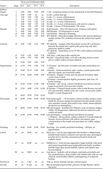
Embryonic development of R. quelen began with fertilization and ended with hatching out from the chorion (stages 1–25; Table 1). The embryonic development was subdivided into seven periods: zygote, cleavage, blastula, gastrula, segmentation, pharyngula, and hatching. The larval development was subdivided into three periods: early larval, mid larval, and late larval (stages 26–28), and is characterized by the presence of the yolk vesicle. Subsequently, after exhaustion of the yolk, the organisms were considered post-larvae (stage 29).
Periods and stages of development of Rhamdia quelen incubated at 21, 24, 27 and 30°C.

hpf = hours postfertilization; dpf = days postfertilization YE = yolk extension, YSL = yolk syncytial layer; na = not analysed.
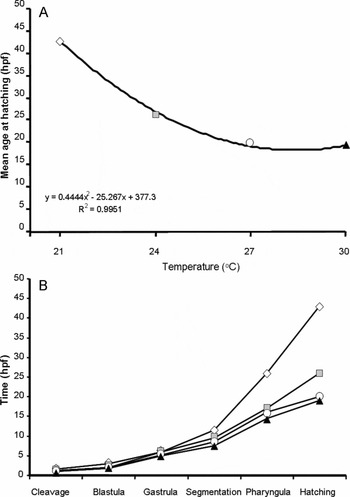
Rhamdia quelen embryos presented a negative relationship (r 2 = 0.9951) between time of development and incubation temperature (Fig. 1A, Table 1). Therefore, at 30°C, larvae hatched after 19 h of incubation, while they spent 43 h to hatch at 21°C. The duration of each period varied according to each test temperature. At 21°C, the time of segmentation and pharyngeal periods was much higher than that at higher temperatures (Fig. 1B).
Effect of incubation temperature on time of hatching for Rhamdia quelen. (A) Age at hatching (hpf; hours post-fertilization) of the embryos incubated in different regimes of temperatures. Mean times (hpf) were fitted to a polynomial function y = 0.4444x2 − 25 267x + 377.3, where y represents time of hatching (hpf) and x represents the temperature (°C). (B) Duration of each period (from cleavage to hatching) in function of the time (hpf) of the embryos of Rhamdia quelen incubated at four different temperatures.

The Table 1 summarizes the main developmental landmarks for R. quelen. The major morphological features in each stage were standardized in spite of temperature, as described below.
Embryonic development
Period 1 – zygote
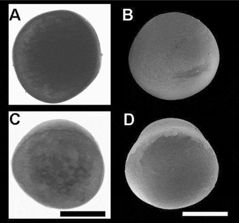
A distinct perivitelline space emerges around the fertilized egg, as the chorion lifts away from the cortex of the yolk sphere. The cytoplasm streams and accumulates on the animal pole, forming the active cytoplasm (blastodisc) (Fig. 2).
Zygote period of Rhamdia quelen. (A, B) Just fertilized. (C, D) The cytoplasm streams and accumulates on the animal pole, forming blastodisc (stage 1). (A) and (C) are light microscope images, and (B) and (C) are scanning electron microscopy images. Scale: 500 μm.

Period 2 – cleavage
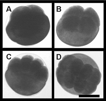
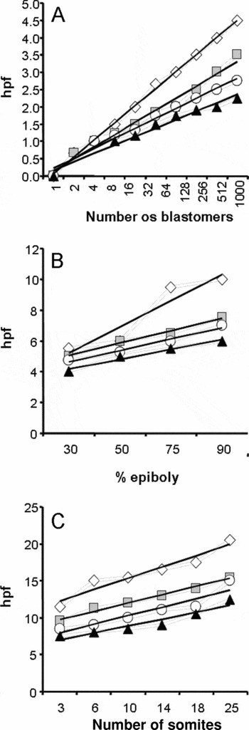
The cleavage period consisted of six stages (stages 2–7; Fig. 3). This period was characterized by a series of mitotic divisions on the active cytoplasm (meroblastic cleavages) that resulted in 64 blastomeres. After each cleavage the blastomeres decrease in size. During cleavage period, embryos formed in average 400 blastomeres per hour at 30°C. At 21°C, this rate decreased to half (200 cells per hour; Fig. 4A).
Cleavage period of Rhamdia quelen. Embryos fixed in 4% paraformaldehyde (light microscopy) with: (A) two (stage 2), (B) four (stage 3), (C) eight (stage 4), and (D) 16 blastomeres (stage 5). Scale: 500 μm.

Development of Rhamdia quelen embryos incubated in four different temperatures (21, 24, 27, and 30°C) as a function of the time (hpf, hour post-fertilization). (A) Number of blastomeres. (B) Per cent of epiboly. (C) Number of somites.

Period 3 – blastula
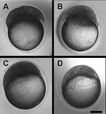
The blastula period consisted of four stages (stages 8–11). The early blastula (stage 8) was characterized by a blastoderm with 128 or 256 cells, forming a half sphere elevated from the yolk cell. The mid-blastula (stage 9) has 512 or few thousands of blastomeres, an outer enveloping layer (EVL) and a peripheral yolk syncytial layer (YSL). The YSL is a syncytial layer located in the intersection of yolk and blastoderm that is perceptible in live embryos under light microscope as a darker region. The edges of the blastoderm are jagged (Fig. 5A, B). During the late-blastula stage (stage 10), the blastoderm flattens due blastomeres intercalation, forming a compact uniform mass (Fig. 5C). At the end of this period, the blastoderm was highly flattened and starts the morphogenetic movement known as epiboly. During the stage 11, the margins of blastoderm cover 30% of the yolk cell (Fig. 5D).
Blastula period of Rhamdia quelen. Life embryos under light microscope. (A) Early blastula (stage 8); (B) Mid blastula (stage 9); (C) Late blastula (stage 10); and (D) 30% epiboly (stage 11). Scale: 200 μm.

Period 4 – gastrula
The gastrula period consisted of four stages (stages 12–15). The stage 12 was characterized by 50% (Fig. 6A) of epiboly and the presence of the germ ring around the margin of the blastoderm (Fig. 6C, D). The germ ring is formed by the epiblast and hypoblast superposition. Subsequently the embryonic shield is formed by thickening of the germ ring in the future dorsal side of the embryo. The shield extends from the germ ring toward the animal pole. As epiboly proceeds, the blastoderm gradually covers the whole yolk cell (Fig. 6E–H). During the neurula stage (stage 15, Fig. 6I, J) the blastoderm fully covers the yolk plug, closing the blastopore; the neural groove is visible; and the embryo became elliptical. The gastrulation, easily visible by per cent of epiboly, lasted about the half of time in higher temperatures (Fig. 4B).
Blastula (late blastula) and gastrula periods of Rhamdia quelen. (A) 50% epiboly (stage 12); (B) late blastula (stage 10); (C) 50% epiboly with germ ring formation (stage 12); (D) 50% epiboly (stage 12); (E, F) 75% epiboly (stage 13); (G, H) yolk plug (90% epiboly; stage 14); and (I, J) neurula stages (stage 15). Embryos of the left column were life (light microscopy), while embryos of the right column were analysed under scanning electron microscope. All embryos are in lateral view, except to (H), that is in view from vegetal pole. Scale: 200 μm.

Period 5 – segmentation
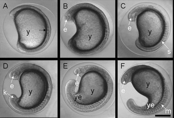
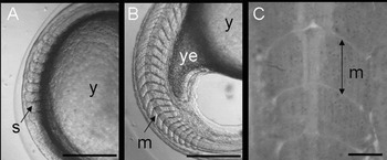
The segmentation period consisted of six stages (stages 16–21; Fig. 7). Somite formation, beginning of organogenesis, and compartmentalization of the brain characterized this period. The mesodermal segmentation begins to form the somites. Together to the development the three first somites, the optic vesicles become conspicuous. The Kupfer's vesicle appears and disappears during this period. The tail bud starts to protrude and the yolk extension (YE) is formed. The first muscular twitches occur in the stage 20 (18 somites). The somites develop into V-shaped myotomes (Fig. 8) that allow side-to-side flexures of the embryo body in the stage 21 (25 somites).
Segmentation period of Rhamdia quelen. Life embryos with: (A) 2–3 somites (stage 16); (B) six somites (stage 17); (C) 10 somites (stage 18); (D) 14 somites (stage 19); (E) 18 somites (stage 20); (F) 25 somites (stage 21). Symbols: e = eye, k = Kupfer's vesicle, m = myomere, s = somite, y = yolk, ye = yolk extension. Scale: 200 μm.

Muscle formation of Rhamdia quelen. (A) Life embryos with 10 somites (stage 18); and (B) 25 somites (stage 21). (C) Histological sagittal section from muscle trunk of just hatching larvae. Symbols: m = myomere, s = somite, y = yolk, ye = yolk extension. Scale: (A, B) 100 μm; (C) 10 μm.

Period 6 – pharyngula
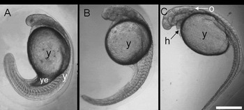
The pharyngula period consisted of three stages (stages 22–24; Fig. 9). The early-pharyngula embryos (stage 22) have about 30 myotomes and present intense motility (Fig. 9A). During this period, the embryo body elongated and the late-pharyngula embryos (stage 24) present about 40 myotome, needing to fold (1/2 trunk) inside the chorion. The blood circulation begins during this period. The weak circulation visible in the stage 23 became strong in the stage 24. The pericardial cavity is visible in the stage 22 and the first heartbeats occur in the mid-pharyngula (stage 23). Remarkable structures such as primordial fins (embryonic fin), midgut, and gaps related to mouth and operculum became visible in the pharyngula period. A pair of otoliths also appears as small granules lying against the inner surface of each otocyst. Blood circulates in all three vitelline veins. The pharyngula period presented greater variation among temperatures, lasting 17 h at 21°C and only 4.5 h at 30°C (Table 1).
Pharyngula period of Rhamdia quelen. Life embryos in stages: (A) Early pharyngula (stage 22); (B) mid pharyngula (stage 23); (C) late pharyngula (stage 24). Symbols: h = heart (pericardial cavity), o = otic cup, y = yolk, ye = yolk extension. Scale: 500 μm.

Period 7 – hatching
The hatching period consisted of one stage (stage 25). The hatching period was characterized by low motility just before hatching. The heart reaches 100 bpm. The hatch larvae do not present any pigmentation. Developmental time was inversely proportional to temperature (Fig. 1A). At 30°C, larvae hatched after 19 h of incubation, while they lasted in average 43 h to hatch at 21°C (Fig. 1B, Table 1).
Larval development
Period 8 – larval
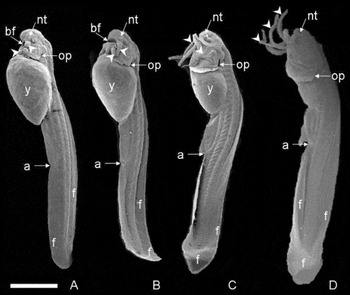
The larval period was divided into three stages (stages 26–28; Fig. 10). Larval development began after the hatching and finished with the complete yolk absorption. The yolk cell is much reduced in stage 28. The eyes became pigmented in the early-larvae (stage 26), but body pigmentation comes into view in the mid-larvae (stage 27). The mouth is an elliptical gap and three pairs of barbells buds grow around it in the early larvae. Inferior and superior lips develop and the jaw became protruded in the mid-larvae. Between stages 27 and 28, the larvae inflate the swim bladder and initiate exogenous feeding.
Larval period of Rhamdia quelen. Scanning electron microscopy of: (A, B) early larvae (stage 26, being A younger than B); (C) mid larvae (stage 27); (D) late larvae (stage 28). Symbols: a = anal pore, bf = buccopharyngeal cavity (oral slit), f = embryonic fin, nt = nostril, op = opercular cavity, y = yolk, head arrows = barbels. Scale: 500 μm.

Organogenesis
Nervous/sensory systems
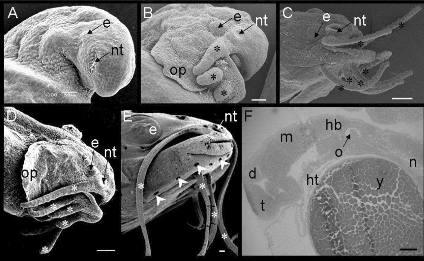
Neural tube forms at the end of gastrulation (stage 15). At the stage 17, the first neuromeres and the buds of some important sensorial structures, as optic vesicles and nostrils are visible (Fig. 11). The otic vesicles (Fig. 12A) appear later, during stage 18. The sensorial epithelium (macula) is located in anterior-ventral of the internal face of the otic cups and the otoliths were observed inside them in stage 21. Forebrain, midbrain and at least three hindbrain neuromeres are clearly visible in stage 20 (Fig. 11F). The early pharyngula (stage 22) presents the forebrain divided in two hemispheres. The eyes develop gradually, became pigmented at the early larval period (stage 26). The lateral line system starts to form before hatching, when the first neuromasts are visible (Fig. 12A). A complex set of pores lateral line system is present in the trunk and cephalic region of the juvenile. The nostrils of the larvae became progressively deeper and covered by ciliated cells (Figs. 11A–E and 12C). The early larvae present nostrils positioned laterally, but these structures displace to a terminal position as the jaw develops. The otic cups (Figs. 11F, 12D) also move to an anterior position. The barbells develop after hatching (Figs. 10 and 11). Three pairs of barbells buds develop around the mouth, being one on maxilla and two mental barbells. In stage 27, small and numerous protuberances (primordial taste buds) appear on the barbells buds (Fig. 12E, F). During the late-larvae stage (stage 28) the barbells elongated, turn into well developed structures, and numerous taste buds cover the barbells and lips (Fig. 12G).
Nervous and sensory systems of Rhamdia quelen. (A) Hatching larvae (stage 25); (B) early larvae (stage 26); (C) late larvae (stage 28); (D) post-larvae (stage 29); (E) juvenile (stage 30); (F) hatching larvae (stage 25). Symbols: d = diencephalon, e = eye, hb = hindbrain, ht = heart (pericardial cavity), m = midbrain, n = notochord, nt = nostril, o = otic cup, op = opercula, t = telencephalon, y = yolk, head arrows = lateral line system pores, * = barbels. (A–E) Scanning electron microscopy; (F) histological sagittal section. Scale: 500 μm.

Sensorial structures and pharyngeal teeth of Rhamdia quelen. (A) Neuromast; (B) longitudinal section of cephalic region; (C) nostril; (D) otic cup; (E, F) barbel; (G) lips; and (H) pharyngeal teeth. Pictures of the left column are obtained by scanning electron microscopy (Scale: 10 μm) and the pictures of the right column are histological sections (Scale: 500 μm). Symbols: ne = neuromast, bn = brain, o = otic cup, g = gill arches, bf = buccopharyngeal cavity, nt = nostril, m = macula, tb = taste buds, th = teeth, arrows = taste buds, head arrows = neuromasts (lateral line system).

Digestive system
During the embryonic and the major part of larval periods, the development depends on the endogenous nutrition (yolk). The YSL is visible in the mid blastula (stage 9). The yolk cell decrease gradually, being completely consumed at the end of the larval period. The foregut is visible at the stage 22, while midgut is distinct at the stage 23. Although, the larvae hatched with an opened mouth (represented by an elliptical slit positioned ventrally in the cephalic region), the anal pore open only after hatching (stage 26). However, the larvae can consume exogenous food even before the yolk exhaustion. The mid larvae (stage 27) have distinct superior and inferior lips that became covered with taste buds in the stage 28. Both taste buds and teeth are present in post-larvae pharynx (Fig. 12H).
Cardiorespiratory system
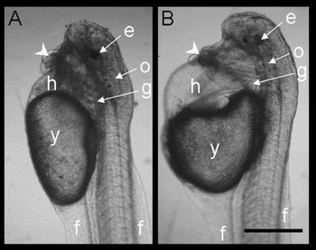
The pericardial cavity appears in the stage 19 and the dorsal aorta is visible at the end of segmentation period. The first heart beats (65 bpm in average) occur in the mid pharyngula (stage 23). In this period the embryo presents weak circulation. Gradually, the heart frequency increase to 100 bpm at the hatching and the blood circulate faster. The operculum open before hatching, but the early larvae (stage 26) present only primordial gill arches, represented by discrete groups of mesoderm cells on pharyngeal region. Some embryo incubated at 30°C presented heart edema (Fig. 13).
Rhamdia quelen early larva incubated at: (A) 24°C (with normal pericardial cavity); and (B) 30°C (presenting heart edema). Symbols: e = eye, f = embryonic fin, g = gill arches, h = heart (pericardial cavity), o = otic cup, y = yolk, head arrows = barbels. Scale: 500 μm.

Locomotory system
The first somites appear in stage 16. During segmentation, about five pairs of somites were formed per hour at 30°C against a rate of less than three somites per hour at 21°C (Fig. 4C). The first muscle contraction occurs during the stage 20, when the embryos have about 18 pair of somites. At the stage 21, the somites become ‘V-shaped’ and the embryo present side-to-side flexures. The hatching embryo presents about 40 myomeres. During the pharyngula period, the embryos form the embryonic fin, characterized by a dorsal fin continuous with a ventral fin (Fig. 10). The definitive fins differentiate during the post-larval period.
Discussion
The knowledge of the embryonic and larval stages, as well as, the control of water temperature under laboratory condition is of great help to optimize time and labour, nevertheless the knowledge of the embryonic and larval development under different temperature regime is important to recognize the best way to produce fingerlings of good quality.
The nomenclature and number of embryonic and larval periods are widely divergent in literature. The classification adopted for the present study was based on Kimmel et al. (Reference Kimmel, Ballard, Kimmel, Ullmann and Schilling1995). We described seven periods for the embryonic development (zygote, cleavage, blastula, gastrula, segmentation, pharyngula and hatching), and three larval periods (early larvae, mid larvae, and late larvae). Moreover, we described the main ontogenetic events during the post-larvae period and the begging of the juvenile phase.
In species with predominant visual sense to food perception, as Cyprinus carpio (Appelbaum e Riehl, Reference Appelbaum and Riehl1997) and Brycon amazonicus (Neumann, Reference Neumann2008), the eyes are well developed even in larvae. The R. quelen eyes became pigmented during early larvae stage. However, like other catfishes (Britzki et al., Reference Britski, Silimon and Lopes1999), R. quelen present relatively reduced eyes and well developed mechanosensory (lateral line) and chemosensory (taste buds and olfactory system) structures. During the larval period, lips and three pairs of barbells develop and are covered with numerous taste buds. Moreover, the neuromasts, present since the late embryonic period, develop in an abundant lateral line system both in trunk and cephalic regions. Efficient lateral line and chemosensory systems is essential to sustain the R. quelen habits. This omnivorous species forages predominantly at night, feeding mainly on benthic preys (Guedes, Reference Guedes1980; Schulz & Leuchtenberger, Reference Schulz and Leuchtenberger2006).
The embryonic fin remained during the overall larval period. Van Snik et al. (Reference Van Snik, Van Den Boogaart and Osse1997) proposed that this fin fold is related not only to locomotion, but also contributes to increase the surface area to gas exchange through the skin. According to Neumann (Reference Neumann2008), the embryonic fin reabsorption marks the end of a period of drastic body changes, when the small fish acquire an efficient gill respiration and the definitive fins are formed, allowing the change of the swimming pattern.
The improvements of structures related to swimming and, mainly, to food detection suggest that R. quelen is potentially able to take exogenous food even before the exhaustion of yolk. Moreover, the presence of abundant chemosensory structures in these early periods suggests that this species is able to select food since the first feeding. The mouth opening of larvae of some fish species occur simultaneously with the beginning of horizontal movements of swimming (Luz et al., Reference Luz, Reynalte-Tataje, Ferreira and Zaniboni-Filho2001) and pigmentation of the eyes (Lasker et al., Reference Lasker, Feder, Theilacker and May1970). The swim bladder of R. quelen inflates during the late larvae stage, allowing best foraging movements coincidently to yolk drop and gut maturation.
During the first days, fish larvae consume the energy reserve from yolk. The period of endogenous feeding is essential to maturation of digestive structures, allowing larvae to reaches the sites of accessible food resources. R. quelen late larvae start exogenous feeding even before total consumption of the yolk. Actually, the mouth starts to open before hatching, during the late pharyngula stage. According to Govoni et al. (Reference Govoni, Boehlert and Watanabe1986) the larvae gut is functional during the first exogenous feeding, although it is structurally and physiologically simpler than the adult gut. In the present study, the late larvae were fed on zooplankton even before the complete yolk exhaustion.
The yolk reserve present in late larvae is probably a guaranty. It diminished the risk of mortality in the case of scarce food in the environment. The beginning of exogenous feeding is a critic period on fish life history as 1-day-fed post-larvae present gut atrophy, having a limited time of survival under starvation in many fish species (Theilacker, Reference Theilackerg1978).
Water temperature is a major determining factor in fish biology. One obvious effect of high temperatures is an increase in the embryonic development rate (Garside, Reference Garside1966; Vernier, Reference Vernier1969; Ojanguren et al., Reference Ojanguren, Reyes-Gavilan and Muñoz1999; Saka et al., Reference Saka, Firat and Çoban2004; Cook, et al., Reference Cook, Kate, Michael and Plesha2005; Martell et al., Reference Martell, Kieffer and Trippel2005; Ninhaus-Silveira et al., Reference Ninhaus-Silveira, Foresti and Azevedo2006). Likewise, we observed a negative relationship between the time of embryonic development and incubation temperature in R. quelen.
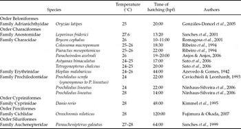
Table 2 presents data from literature concerning the duration of embryonic development in a number of teleost species. From analysis of these data it is possible to conclude that time of hatching is not related to the taxonomic organization of the species. With few exceptions, fish embryos kept under 24–26°C hatch up to 24 h. Higher temperatures usually accelerate, while lower temperatures reduce, the duration of embryonic development. The exceptions to this pattern are probably related to the ecological and/or behavioural features of each species, as the occurrence of parental care or egg adhesiveness, allowing a slower development.
Previous studies of teleost development: embryonic development (time of hatching) in hours postfertilization (hpf).

Gomes et al. (Reference Gomes, Golombieski, Chippari-Gomes and Baldisserotto2000) showed that R. quelen fingerlings acclimated at 31°C tolerate temperatures between 15 and 34°C. All temperatures tested in the present study (21, 24, 27 and 30°C) were found to support successful embryonic and larval development in this species.
R. quelen hatches faster at 30°C compared with the other lower temperatures. Nevertheless, some anatomical defects, like heart edema, were more frequent at 30°C. The presence of malformations, however, were not quantified nor analysed statistically, needing further studies. Arenzon et al. (Reference Arenzon, Lemos and Bohrer2002) showed similar results in Cynopoecilus melanotaenia. In this species the larvae incubated in higher temperatures had hatched with morphologic anomalies and remained immovable, making impossible its survival. Additionally, the high metabolic rates under high temperatures lead to faster yolk consumption and may be hazardous in natural environments, as the yolk could be exhausted before complete maturation of the structures related to catching and processing of the exogenous food and/or before the post-larvae reaches the sites with high food concentration.
Acknowledgements
We thank Dr Dorly de Freitas Buchi (UFPR, Brazil) for kind donation of some reagents used in our experiment (resources from project supported by Paraná Government–SETI).