Introduction
Cervical cancer is one of the most common female cancers worldwide. Reference Arbyn, Weiderpass and Bruni1 In 2017, new cases of cervical cancer in Thailand were more than 8,000· Reference Phoolcharoen, Kantathavorn and Sricharunrat2 The treatments for cervical cancer are surgery, radiotherapy or systemic treatment. For radical radiotherapy with chemotherapy, the combination of external beam radiotherapy (45–50·4 Gy in 23–28 fractions) and brachytherapy (3–5 fractions) is the standard treatment for locally advanced disease to give a dose of at least 80 Gy in EQD210 to the target. Reference Viswanathan, Beriwal and Jennifer3
Brachytherapy has been used for a long time to escalate the dose to the cervix to achieve the curative goal. 4 Nowadays, brachytherapy has been transformed from a point-based plan (2D) to a volume-based plan (3D), according to many promising publications from the GEC-ESTRO working group. In the volume-based plan, or image-guided adaptive brachytherapy (IGABT), magnetic resonance imaging (MRI) is the standard imaging technique as it provides the best soft tissue discrimination. Reference Haie-Meder, Pötter and Van Limbergen5,Reference Pötter, Haie-Meder and Van Limbergen6 Many international publications have reported promising results from MRI-guided brachytherapy. Reference Pötter, Dimopoulos and Georg7–Reference Pötter, Tanderup and Schmid15
Computed tomography (CT)-based IGABT was developed in practice because of the difficulties in using MRI for brachytherapy. Although CT showed poorer tissue discrimination than MRI, CT can support treatment in the volume-based plan as reported in many international publications. Reference Hegazy, Pötter and Kirisits16,Reference Hellebust17 Promising results of CT-based brachytherapy for cervical cancer have been published internationally. Reference Tan, Coles and Hart18–Reference Ohno, Noda and Okonogi24 Another method, point-based adaptive planning by transabdominal ultrasound (TAUS), was developed for use in brachytherapy. A few studies have reported the use of a TAUS-based plan and have shown a clinical benefit. Reference Tharavichitkul, Chakrabandhu and Wanwilairat21,Reference van Dyk and Bernshaw25–Reference Tharavichitkul, Chakrabandhu and Klunklin31
At our Institute, transformation from 2D (point) to 3D (volume) began in 2008 with CT-based brachytherapy. Unfortunately, the total transformation could not be performed immediately due to our workloads and equipment. In 2011, ultrasound for brachytherapy was installed at our Institute. In 2012, a project to improve the quality of 2D planning using TAUS-guided brachytherapy as 2·5D planning was initiated. From these projects, intermediate-term results were reported in 2013 and 2018, respectively. Reference Tharavichitkul, Chakrabandhu and Wanwilairat21,Reference Tharavichitkul, Chakrabandhu and Klunklin31 The total transformation to a volume-based approach (CT-based plan) succeeded in 2019 when a 4-slice CT instrument was installed in our brachytherapy unit. After that point, our clinical practice changed to volume-based treatment by CT, and TAUS was utilised to prevent uterine perforation and to support the CT-based contouring. However, dose evaluation or correlation between these two approaches was not performed.
With this in mind, we performed the present study to evaluate comparative dosimetry of brachytherapy treatment planning between a volume-based plan by CT, and a point-based plan by TAUS, in CT datasets for brachytherapy.
Materials and Methods
This study was a case–control study approved by the Institutional Review Board of the Faculty of Medicine, Chiang Mai University, with the code RAD-2563–07479.
CT datasets
From January 2019 to January 2021, 59 CT image datasets with 3 mm slice thickness (Alexion, Toshiba Medical Systems, Tochigi, Japan) were collected from 38 patients with locally advanced cervical cancer treated by intracavitary CT-based brachytherapy. High-risk clinical target volume (HR-CTV), intermediate-risk clinical target volume (IR-CTV) and OARs were contoured by experienced radiation oncologists during treatment according to GEC-ESTRO recommendations and the work of Viswanathan et al. Reference Haie-Meder, Pötter and Van Limbergen5,Reference Pötter, Haie-Meder and Van Limbergen6,Reference Viswanathan, Erickson and Gaffney32 All fractions were treated by CT-based brachytherapy (volume-based approach) to give a dose of at least 7 Gy to D90 of HR-CTV. In these fractions, TAUS (Flex Focus 400, BK medical, Harlev, Denmark) was performed by a radiation oncologist to prevent uterine perforation during applicator placement.
Study methods
To perform this study, all CT datasets and structures were imported into Oncentra Master Plan version 4.5.3 (Elekta Brachytherapy, Veenendaal, the Netherlands). Applicator reconstruction was performed by manual reconstruction or library-based registration according to the applicator type. Two planning approaches were performed, and sources were applied with the same dwell positions in both approaches. The dwell times in each dwell position were optimised as the planning aim of each approach.
Approach 1: volume approach
In the volume-based plan by CT, the target of the treatment was HR-CTV, and planning aims were performed to keep the dose at 90% of HR-CTV to give a dose of at least 7 Gy.
Approach 2: point approach
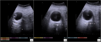
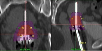
In the point-based plan by TAUS, eight measurements of cervix dimensions (from intrauterine tandem to uterine wall) were performed at the level of the cervical os, and 2 cm cranially to the cervical os, based on previous work by van Dyk et al. and Tharavichitkul et al. Reference van Dyk and Bernshaw25,Reference Tharavichitkul, Tippanya and Jayavasti28 Eight cervix reference points were generated from cervix dimensions in sagittal and axial views. Planning aims were performed to keep the minimum dose to the cervix reference points to give a dose of at least 7 Gy.
In both approaches, doses at selected parameters were recorded as follows. 4
-
HR-CTV: D50, D90, D95, D98 and D100
-
IR-CTV: D90 and D98
-
OARs (bladder, rectum, sigmoid colon): D0.1cc, D1cc and D2cc
-
Point A
-
Bladder ICRU, Rectal ICRU and Vaginal dose point (5 mm)
-
Vaginal reference point by PIBS (Westerveld et al.) Reference Westerveld, Pötter and Berger33
-
Conformity index (COIN) Reference Poddar, Sharma and Suryanarayan34
 $${\rm{COIN\; = \;}}{{{{{\rm{V}}_{{\rm{ref,TV}}}}}\over{{\rm{TV}}}}}{\rm{ \times }}{{{{{\rm{V}}_{{\rm{ref,TV}}}}}\over{{{\rm{V}}}_{{\rm{ref}}}}}}$$
-
Homogeneity index (DHI) Reference Poddar, Sharma and Suryanarayan34
 $${\rm{DHI\; = \;}}{{{{\rm{(TV}}{{\rm{D}}_{{\rm{ref\;}}}}{\rm{ - \;TV1}}{\rm{.5}}{{\rm{D}}_{{\rm{ref}}}}{\rm{)}}}\over{{\rm{TV}}{{\rm{D}}_{{\rm{ref}}}}}}}$$
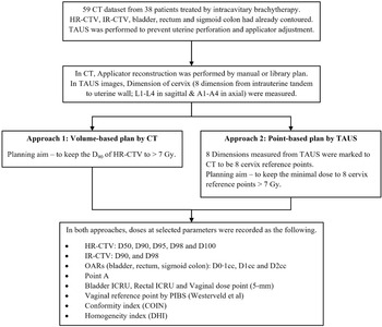
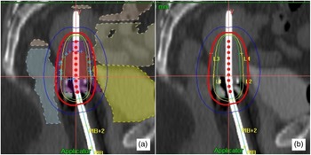
Images of the entire study process, measurements of eight cervix dimensions by TAUS, targets (HR-CTV & eight cervix reference points) and isodose distributions of both approaches are shown in Figures 1–4.
Study process.

Measurements of eight cervix dimensions by TAUS: (a) sagittal view of TAUS, (b) axial view of TAUS at the cervical os and (c) axial view of TAUS at 2 cm cranially from the cervical os.

HR-CTV, IR-CTV and eight cervix reference points (A1-A4 & L1-L4) in the CT datasets.

Isodose distributions of (a) volume-based plan by CT and (b) point-based plan by TAUS.

Statistical analysis
All dose parameters were compared for the volume-based plan by CT and the point-based plan by TAUS. A Wilcoxon sign-ranked test was used to evaluate the comparison.
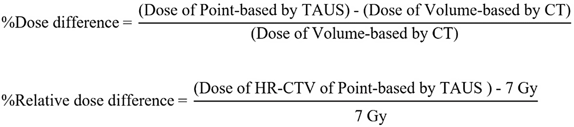
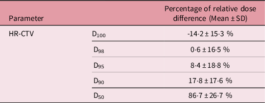
Percentage dose difference was evaluated from the dose difference to the targets or OARs in the volume-based plan by CT versus the point-based plan by TAUS. Percentage of relative dose difference was evaluated as the difference of the goal dose to the cervix reference points (at least 7 Gy) by TAUS to the dose to HR-CTV (contoured by CT) in terms of D50, D90, D95, D98 and D100. (Figure 5) IBM SPSS version 22 (IBM SPSS, Chicago, IL, USA) was used to evaluate the results.
Percentage dose difference and relative dose difference formulae.

Results
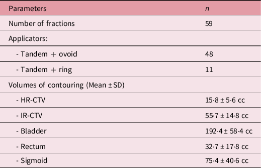
Of 59 fractions, a tandem ovoid applicator was used in 48 fractions. The mean volume of HR-CTV was 15·8 cc. Table 1 shows the characteristics of the studied datasets.
Characteristics data

Note: HR-CTV = high-risk clinical target volume, IR-CTV = intermediate-risk clinical target volume.
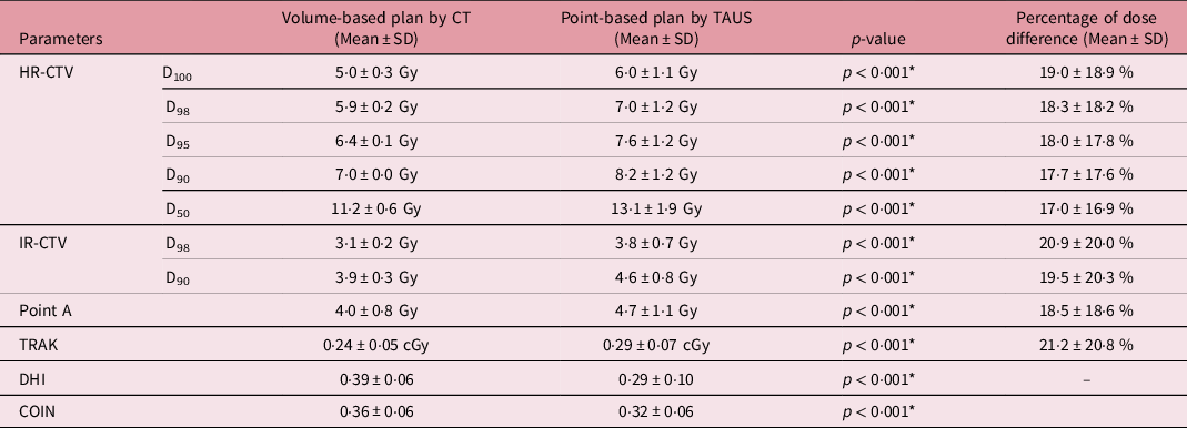
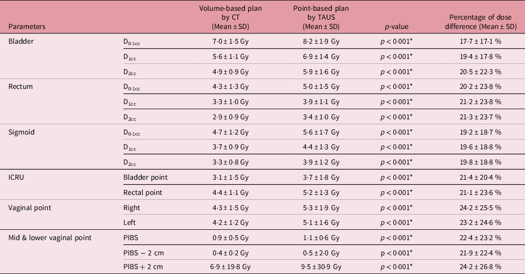
In the targets, generally, the point-based plan by TAUS yielded a significantly higher dose to D90 of HR-CTV, D90 of IR-CTV, point A, TRAK, DHI and COIN. The percentage of dose difference between the point-based plan by TAUS and the volume-based plan by CT ranged from 17·0 to 21·2% (see Table 2). Interestingly, when we further evaluated the percentage of relative dose difference between the planning aim of the point-based plan by TAUS (at least 7 Gy at cervix reference points) to volume parameters of HR-CTV (from D50 to D100), the TAUS-based planning aim was close to D98 of HR-CTV with a mean of 0·6%. Tables 2 and 3 show the dose parameters, % of dose difference and % of relative dose difference, for the volume-based plan by CT and the point-based plan by TAUS relative to target dose. In normal tissues, the point-based plan by TAUS yielded a significantly higher dose to the bladder, rectum, sigmoid, ICRU points and vaginal points. The percentage of dose difference between the point-based plan by TAUS and the volume-based plan by CT ranged from 17·7 to 24·2% (see Table 4). Table 4 shows the dose parameters and % of dose difference for the volume-based plan by CT and the point-based plan by TAUS in OARs.
Doses to target for volume-based plan by CT and point-based plan by TAUS plans

Note: COIN = conformity index, CT = computed Tomography, D100 = the minimum dose covering 100% of volume, D98 = the minimum dose covering 98% of volume, D95 = the minimum dose covering 95% of volume, D90 = the minimum dose covering 90% of volume, D50 = the minimum dose covering 50% of volume, DHI = dose homogeneity index, HR-CTV = high-risk clinical target volume, IR-CTV = intermediate-risk clinical target volume, TAUS = transabdominal ultrasound, TRAK = total reference air kerma, SD = standard deviation.
* Significant difference.
Percentage relative dose difference of HR-CTV in point-based plan by TAUS plan

Note: D100 = the minimum dose covering 100% of volume, D98 = the minimum dose covering 98% of volume, D95 = the minimum dose covering 95% of volume, D90 = the minimum dose covering 90% of volume, D50 = the minimum dose covering 50% of volume, HR-CTV = high-risk clinical target volume, SD = standard deviation.
Doses to organs at risk for volume-based plan by CT and point-based plan by TAUS plans.

Note: CT = computed tomography, D0.1cc = dose at 0.1 cc of volume, D1cc = dose at 1 cc of volume, D2cc = dose at 2 cc of volume, ICRU = The International Commission on Radiation Units and Measurements, PIBS = Posterior-Inferior Border of Symphysis, TAUS = transabdominal ultrasound, SD = standard deviation.
* Significant difference.
Discussion
In clinical practice, although the trend in brachytherapy has moved to volume-based planning, some centres still perform point-based planning. Here, TAUS supports reduction of overdose in the bladder and rectal points in comparison to the conventional prescription. Reference Tharavichitkul, Chakrabandhu and Wanwilairat21,Reference Tharavichitkul, Tippanya and Jayavasti28 Our results showed that the point-based prescription to the cervix reference points by TAUS caused a higher dose in comparison to the volume-based prescription to D90 of HR-CTV. Moreover, when we explored the 7 Gy prescription to the cervix reference points, we found the dose to be nearly 7 Gy, close to D98 of HR-CTV. This may support the finding that when we treat by cervix reference points measured by TAUS, the cumulative dose from EBRT plus brachytherapy should focus to at least 75 Gy in EQD210 (the planning aim of D98 of HR-CTV published in the EMBRACE II protocol). Reference Pötter, Tanderup and Kirisits35 For example, if we treated EBRT 46 Gy in 23 fractions, the BT dose by ≥ 29 Gy in EQD210 (such as 21 Gy in 3 fractions or 24 Gy in 4 fractions) will be prescribed to the cervix reference points to get the cumulative dose by at least 75 Gy in EQD210.
This study has some limitations. First, as mentioned above, the cumulative dose of EBRT and BT doses were not evaluated. Second, this study is a case–control study in which we performed two planned approaches on CT datasets of previous treatment. Third, only a single fraction was evaluated. Fourth, these findings were obtained using intracavitary brachytherapy only. Finally, MRI was not performed in these patients. Although there were some limitations, the results of our study revealed that the doses to targets and OARs when we prescribed 7 Gy to the cervix reference points by TAUS were higher than the volume-based prescription to D90 of HR-CTV with an average 20% difference. When we evaluated the correlation of planning aims of 7 Gy to the cervix reference points to the volume of HR-CTV, it showed close correlation to D98 of HR-CTV with a mean percentage of difference of 0·6%.
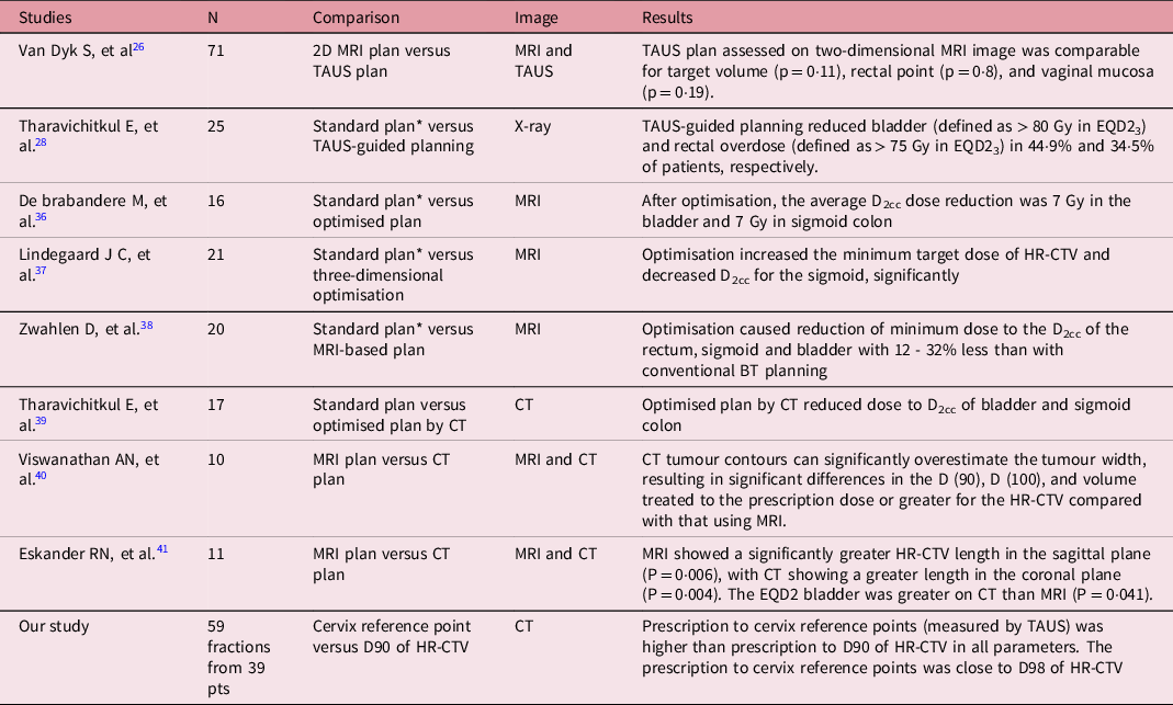
Based on the literature, three types of dose comparison studies were performed. First, comparative studies performed to compare between point A and image-guided treatment in the same type of image showed that the image-guided plan improved the dose to targets or reduced overdosage to normal tissues (in comparison to the point A plan). Reference Tharavichitkul, Tippanya and Jayavasti28,Reference De Brabandere, Mousa and Nulens36–Reference Tharavichitkul, Mayurasakorn and Lorvidhaya39 Second, comparative studies were performed to compare MRI- versus CT-based contouring. All studies showed that CT-based contouring gave an overestimation and a higher dose to normal tissues in comparison to MRI contouring. Reference Viswanathan, Dimopoulos, Kirisits, Berger and Pötter40,Reference Eskander, Scanderbeg, Saenz, Brown and Yashar41 Third, comparative studies were performed to compare MRI- versus TAUS-based planning. A report by van Dyk et al. has shown the methods to be comparable. Reference van Dyk, Narayan and Fisher26 Table 5 shows selected studies of dose comparisons. Our study is categorised in the first type, but this is modified to compare between cervix reference points versus D90 of HR-CTV in CT images. Ultrasound showed correlation to MRI in dimensional evaluation of the cervix for brachytherapy. The comparison studies mostly compared measurements between TAUS and MRI and showed similar results in measurement. Reference van Dyk and Bernshaw25,Reference Mahantshetty, Khanna and Swamidas42 Moreover, the use of transrectal ultrasound (TRUS)-guided CT contouring showed correlation to MRI in HR-CTV evaluation. Reference Schmid, Nesvacil and Pötter43,Reference Mahantshetty, Khadanga and Gudi44
Selected studies of dose comparison.

Note: BT = brachytherapy, CT = computed tomography, D2cc = dose at 2 cc of volume, EQD2 = equivalent dose of 2 Gy, HR-CTV = high-risk clinical target volume, MRI = magnetic resonance imaging, OARs = organs at risk, TAUS = transabdominal ultrasound.
* Standard plan means prescription to point A.
Nowadays, improving treatment quality in brachytherapy for cervical cancer depends on equipment, manpower and workload. At our Institute, we have CT and TAUS for brachytherapy. The use of TAUS-guided planning or CT contouring supported by US (TAUS or TRUS) will be the future approach to improve IGBT for cervical cancer at our institute.
Conclusions
In conclusion, this study has shown that the point-based plan by TAUS yielded a higher dose to targets and OARS in all parameters, with a mean percentage of difference of approximately 20%. In the additional analysis of percentage of relative dose difference, the dose of 7 Gy to the cervix reference points was close to D98 of HR-CTV (with a mean percentage of difference of 0·6%).
Conflicts of Interest
None.