Introduction
Preterm newborn at 36 weeks, birth weight 2000 g, only child, healthy parents. Pregnancy was well controlled, with negative maternal serologies. During screening for prematurity, an abdominal ultrasound detected the presence of multiple echogenic solid images distributed throughout the liver parenchyma, predominantly in the right lobe. These images showed significant vascularisation on colour Doppler examination, with turbulent arterial and venous flow. These findings were likely related to hepatic hemangiomas.
The hepatology service decided to start treatment with propranolol, and a prior cardiology evaluation before starting medication revealed on echocardiogram a well-circumscribed, homogeneous mass on the posterosuperior wall of the right atrium, near the superior vena cava, and another smaller mass near the tricuspid annulus, which fills with colour Doppler. There was also a grade II pericardial effusion (Figure 1a, 1b, 1c, 1d).
Transthoracic echocardiogram and cardiac MRI images. 1(a) Four-chamber view. A well-circumscribed, homogeneous mass on the posterosuperior wall of the right atrium, near the superior vena cava, and another smaller mass near the tricuspid annulus (blue arrows). Anterior and posterior pericardial effusion (purple arrow). 1(b) The tumor fills with color Doppler (blue arrow). 1(c) Tumor size: 10 × 9 mm. 1(d) Tumor area: 0.94 cm². 1(e) Cardiac MRI. T2 STIR sequence, four-chamber view. Hyperintense lesions in T2 in the right atrium and atrioventricular groove (blue arrows). 1(f) Cine SSFP sequence, four-chamber view. Hyperintense lesions in T2 in the right atrium and atrioventricular groove (blue arrows).

A cardiac MRI was performed (Figure 1e, 1f): On the posterior wall of the right atrium, from the entrance of the superior vena cava, a heterogeneous, lobulated, irregular image was seen measuring 24 mm in longitudinal axis x 10 mm anteroposterior x 15 mm laterolateral, fixed. Another heterogeneous image was observed on the anterior face of the right atrioventricular groove measuring 13 mm x 9 mm x 12 mm, which did not cause obstruction to the entry or valve dysfunction. These structures were isointense in T1, hyperintense in T2, with positive perfusion and negative late enhancement. They were compatible with a vascular tumour. It was not possible to define whether these were two separate structures or whether they were connected.
The child also had a capillary vascular lesion along the midline of the forehead (hemangioma).
Ophthalmologic evaluation was normal, and the genetic service did not observe any significant dysmorphisms.
During the hospitalisation, the patient progressed with severe pericardial effusion, which did not respond to diuretics or corticosteroids. Pericardiocentesis was performed, yielding inflammatory fluid without tumour cells. Due to the persistence of severe pericardial effusion, an evacuative drainage and pleuropericardial window were performed. Subsequently, the pericardial effusion did not recur.
The patient continued with outpatient follow-up, including echocardiographic monitoring of tumour size. She never presented arrhythmias or obstruction to the inflow or outflow tracts of the ventricles, with preserved ventricular function at all times.
In several successive follow-ups, the size of the cardiac tumour and hepatic hemangiomas remained the same. She continued being treated with propranolol as recommended by the hepatology service.
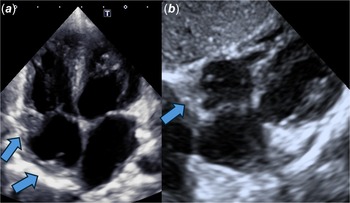
In the last follow-up at 3 years of age, a marked reduction in the cardiac hemangioma was observed on echocardiogram (Fig 2). The most visible portion was located on the posterior side, homogeneous, and did not cause obstruction in either the inflow tract or the superior vena cava, with which it had the closest relation. It measured 4 mm x 3 mm
Last echocardiogram. 2(a) Four-chamber view. Small residual tumor on the roof of the right atrium and another one near the tricuspid annulus (blue arrows). 2(b) Subcostal view. Residual tumor (blue arrow).

Discussion
Cardiac vascular tumours in children, including cardiac hemangiomas, are extremely rare. The medical literature indicates that primary cardiac tumours in the paediatric population are uncommon and are usually benign, with rhabdomyoma being the most common type. Reference Morka, Kohut and Radzymińska-Chruściel1,Reference Padalino, Vida and Boccuzzo2 Cardiac hemangiomas are even less common, representing only a small fraction of paediatric cardiac tumours. Reference Placidi, Calcagni and Lioncino3
The exact incidence of cardiac hemangiomas in children is not well documented due to their rarity. However, studies on paediatric cardiac tumours show that hemangiomas constitute a minority of cases. Reference Placidi, Calcagni and Lioncino3 In terms of prognosis, benign cardiac tumours in children, including hemangiomas, generally have a good outlook if managed appropriately. Most of these tumours are non-malignant and can be successfully treated through observation or surgical resection, depending on their size and location. Reference Delmo Walter, Javier and Sander4,Reference Padalino, Vida and Boccuzzo2
The progression of cardiac hemangiomas can vary. Some may remain stable without causing significant symptoms, while others may require intervention if they lead to haemodynamic obstruction or arrhythmia. Reference Delmo Walter, Javier and Sander4,Reference Pucci, Botta and Sina5 Surgical resection is an option in cases where the tumour causes significant symptoms, and long-term outcomes are usually favourable, with most patients remaining symptom-free following complete or partial resection. Reference Delmo Walter, Javier and Sander4,Reference Padalino, Vida and Boccuzzo2
In summary, although cardiac hemangiomas in children are rare, their prognosis is generally positive when diagnosed and managed appropriately. Medical literature suggests that most benign cardiac tumours in children have good long-term outcomes, especially when complete or partial resection is performed. Reference Delmo Walter, Javier and Sander4,Reference Padalino, Vida and Boccuzzo2
The association of hepatic hemangiomas and cardiac hemangiomas in children is uncommon. Infantile hemangiomas are the most frequent vascular tumours in paediatrics, and while they can occur in multiple locations, most cases involve the skin and, to a lesser extent, the liver Reference Rodríguez Bandera, Sebaratnam, Wargon and Wong6,Reference Krowchuk, Frieden and Mancini7 Hepatic hemangiomas can be multifocal or diffuse and, in rare cases, may be associated with high-output heart failure due to macrovascular shunts. Reference Krowchuk, Frieden and Mancini7,Reference Northup, Garcia-Pagan and Garcia-Tsao8 However, the medical literature does not specifically mention the coexistence of hepatic and cardiac hemangiomas as a common or recognised presentation in children.
It is important to highlight that hepatic hemangiomas can be part of complex syndromes such as PHACE syndrome, which includes posterior fossa malformations, arterial anomalies, cardiac defects, among others. Reference Rodríguez Bandera, Sebaratnam, Wargon and Wong6 However, the presence of cardiac hemangiomas as part of this syndrome is not a typical feature.
In summary, although hemangiomas can occur in multiple organs, the specific association of hepatic and cardiac hemangiomas in children is not documented as a common occurrence in current medical literature.
Conclusion
Cardiac hemangiomas in children are extremely rare and represent a small fraction of paediatric cardiac tumours, most are benign and can be effectively treated with observation or surgical intervention if necessary. In this case, the progression of the cardiac hemangioma was monitored closely with successful outcomes, including a marked reduction in size during follow-up.
The coexistence of hepatic and cardiac hemangiomas in a paediatric patient is uncommon and not well-documented in current medical literature. This case adds to the limited knowledge about the potential relationship between these two types of hemangiomas and highlights the importance of continuous monitoring and multidisciplinary management for optimal outcomes.
Further research and case studies are needed to better understand the potential interactions between these two types of vascular tumours in paediatric patients.
Supplementary material
For supplementary material accompanying this paper visit https://doi.org/10.1017/S1047951125100656
Financial support
This research received no specific grant from any funding agency, commercial, or not-for-profit sectors.
Competing interests
None.
Ethical standards
None.