I. INTRODUCTION
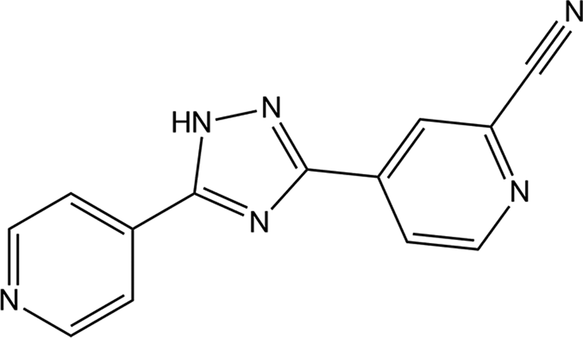
Topiroxostat (4-(5-(pyridine-4-yl)-1H-1,2,4-triazole-3-yl)pyridine-2-carbonitrile) is a selective xanthine oxidoreductase inhibitor, which is effective in decreasing urinary albumin excretion and reducing the level of serum urate in the human body (Hosoya et al., Reference Hosoya, Ohno, Nomura, Hisatome, Uchida, Fujimori, Yamamoto and Hara2014). Thus, topiroxostat (TOPI) was approved in Japan in June 2013 for the treatment of gout and hyperuricemia (Hosoya et al., Reference Hosoya, Sasaki, Hashimoto, Sakamoto and Ohashi2016). The chemical structure of topiroxostat is shown in Figure 1. As is known that more than half of the pharmaceutical compounds exhibit solid-state polymorphism, and is of great importance as different crystal forms of the drug can show different stability, solubility, dissolution rate and bioavailability, especially for poorly soluble drugs. TOPI has been reported that there are five different crystal forms, such as form I, form II, form III, monohydrate and form A (Lee et al., Reference Lee, Erdemir and Myerson2011; Iwabuchi et al., Reference Iwabuchi, Miyata, Sato, Uda, Kandou, Inoue and Nakano2014), but their crystal structures have not been reported yet.
Chemical structure of 4-(5-(pyridine-4-yl)-1H-1,2,4-triazole-3-yl)pyridine-2-carbonitrile.

We have inspected the Cambridge Structural Database (Groom et al., Reference Groom, Bruno, Lightfoot and Ward2016) and the PDF4 + database (Gates-Rector and Blanton, Reference Gates-Rector and Blanton2019) and have not found any entries for the topiroxostat form II (TOPI-II) and the topiroxostat monohydrate (TOPI-H2O) in the mentioned databases. Therefore, we have decided to characterize these compounds by X-ray powder diffraction and X-ray single-crystal diffraction techniques.
II. EXPERIMENTAL
A. Sample preparations
The sample was supplied by Zhejiang Jingxin Pharmaceutical Co., Ltd (purity >99.9%) and used without further purification. Dissolving topiroxostat (400 mg) in anhydrous ethanol (250 ml) and ethanol–water (300 ml, 9:1 v/v), respectively, at reflux temperature and slow cooling of the solutions yielded crystals of TOPI-II and TOPI-H2O. Then, the crystals were dried, smashed and mounted on a flat zero-background plate.
B. Powder diffraction data collection
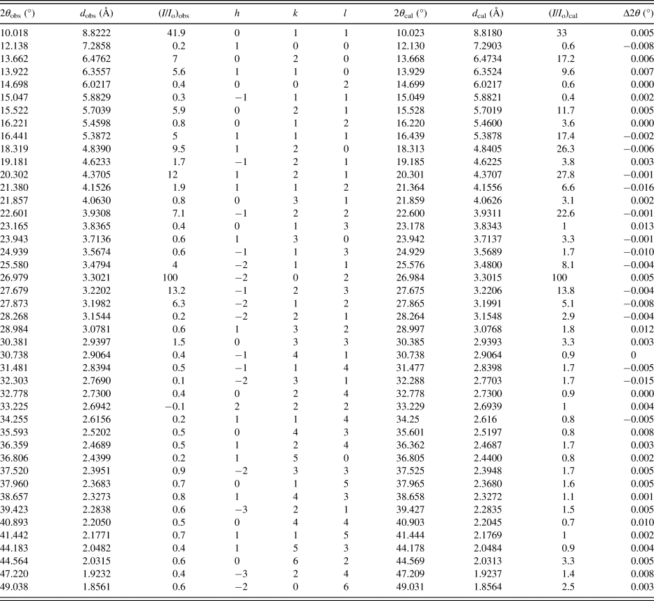
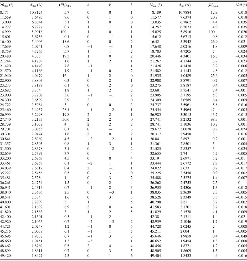
X-ray powder diffraction data were collected at room temperature with a SmartLab diffractometer with parafocusing Bragg-Brentano geometry using a Cu Kα radiation (λ = 1.5418 Å) and operated at 40 kV and 180 mA. The D/tex Ultra 250 detector was employed to collect XRD data over the 2θ range from 3° to 50° with a step size of 0.02° and a counting time of 1.2 s step−1. The software package MDI-Jade version 7.5 (Materials Data Inc., USA) was used to smooth the data, fit the background, strip off the Kα2 component and obtain the peak positions and intensities (Tables I and II). The Kα1was used in converting observed 2θ to d-spacing.
X-ray powder diffraction data of TOPI-II.

X-ray powder diffraction data of TOPI-H2O.

C. Single-crystal diffraction data collection
X-ray single-crystal diffraction data were collected at room temperature with a Bruker D8 Venture diffractometer with Mo Kα radiation (λ = 0.71073 Å) for cell determination and subsequent data collection. Data reduction was performed by APEX3 software and multi-scan absorption correction was applied. Using Olex2 (Dolomanov et al., Reference Dolomanov, Bourhis, Gildea, Howard and Puschmann2009), the structures were solved with the ShelXT (Sheldrick, Reference Sheldrick2015a) structure solution program using intrinsic-phasing and refined with the ShelXL (Sheldrick, Reference Sheldrick2015b) refinement package using Least Squares minimization.
III. RESULTS AND DISCUSSION
Indexing of the experimental X-ray diffraction patterns and unit-cell refinements were done using MDI-Jade (Materials Data Inc., 2002). The cell refinement results confirmed that TOPI-II is monoclinic with the space group P21/c and unit-cell parameters: a = 7.344(9) Å, b = 12.946(7) Å, c = 12.133(5) Å, β = 96.99(3)°, unit-cell volume V = 1145.2(4) Å3, Z = 4. The figure of merit is F 30 = 184.8(0.0053,30) (Smith and Snyder, Reference Smith and Snyder1979). TOPI-H2O is also triclinic with the space group P-1 and unit-cell parameters: a = 7.422(9) Å, b = 8.552(1) Å, c = 11.193(5) Å, α = 74.85(1)°, β = 81.17(1)°, γ = 66.29(1)°, unit-cell volume V = 627.0(6) Å3 and Z = 2. The figure of merit is F 30 = 72.5 (0.013,30) (Smith and Snyder, Reference Smith and Snyder1979). The values of 2θ obs, d obs, I obs, h, k, l, 2θ cal, d cal, I cal and Δ2θ are listed in Tables I and II. Because the morphology of TOPI-II and TOPI-H2O was plate crystals with preferred orientations, there is a minor difference in the relative intensities of the diffraction peaks between the experimental X-ray diffraction patterns and the calculated XRD patterns.
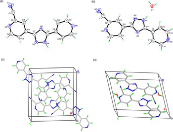
Based on the single-crystal data, the structures of TOPI-II and TOPI-H2O were solved and refined. The detailed crystallographic information is summarized in Supplementary Tables SI–SIII, and the asymmetric units of both forms with the corresponding atom labeling scheme are illustrated in Figure 2. In the crystal structure of TOPI-II, there is one topiroxostat molecule in the asymmetric unit, which is connected by hydrogen bond N2–H4⋯N6i [symmetric code: (i) 2−x, 1/2+y, 3/2−z] to form an infinite “zigzag” chain along the b-axis. The asymmetric unit of TOPI-H2O contains a topiroxostat molecule and an H2O molecule. In the crystal structure, the H2O molecule was involved in hydrogen bonding (O1–H1B⋯N2ii, O1–H1A⋯N6iii, N4–H4⋯O1, [symmetric code: (ii) x, y, –1 + z; (iii) −1 + x, 1 + y, z]) with the topiroxostat molecule forming hydrogen two-dimensional hydrogen-bond networks.
Asymmetric unit of TOPI-II (a) and TOPI-H2O (b) shown in the thermal ellipsoid model with 50% probability. The crystal packing of TOPI-II (c) and TOPI-H2O (d) in the unit cell.

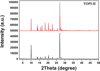
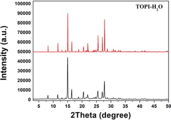
In addition, there are good agreements between the experimental powder diffraction pattern and the simulated pattern derived from the single-crystal data (Figures 3 and 4). The deviations of the unit-cell parameters and unit-cell volume of TOPI-II were between 0.07% and 0.08%. The deviations of the unit-cell parameters and unit-cell volume of TOPI-H2O were between 0.23% and 0.37%.
X-ray powder diffraction pattern (black line) and the simulated pattern of the crystal structure (red line) of TOPI-II.

X-ray powder diffraction pattern (black line) and the simulated pattern of the crystal structure (red line) of TOPI-H2O.

SUPPLEMENTARY MATERIAL
The supplementary material for this article can be found at https://doi.org/10.1017/S088571562200029X.
FUNDING INFORMATION
This work was financially supported from Zhejiang University Experimental Technology Research (SYB202103).