In developing countries, rheumatic heart disease continues to be a major public health problem and contributes to significant cardiac morbidity and mortality.Reference Rajamannan, Antonini-Canterin and Moura1
It is convincingly demonstrated that molecular mimicry between streptococcus pyogenes antigen and human proteins leads to autoimmune reactions, both humoral and cell mediated, causing rheumatic fever and rheumatic heart disease.Reference Essop and Nkomo2
Presence of inflammatory cells and increased expression of several cytokines in cases of end-stage rheumatic heart disease reflects a possible sub-clinical, ongoing insult, or inflammation to some unrecognised antigenic stimulus by beta haemolytic streptococcal antigens that have sensitised/primed the various target tissues and that further culminate in permanent valve damage.Reference Chopra and Gulwani3
There is a hypothesis that low-grade ongoing inflammation is a marker for subsequent cardiovascular disease. It leads to lipid peroxidation and increases the risk of atherosclerosis.Reference Ridker, Buring, Shih, Matias and Hnnekens4 But whether this will apply to children with rheumatic heart disease remains to be elucidated.
Aim of the work
To study the possibility of the presence of ongoing inflammation as detected by the high-sensitivity C-reactive protein in patients with chronic rheumatic heart disease, its relation to the degree of valvular involvement, and whether it imposes an extra risk for atherosclerosis in those patients.
Subjects and methods
This cross-sectional randomised study was conducted on patients attending the paediatric cardiology clinic, or on those admitted to the inpatient department, children's hospital, Ain Shams University. The patients were classified into the following groups:
Group one: 30 patients with chronic rheumatic heart disease, not in activity, excluding any patient having any current clinically detectable inflammatory condition. The mean and standard deviation of their ages were 12.63 and 3.12 years, respectively.
Group two: six patients with acute rheumatic carditis, diagnosed according to John's criteria.Reference Ferrieri5 The mean and standard deviation of their ages were 10.5 and 2.59 years, respectively.
Group three: 15 age- and sex-matched children served as a control group. The mean and standard deviation of their ages were 11.34 and 3.75 years, respectively.
All patients underwent a full history taking and thorough clinical examination laying stress on cardiac examination. Cardiac status was assessed by echocardiography to obtain the degree of valvular affection, left ventricular diameter as well as left ventricular systolic function by estimation of the ejection fraction (the normal value is 50–80%) and fractional shortening (normal value is 28–44%), using Vivid 7 Dimension, General Electric (Vingmed ultrasound AS N-3190 Horten, Norway). Laboratory work-up comprised the lipid profile (cholesterol, low-density lipoprotein, and high-density lipoprotein; Stanbio Laboratory, Texas, United States of America), assay of high-sensitivity C-reactive protein by enzyme-linked immune sorbent assay technique (Hyphen, Biomed, France; normal value ranges from 0.2 to 10 micrograms per millilitre), and homocysteine.
Homocysteine level in the serum samples was detected by the enzyme immunoassay kit provided by Axis (IBL, Hamburg, Germany). Protein-bound homocysteine was reduced to free homocysteine and enzymatically converted to S-adenosyl l-homocysteine in a separate procedure. The following solid phase enzyme immunoassay was based on competition between S-adenosyl l-homocysteine in the sample and immobilised S-adenosyl l-homocysteine bound to the wall of the microtitre plate for binding sites on the monoclonal anti S-adenosyl l-homocysteine antibody. After removal of the unbound anti S-adenosyl l-homocysteine antibody, a secondary rabbit anti-mouse antibody labelled with the enzyme horseradish peroxidase was added, in which its activity was measured spectrophotometrically. The absorbance is inversely proportional to the concentration of homocysteine. Levels of homocysteine were expressed in nanogram per decilitre.
Data collected and analysed using appropriate statistical methods, mean, and standard deviation, were used for parametric data and median and inter-quartile range were used for non-parametric data. Results were identified as significant when p-value less than 0.05 was reported.
Informed written consent was taken from the parents of all patients before enrolment in the study. The study was approved by the local ethics committee of Ain Shams University hospitals.
Results
This study was conducted on 36 patients whose mean and standard deviation of their ages were 12.63 years and 3.12 years, respectively, and whose mean and standard deviation of their duration of illness were 4.11 years and 2.86 years. None of the involved patients had a history suggestive of myocardial ischaemia.
Classification
Group one: 30 patients with chronic rheumatic heart disease, whose mean and standard deviation of their ages were 13.33 years and 3.01 years; 17 were females and 13 were males. The mean duration of their illness was 4.6 years. Echocardiography revealed the presence of mitral regurge in all of them, with a median degree of two and an inter-quartile range of 1.75. There were 10 of them who had aortic regurge as well with a median of two and an inter-quartile range of 0.75. Their left ventricular assessment revealed a mean and standard deviation of end-diastolic diameter of 4.59 centimetres and 0.57 centimetres, respectively, mean ejection fraction of 69.89%, and a mean fractional shortening of 39.72%. The pulmonary artery pressure measurement showed a mean and standard deviation of 33.61 and 8.15 millimetres of mercury, respectively.
Group two: six patients with active rheumatic carditis, whose mean and standard deviation of their ages were 10.5 and 2.59 years; four were females and two were males. The mean duration of their illness was 2.67 years. The echocardiographic study revealed mitral valve regurge in all of them, with a median grade of 3 and an inter-quartile range of 1, aortic valve regurge in all of them with a median of 1 and an inter-quartile range of 1, and significant tricuspid regurge in four of them with a median grade of 1 and an inter-quartile range of 0.75. The mean and standard deviation of their left ventricular end-diastolic diameter were 4.5 and 0.57 centimetres, respectively. Assessment of their left ventricular systolic function revealed a mean ejection fraction of 64.33% and a mean fractional shortening of 39.72%. The mean and the standard deviation values of their estimated systolic pulmonary artery pressure were 41.50 and 3.62 millimetres of mercury, respectively.
Group three: 15 age- and sex-matched children served as a control group. The mean and standard deviation of their left ventricular end-diastolic diameter were 3.6 and 0.67 centimetres, respectively; the mean of their left ventricular ejection fraction was 67.3% and fractional shortening was 30%; no valvular affection was detected in any of them.
None of the studied patients showed ventricular wall motion abnormality.
A comparison between patients with rheumatic carditis and patients with rheumatic heart disease showed significant elevation of systolic pulmonary artery pressure (t-value equals −2.270, p-value less than 0.05), grade of mitral valve regurge (z-value equal to −3.240, p-value less than 0.001), and significant decrease in fractional shortening (t-value equals 2.157, p-value less than 0.05) in patients with carditis.
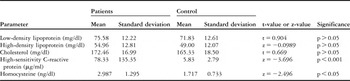
The parameters of lipid profile, as well as the levels of high-sensitivity C-reactive protein and homocysteine of whole patients were compared with those of the control group (Table 1).
Comparison between patients and controls as regards the lipid profile, high-sensitivity C-reactive protein, and homocysteine.

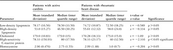
The lipid profile, as well as the levels of high-sensitivity C-reactive protein and homocysteine of patients with chronic rheumatic heart disease and acute rheumatic fever, is presented in Table 2.
Comparison between patients with active carditis and patients with chronic rheumatic as regards the lipid profile, high-sensitivity C-reactive protein, and homocysteine.

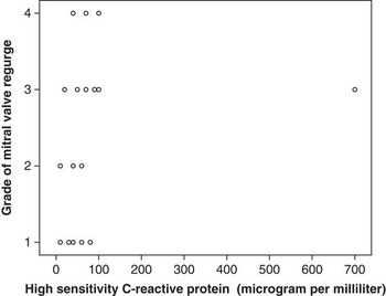
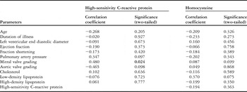
In all patients, the degree of aortic regurge correlated with the age (correlation coefficient equals 0.580, p-value less than 0.05), while the degree of mitral regurge correlated negatively with the ejection fraction (correlation coefficient equals −0.444, p-value less than 0.05), fractional shortening (correlation coefficient equals −0.484, p-value less than 0.05), and positively with the pulmonary artery pressure (correlation coefficient equals 0.42, p-value less than 0.05) and high-sensitivity C-reactive protein (correlation coefficient equals 0.480, p-value less than 0.05; Fig 1). The degree of valvular involvement did not show significant correlation with any of the parameters of the lipid profile or the level of homocysteine.
Correlation between high-sensitivity C-reactive protein and grade of mitral valve regurge.

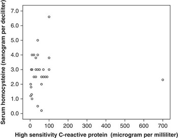
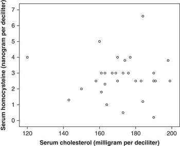
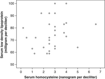
Correlation between high-sensitivity C-reactive protein, homocysteine, and all the studied parameters in patients will be presented in Table 3, and in Figures 2, 3, and 4.
Correlation between high-sensitivity C-reactive protein, homocysteine, and all other studied parameters.

Correlation by Spearman's test, correlation is significant at the 0.05 level (two-tailed)
Significant value is indicated in bold
Correlation between high-sensitivity C-reactive protein and homocysteine.

Correlation between serum cholesterol and serum homocysteine.

Correlation between homocysteine and low-density lipoprotein.

Discussion
This study was designed to assess the existence and extent of inflammation, lipid profile, and possible atherosclerotic risk in children with chronic rheumatic heart disease in reference to those in acute rheumatic activity as well as healthy children. That would evaluate the hypothesis of ongoing inflammation and subsequent premature atherosclerosis in children with chronic rheumatic heart disease and would give an idea about the need for long-term treatment.
The hepatocyte-derived acute phase reactant C-reactive protein was the subject of intense researches over the last two decades. The high-sensitivity CRP was found to be significantly increased in our studied patients with chronic rheumatic heart disease but to a lower extent than those with rheumatic activity.
Many authors reported high levels of high-sensitivity C-reactive protein either in acute or chronic rheumatic heart disease.Reference Chopra and Gulwani3, Reference Golbasi, Ucar and Keles6–Reference Davutoglu, Celik and Aksoy8 Golbasi et alReference Golbasi, Ucar and Keles6 reported that the levels of high-sensitivity C-reactive protein were higher in patients with chronic rheumatic heart disease than in patients with prosthetic valves and healthy participants, which may indicate that the inflammatory response persists in the chronic phase of rheumatic heart disease.
Furthermore, recent studies demonstrated the histological features of angiogenesis, inflammation, and bone formation within rheumatic valves explanted at surgical valve replacement from patients with rheumatic heart disease.Reference Rajamannan, Antonini-Canterin and Moura1, Reference Krasuski, Bush and Kay9
The levels of high-sensitivity C-reactive protein correlated with the degree of mitral regurge of our studied patients. Davutoglu et alReference Davutoglu, Celik and Aksoy8 reported that the chronic phase of rheumatic heart disease is associated with ongoing serum inflammatory mediators, which correlate strongly with the severity of valve involvement, valve scarring, and subsequent valve calcification.
C-reactive protein as a marker of ongoing inflammation appears to predict cardiovascular events. High levels of C-reactive protein was associated with a fourfold increase in risk of stroke and threefold increase in risk of myocardial infarction.Reference Hingorani, Shah, Casas, Humphries and Talmud10 Some studies also suggested that C-reactive protein might directly contribute to vascular damage due to its ability to stimulate tissue factor production by macrophages,Reference Zhang, Cliff, Schoefi and Higgins11 and its enhancement of complement activation after binding partly degraded non-oxidised low-density lipoprotein cholesterol.Reference de Beer, Soutar, Baltz, Trayner, Feinstein and Pepys12, Reference Bhakdi, Torzewski, Klouche and Hemmes13 Furthermore, the prevalence of coronary artery disease in adults with rheumatic valvular disease was found to be as high as 10.9%.Reference Li, Li and Hou14
In order to investigate the susceptibility of our studied patients to premature atherosclerosis, they were subjected to measurement of the parameters of lipid profile as well as homocysteine.
All the parameters of lipid profile of our patients did not differ significantly from the controls and they were not correlated with the levels of high-sensitivity C-reactive protein or with the level of homocysteine.
Panamonta et alReference Panamonta, Settasatinan, Kaplan and Chaikitpinyo15 reported normal parameters of lipid profile in patients with chronic rheumatic heart disease and those with acute rheumatic fever, except for the low-density lipoprotein cholesterol, which was significantly high in patients with acute rheumatic fever and correlated with the severity of carditis.
In adults with rheumatic heart disease, Yilmaz et alReference Yilmaz, Demirkan and Caldir16 found a significant correlation between total- to high-density lipoprotein cholesterol and the Wilkins score of the mitral valve. Furthermore, Antonini-Canteri et alReference Antonini-Canteri, Leiballi and Enache17 observed a positive effect of statin treatment in reducing the progression of rheumatic valvular affection but the underlying mechanism remains to be elucidated.
In this study, homocysteine was significantly elevated and to the same extent in patients with acute rheumatic fever and those with chronic rheumatic heart disease as compared to the controls. This result denoted that the atherosclerotic risk was not related only to the episode of acute inflammation but that it would continue and to the same extent even in the chronic quiescent state.
There are no previous data addressing how the levels of homocysteine would differ from children with acute rheumatic fever to those with chronic rheumatic heart disease.
Kurban et alReference Kurban, Mehmetoglu, Oran and Kiyici18 observed significantly high serum homocysteine levels in patients with acute rheumatic fever. They concluded that those increased levels could be considered as a sign of increased inflammation and oxidative stress in those patients.
The results of the study conducted by Wald et alReference Wald, Law and Marris19 strengthen the evidence that raised serum homocysteine is a cause of atherosclerosis and that a lowering of serum homocysteine can reduce the risk. There are several proposed mechanisms underlying the atherogenic effect of homocysteine, as a result of direct injury to endothelial cells, platelet activation, and affection of clotting factors.Reference Cleophas, Hornstra, van Hoogstraten and van der Meulen20
The results of this study signify that children with rheumatic heart disease who have evidence of chronic inflammation might be at risk of premature atherosclerosis, which could be preventable, but further follow-up of those patients for detection of any event out of atherosclerosis is needed. The main limitation of this study was the small sample size of the acute rheumatic fever group.
Conclusion
The inflammatory reaction which occurs in acute rheumatic fever persists in a lesser degree in the chronic phase of the disease and may precipitate the progression of valvular involvement, necessitating a more prolonged anti-inflammatory regimen. This ongoing cardiovascular inflammation may put those children at risk for premature atherosclerosis, which may indicate targeted therapy to lower the levels of homocysteine.