Introduction
Plagiorchis Lühe, 1899 is one of the most speciose genera in the family Plagiorchiidae Lühe, 1901. This genus comprises trematodes parasitising tetrapods, including humans. In most studies to date, the species identification of trematodes of the genus Plagiorchis is based on morphological characteristics. Due to the similarity of morphological characteristics of representatives of the genus from geographically remote regions, many Plagiorchis species are considered cosmopolitan (Besprozvannykh et al. Reference Besprozvannykh, Ermolenko and Nadtochy2012; Le et al. Reference Le, Ngo and Ermolenko2013; Skrjabin Reference Skrjabin1958).
More than 26 species of the genus have been identified from East Asia, with type localities of six of them located in this region (Besprozvannykh et al. Reference Besprozvannykh, Ermolenko and Nadtochy2012; Le et al. Reference Le, Ngo and Ermolenko2013; Oshmarin Reference Oshmarin1963; Skrjabin Reference Skrjabin1958). The species identification of all Plagiorchis representatives found in East Asia is based only on morphometric characteristics. In general, molecular data have been obtained for a very limited number of species despite the high species richness of the genus and the importance of using not only morphological data but also molecular characteristics for species identification of Plagiorchis, as emphasised by Boyce et al. (Reference Boyce, Hide, Craig, Reynolds, Hussain, Bodell, Bradshaw, Pickles and Rogan2014). By the time of our study, data for only eight species were available in the National Center for Biotechnology Information (NCBI) database. For six of them (Plagiorchis neomidis Brendow, 1970, Plagiorchis multiglandularis Semenov, 1927, Plagiorchis elegans (Rudolphi, 1802) Braun, 1902, Plagiorchis maculosus (Rudolphi, 1802) Lühe, 1899, Plagiorchis muelleri Tkach & Sharpilo, 1990, and Plagiorchis mordovii Schaldybin in Skrjabin & Antipin, 1958), nucleotide sequences were obtained from specimens collected at their type localities. For the remaining two species, Plagiorchis vespertilionis (Müller, 1780) Braun, 1900 and Plagiorchis koreanus Ogata, 1938, specimens were collected in an untypical region and were used to obtain molecular data (Kirillova et al. Reference Kirillova, Kirillov, Shchenkov, Knyazev and Vekhnik2024; Kundid et al. Reference Kundid, Pantoja, Janovcová and Soldánová2024; Sharifdini et al. Reference Sharifdini, Amin, Ashrafi, Karamzadeh, Mobedi, Rahmati and Hesari2020; Tkach et al. Reference Tkach, Pawlowski and Mariaux2000; Zikmundová et al. Reference Zikmundová, Georgieva, Faltýnková, Soldánová and Kostadinova2014).
We found adult trematodes of two species morphologically assignable to the genus Plagiorchis in the southern Far East of Russia. Specimens of one of them were found in the intestine of a Japanese large-footed bat (Myotis macrodactylus (Temminck, 1840)). Specimens of the other species were obtained through experimental infection of a golden hamster (Mesocricetus auratus (Waterhouse, 1839)) with metacercariae collected from a naturally infected stonefly.
Below are the results of morphological and molecular studies of the found trematodes.
Material and methods
Morphology data
Metacercariae (10 specimens) morphologically similar to those of the family Plagiorchiidae were found in a nymph of Paragnetina flavotincta (McLachlan, 1872) from the Pavlovka River (Ussuri River Basin) (44.16° N, 134.95° E) and fed to a golden hamster, M. auratus. On day 10 post-infection, three sexually mature trematodes were found in the hamster’s intestine. In addition, 10 sexually mature trematodes were found in the intestine of a Japanese large-footed bat, M. macrodactylus, captured in the Serafimovskaya Cave (43.52° N, 135.15° E). The adult trematodes were fixed in 70% ethanol and then transferred to 96% ethanol for storage. Whole mounts were using from two trematode specimens from the hamster and five specimens from the bat. One of the three trematodes experimentally reared in the golden hamster and one of the 10 sexually mature trematodes from the naturally infected bat were used for molecular studies. The whole mounts of adult specimens were prepared by staining with alum carmine, dehydration in a graded ethanol series, clearing in clove oil, and embedding in Canada balsam. All measurements were in micrometres (μm).
All applicable international, national, and institutional ethical guidelines for the care and use of animals in research were followed. Approval was granted by the Commission for the Regulation of Experimental Research (Bioethics Commissions) of the Federal Scientific Center of the East Asia Terrestrial Biodiversity (FSCEATB), Far Eastern Branch, Russian Academy of Sciences (FEB RAS) (dated March 12, 2025, No. 5).
Euthanasia of the hamster was carried out in accordance with the decision of the Committee on the Ethics of Animal Experiments, FSCEATB FEB RAS (Permit No. 1 of April 25, 2022).
Molecular data
DNA extraction, amplification, and sequencing
Genomic DNA was extracted from the trematode specimens by the HotSHOT technique (Truett et al. Reference Truett, Heeger, Mynatt, Truett, Walker and Warman2000). Partial sequences of the 28S rRNA gene (28S) nuclear DNA and partial sequences of the cox1 gene (cox1) of mitochondrial DNA were amplified by polymerase chain reaction (PCR) using specific primers (Table S1, Supplementary materials). Both external and internal primers were used for sequencing (Table S1, Supplementary materials). The annealing temperature was 57°C for the 28S marker and 51°C for the cox1 marker. The efficiency and contamination of PCR was tested by setting positive and negative controls, respectively. The PCR products were sequenced by the Sanger method using a BigDye Terminator Cycle Sequencing kit (Applied Biosystems, USA). Nucleotide sequences were determined on an ABI 3500 genetic analyser (Applied Biosystems, USA) at the FSCEATB FEB RAS, Russia.
Analysis of genetic data
Processing and alignment of consensus sequences were carried out using the FinchTV 1.4 and MEGA 5.0 programs (Tamura et al. Reference Tamura, Peterson, Peterson, Stecher, Nei and Kumar2011). Sequences for the 28S marker that were less than 1,000 bp in length were removed from the analysis. The p-distances between and within species were analysed in the MEGA software without including indels. Phylogenetic relationships within the family Plagiorchiidae were assessed independently for both markers using newly obtained sequences and data for all species deposited in GenBank. The members of the trematode families located left (basal) of the Plagiorchiidae in the phylogenetic tree constructed by Olson et al. (Reference Olson, Cribb, Tkach, Bray and Littlewood2003) using nucleotide sequences of the 28S and 18S rRNA genes were selected as outgroup in our study. The list of samples used in the study is provided in Supplementary materials, Tables S2–S3.
Phylogenetic relationships were reconstructed using the Bayesian Inference (BI) algorithm in the MrBayes 3.1.2. program (Ronquist and Huelsenbeck Reference Ronquist and Huelsenbeck2003) by applying a model selected as optimal on the basis of the Akaike information criterion (AIC) in the jModeltest 2.1.7 program (Darriba et al. Reference Darriba, Taboada, Doallo and Posada2012): TVM+G for 28S and TrN+I+G for cox1. The method of posterior probabilities was used for the trees constructed by the BI algorithm. In the BI analysis, 600,000 and 7,500,000 generations of the Markov chain Monte Carlo posterior part third (MCMC) were simulated for 28S and cox1, respectively. The number of generations was determined as sufficient, since the SD value calculated was ˂0.01. The chain was sampled every 100 generations. Of the samples assessed, 25% were excluded to permit the construction of consensus trees.
Results
Morphological data
Plagiorchis koreanus Ogata, 1938
Definitive host: Myotis macrodactylus.
Site: small intestine.
Locality: 43.52° N, 135.15° E, Primorsky Krai, southern Far East of Russia.
Adult (based on five specimens) (Figure 1A, Table 1). Body elongated, with tapered anterior end. Tegument with spines. Oral sucker subterminal, round. Ventral sucker rounded, smaller than oral sucker. Prepharynx absent. Pharynx well-developed, rounded, adjacent to oral sucker. Oesophagus short. Intestinal bifurcation in middle between pharynx and ventral sucker. Caeca slightly not reaching posterior end of body. Testes two, oval, located in second half of body, tandem, separated. Cirrus-sac elongated, extending from level of ovary to middle of distance between intestinal bifurcation and ventral sucker. Internal seminal vesicle elongated-saccular. Genital pore medial. Cirrus long, unarmed. Ovary rounded, at end of anterior half of body, separated from ventral sucker and anterior testis. Uterus reaching posterior end of body and partially overlapping anterior and posterior testes. Eggs oval, small. Vitellarium from two lateral fields, overlapping caeca. Vitellarium fields extending from level of posterior edge of ventral sucker and slightly not reaching level of posterior ends of caeca.
Measurements of adult Plagiorchis (μm)

Adult worm of A, Plagiorchis korеanus; and B, Plagiorchis cf. eutamiatis. Scale-bars: μm.

Plagiorchis cf. eutamiatis Schulz, 1932
Definitive host: Mesocricetus auratus (experimentally).
Site: small intestine.
Second intermediate host: Paragnetina flavotincta.
Site: visceral muscle tissue.
Locality: Pavlovka River, 44.16° N, 134.95° E, Primorsky Krai, southern Far East of Russia.
Adult worm (based on two specimens) (Figure 1B, Table 1). Body flat, elongated, with rounded anterior and posterior ends. Tegument with spines. Oral sucker subterminal, rounded. Ventral sucker rounded, smaller than oral sucker. Prepharynx and oesophagus absent. Pharynx rounded, adjacent to oral sucker. Intestinal bifurcation immediately posteriorly to pharynx. Caeca slightly not reaching posterior end of body. Testes two, rounded or oval, in posterior half of body, tandem, separated. Cirrus-sac elongated, curved, running around ventral sucker on right and partially covered by ventral sucker, with distal end located at level of ovary, sinisterly to it. Internal seminal vesicle elongate-saccular. Genital pore immediately anteriorly of ventral sucker. Cirrus long, unarmed. Ovary rounded, between ventral sucker and anterior testis, dextrally to ventral sucker, separated from ventral sucker and anteriorly of testis. Uterus reaches posterior end of body and partially covers anterior and posterior testes. Eggs oval, small. Vitellarium from two lateral fields, overlapping caeca. Vitellarium fields extending from level of oral sucker to posterior body end. Vitellarium fields merge at level of prepharynx and intestinal bifurcation and post-testicularly.
Molecular data
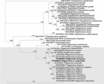
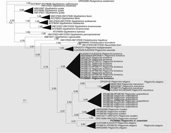
For the reconstruction of phylogenetic trees, aligned sequences of 1,085 and 300 bp were used for 28S and cox1, respectively. In the phylogenetic reconstruction based on both nuclear and mitochondrial markers, the studied trematodes included on a branch combining trematodes of the genus Plagiorchis (Figures 2 and 3). The nucleotide sequence we obtained from a trematode from the naturally infected bat, M. macrodactylus, was identical to the sequences of P. koreanus from the NCBI database for the nuclear marker (Table 2). The genetic distances between the nucleotide sequence from the trematode from the bat and the sequence of P. koreanus from NCBI were at an intraspecific level for the mitochondrial marker (Table 3). The nucleotide sequence from the trematode from the experimentally infected hamster, M. auratus, was formed a separate branch in both reconstructions (Figures 2 and 3). The genetic distances between the specimen from the hamster and those of other Plagiorchis members represented in the reconstruction were at an interspecific level (Tables 2 and 3).
Genetic distances between and within species of the genus Plagiorchis based on 28S sequences

Notes: Interspecific distances of 0.4–2.1% were obtained in the present study. Standard error values are highlighted in bold.
Genetic distances between and within species of the genus Plagiorchis based on cox1 sequences

Notes: Interspecific distances of 8.5–17.8% were obtained in the present study. Standard error values are highlighted in bold.
Phylogeny of the family Plagiorchiidae based on 28S sequences using the Bayesian algorithm. Numerals at the nodes are a posterior probability values of ≥50. The scale bar indicates the number of substitutions per site.

Phylogeny of the family Plagiorchiidae based on cox1 sequences using the Bayesian algorithm. Numerals at the nodes are a posterior probability values of ≥50. The scale bar indicates the number of substitutions per site.

Discussion
Cosmopolitan of P. koreanus species
The trematodes obtained from the bat, M. macrodactylus, have appeared to be morphometrically identical to P. koreanus (Table 1). This species was first discovered in the East Asian region, specifically in North Korea (Ogata, 1938, cited by Skrjabin Reference Skrjabin1958). Subsequently, trematodes morphologically similar to those described by Ogata (1938), cited by Skrjabin (Reference Skrjabin1958), and designated as P. koreanus were repeatedly recorded both from the European region (Kirillov et al. Reference Kirillov, Kirillova and Vehknik2012; Kirillova et al. Reference Kirillova, Kirillov and Vehknik2007; Krasnolobova Reference Krasnolobova1987) and from East Asia (Besprozvannykh et al. Reference Besprozvannykh, Ermolenko and Nadtochy2012; Orlovskaya et al. Reference Orlovskaya, Dokuchaev, Atrashkevich and Lazutkin2022). The morphological similarity of the specimens identified as P. koreanus and found in different parts of Eurasia allows considering this trematode as a cosmopolitan species. However, the great distance between the localities where these specimens were sampled and identified as P. koreanus (Europe and East Asia) may be strong evidence for their different species. The present study has shown that the morphological similarity does not guarantee that trematodes can be assigned to the same species (Besprozvannykh et al. Reference Besprozvannykh, Atopkin, Ngo, Beloded, Ermolenko, Ha and Tang2015; Izrailskaia et al. Reference Izrailskaia, Besprozvannykh and Tatonova2021; Shumenko et al. Reference Shumenko, Tatonova and Besprozvannykh2017; Tatonova et al. Reference Tatonova, Shumenko and Besprozvannykh2018). Thus, the question as to the correctness of species identification of the European trematodes listed as P. koreanus remains open, and the answer may be addressed by studying molecular characteristics of both European and East Asian trematodes. Until the present study, molecular data have been obtained only for European specimens (Tables S1, S2, Supplementary materials), which, in the absence of such data for East Asian trematodes, does not clarify whether they belong to the same or different species. According to our phylogenetic reconstructions based on nuclear and mitochondrial markers, the specimens we obtained in the present study are combined with other specimens from NCBI referred to as P. koreanus into separate clades with a high a posteriori probability (Figures 2 and 3). The genetic distances between these specimens are minor absent for the 28S marker and are 0.2% for the cox1 marker, which indicates intraspecific variability within genus (Tables S2 and S3, Supplementary materials). Thus, our molecular data confirm the hypothesis, based on morphological characteristics, that P. koreanus is a cosmopolitan species distributed across Eurasia.
Taxonomic identification and phylogenetic relationships of P. cf. eutamiatis
The adult trematodes that we obtained from metacercariae - naturally infected stonefly, in terms of their morphometric characteristics, have the greatest similarity with P. multiglandularis and Plagiorchis eutamiatis Schulz, 1932 (Table 1). The trematodes from our material almost do not differ from the above-mentioned species in morphometric characteristics. However, trematodes P. multiglandularis were first recorded from birds (two species of woodpeckers and the lesser grey shrike) in Belarus (Semenov, 1927, cited in Krasnolobova Reference Krasnolobova1987; Skrjabin Reference Skrjabin1958), whereas P. eutamiatis was described from the Siberian chipmunk, Eutamias asiaticus (Laxmann, 1769), in the European region (Skrjabin Reference Skrjabin1958). These trematodes have subsequently been found in birds and chipmunks within the East Asian region (Besprozvannykh et al. Reference Besprozvannykh, Ermolenko and Nadtochy2012; Orlovskaya and Dokuchaev Reference Orlovskaya and Dokuchaev2021). Thus, the preference for particular taxonomic groups of definitive hosts shown by the trematodes P. multiglandularis and P. eutamiatis is the only characteristic (in the absence of molecular data for P. eutamiatis) to differentiate these species, in our opinion. The ability of the trematodes we studied to infect mammals may indicate that they belong to P. eutamiatis. Taking into account this fact and the possible cosmopolitan distribution of P. eutamiatis (similar to P. koreanus), in the absence of molecular evidence for trematodes of this species from the type locality, we refer to the trematodes from our material as Plagiorchis cf. eutamiatis. The genetic distances between Plagiorchis cf. eutamiatis and other Plagiorchis species (including P. multiglandularis) in nuclear and mitochondrial markers fit into the interspecific range (0.8–1.7 and 14.0–17.8%, respectively), thus, confirming the independence of the trematodes found (Tables S2 and S3, Supplementary materials).
Taxonomic identification of Plagiorchis based on cercariae
It is important to note that in the phylogenetic reconstruction based on the cox1 marker, the specimen designated as P. multiglandularis forms a separate clade with the specimen identified as P. neomidis (Figure 3). The genetic distance between the specimens within the clade is at an intraspecific level (0.6%). Nucleotide sequences for P. multiglandularis and P. neomidis were obtained from cercariae (Gacad et al. Reference Gacad, Yurlova, Ponomareva and Urabe2023; Zikmundová et al. Reference Zikmundová, Georgieva, Faltýnková, Soldánová and Kostadinova2014). Gacad et al. (Reference Gacad, Yurlova, Ponomareva and Urabe2023) differentiated P. multiglandularis and P. neomidis on the basis of number of pairs of penetration gland in cercariae: six pairs vs. seven pairs, respectively. However, the identification of trematode species based solely on cercariae morphology is not always reliable, because morphologically similar cercariae may appear belonging to different species. In addition, it is challenging to count the number of penetration glands in cercariae. The results may not be correct, and it may appear that the specimens identified as P. multiglandularis and P. neomidis are actually from the same species. The suggestions above are supported by molecular data obtained from cercariae identified as P. elegans. In the phylogenetic reconstruction based on mitochondrial marker, the majority of P. elegans specimens form a separate clade with a high a posteriori probability (Figure 3). Except for the specimen OP429100 of P. elegans, it forms a separate branch. The genetic distance between specimen OP429100 of P. elegans and the other P. elegans specimens in the reconstruction is 11.3%. The latter indicates the trematodes from different species, and, accordingly, the question as to which of the specimens referred to as P. elegans belong to this species remains open.
Conclusion
In this study, we have obtained, for the first time, morphological and molecular characteristics for P. koreanus from the type region. Our results confirm that the trematodes belong to a cosmopolitan species. Based on the morphological similarity, the definitive host (mammal), and the differentiation of the East Asian trematode from other Plagiorchis species by nuclear and mitochondrial markers, we have referred to the trematodes from the hamster experimentally infected with metacercariae from a naturally infected stonefly as Plagiorchis cf. eutamiatis. The question of species identification of these specimens can be addressed only by obtaining molecular data for P. eutamiatis from the type locality.
Supplementary material
The supplementary material for this article can be found at http://doi.org/10.1017/S0022149X25100527.
Financial support
The research was carried out within the state assignment of Ministry of Science and Higher Education of the Russian Federation (themes No. 121031000154-4 and 0207-2024-0020) and government assignment no. 123022200035-0 ‘The structure of natural foci of parasitic diseases in the south of the Russian Far East’.
Competing interests
The authors declare no conflict of interest.
Ethical standard
The authors state that all procedures contributing to this work comply with the ethical standards of the relevant national and institutional guides on the care and use of laboratory animals including birds and mammals.
Data availability
The data supporting the findings of this study are available within the article. All newly generated sequences were deposited in the GenBank database under the following accession numbers: PV299262, PV299263, PV300861, and PV300862.